Premium Bangalore Call Girls Jigani Dail 6378878445 Escort Service For Hot Ma...
Article of Reconstruction of cleft lip and palate defect
1. Reconstruction of Cleft Lip and Palate Defect
Amin Abusallamah
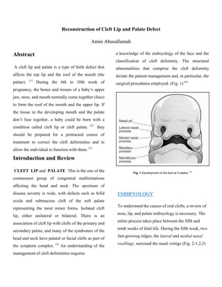
Abstract a knowledge of the embryology of the face and the
classification of cleft deformity. The structural
A cleft lip and palate is a type of birth defect that abnormalities that comprise the cleft deformity
affects the top lip and the roof of the mouth (the dictate the patient management and, in particular, the
(1)
palate). During the 6th to 10th week of surgical procedures employed. (Fig. 1) (4).
pregnancy, the bones and tissues of a baby’s upper
jaw, nose, and mouth normally come together (fuse)
to form the roof of the mouth and the upper lip. If
the tissue in the developing mouth and the palate
don’t fuse together, a baby could be born with a
(2)
condition called cleft lip or cleft palate. they
should be prepared for a protracted course of
treatment to correct the cleft deformities and to
allow the individual to function with them. (3)
Introduction and Review
CLEFT LIP and PALATE This is the one of the Fig. 1 Development of the face at 5 weeks. (4)
commonest group of congential malformations
affecting the head and neck. The spectrum of
disease seventy is wide, with defects such as bifid EMBRYOLOGY
uvula and submucous cleft of the soft palate
To understand the causes of oral clefts, a review of
representing the most minor forms. Isolated cleft
nose, lip, and palate embryology is necessary. The
lip, either unilateral or bilateral. There is an
entire process takes place between the fifth and
association of cleft lip with clefts of the primary and
tenth weeks of fetal life. During the fifth week, two
secondary palate, and many of the syndromes of the
fast-growing ridges, the lateral and medial nasal
head and neck have palatal or facial clefts as part of
(3) swellings, surround the nasal vestige (Fig. 2-1,2,3)
the symptom complex. An understanding of the
management of cleft deformities requires
2. The lateral swellings will form the alae of the nose;
the medial swellings will give rise to four areas: (1)
the middle portion of the nose, (2) the middle
portion of the upper lip, (3) the middle portion of
the maxilla, and (4) the entire primary palate.
Simultaneously the maxillary swellings will
approach the medial and lateral nasal swellings bul
remain separated from them by wellmarked Fig. 2.3: Scanning electron micrograph of a embryo at A. 6-
week embryo. The nasal prominences are gradually separated
grooves.
from the maxillary prominence by deep furrows.. B. 7-week
embryo. Maxillary prominences have fused with the medial
nasal prominences. (5)
During the next 2 weeks, the appearance of the face
changes considerably. The maxillary swellings
continue to grow in a medial direction and compress
the medial nasal swellings toward the midline.
Subsequently these swellings simultaneously merge
Fig. 2.1: Frontal aspect of the face. A. 5-week embryo. B. 6- with each other and with the maxillary swellings
week embryo. The nasal prominences are gradually separated
from the maxillary prominence by deep furrows. (5) laterally. Hence the upper lip is formed by the two
medial nasal swellings and the two maxillary
swellings. The two medial swellings merge not only
at the surface but also at the deeper level. The
structures formed by the two merged swellings are
known together as the intermaxillary segament (Fig.
3) which is comprised of three components: (1) a
labial component, which forms the philtrum of the
upper lip; (2) an upper jaw component, which
Fig. 2.2: Frontal aspect of the face. A. 7-week embryo.
Maxillary prominences have fused with the medial nasal carries the- four incisor teeth; and (3) a palatal
prominences. B. 10-week embryo. (5) component, which forms the triangular primary
palate. Above, the intermaxillary segment is
continuous with the nasal septum, which is formed
by the frontal prominence.
3. Two shelflike outgrowths from the maxillary.
swellings form the secondary palate. These palatine
shelves appear in the sixth week of development
and are directed obliquely downward on either side
of the tongue. In the seventh week, however, the
palatine shelves ascend to attain a horizontal
position above the tongue and fuse with each other,
thereby forming the secondary palate. Anteriorly
the shelves fuse with the triangular primary palate,
and the incisive foramen is formed at this junction.
At the same time, the nasal septum grows down and
joins the superior surface of the newly formed
palate. The palatine shelves fuse with each other
and with the primary palate between the seventh
and tenth weeks of development. Clefts of the FIG.3: A, Frontal section through head ( 61 /a-weeK-old
primary palate result from a failure of me.soderm to embryo. Palatine shelves are located fn vertical position
on each side of tongue, B, Ventral view of same, Note
penetrate into the grooves between the medial nasal clefls between primary triangular palate and palatine
ihelves, which arc itill in vertical position. C, Frontal
and maxillary processes, which prohibits their
section through head of 7V2-week-old embryo, Tongue
merging with one another. Clefts of the secondary has rtiovrd downward, and palatine shelves have
reached horizontal position. D, Ventral view of same.
palate are caused by a failure of the palatine shelves Shelves are in horizontal position, E, Frontal section
to fuse with one another. The causes for this arc? through head of 10-week-old embryo. Two paiatine
shelves have fused with each other and with nasal
Speculative and include failure of the tongue to septum. F, Ventral view of 5ame.
(5)
descend into the oral cavity. (5)
INCIDENCE
Cleft lip = 1: l000,
Cleft palate (isolated) = 0.45: l000,
Complete cleft = 1:1800,
Submucous cleft = 1:1200, and
Bifid uvula = 1: l00. (3)
4. GENETICS AND ETIOLOGY CLASSIFICATION
Clefts of the upper lip and palate are the most The typical classification system used clinically to
common major congenital craniofacial abnormality describe standard clefts of the lip and palate is
and are present in approximately 1 in 700 live based on careful anatomic description. Clefts can be
births. (6)
The causes of facial clefting have been unilateral or bilateral; microform, incomplete, or
extensively investigated. The exact cause of clefting complete; and may involve the lip, nose, primary
is unknown in most cases. For most cleft conditions, palate, and/or secondary palates (Fig. 5). (12)
no single factor can be identified as the cause.
However, it is important to distinguish between
isolated clefts (in which the patient has no other
related health problem) and clefts associated with
other birth disorders or syndromes. (7) Instead clefts
are thought to be of a multifactorial etiology with a
number of potential contributing factors. These
factors may include chemical exposures, radiation,
maternal hypoxia, teratogenic drugs, nutritional
deficiencies, physical obstruction, or genetic
influences. One prevailing theory relates the process
of clefting as a threshold in which multiple factors
come together to raise the individual above a FUNCTIONAL CONSEQUENCES
threshold at which time the mechanism of fusion
To regard a cleft as a deformity only is inadequate.
fails.# Recently multiple genes have been
This malformation causes of functional disturbances
implicated in the etiology of clefting. Some of these
that indispensably should be taken into
genes include the MSX, LHX, goosecoid, and DLX
(8) (9) (10)
consideration in determining appropriate treatment.
genes. some Syndromes Associated with (13)
Cleft Palate like: Marfan syndrome , Apert
1. Nutrition Difficulties: Nutrition in cleft infants
syndrome , Downs syndrome , Digeorge syndrome ,
has often been described as difficult because of
Edward syndrome , Treacher collins , Pierre Robin
their incompetence in sucking. In reality, babies
and Stickler syndrome. (11)
need to suck only to position the nipple, and for
5. drinking they “milk” the nipple with their tongue. tongue position but malposition of teeth and gaps in
Nutrition difficulties are caused by the tongue the dental arches may contribute. (13) Voice diseases
position in the cleft palate, occluding the nasal may occur accidentally or following attempts to
airway. Therefore, cleft babies are unable to drink compensate for speech disturbances. Myofunctional
and breathe at the same time. (13) imbalances are the result of the displaced tongue
2. Hearing Disorders: By swallowing , the with its dislocated functional pressure and the
Eustachian tubes are opened and the secretions of dysharmonious interaction of tongue and orofacial
the middle ear can flow off. In the presence of a musculature. (20) Mimic movements are the attempt
cleft palate, however, the velopharyngeal ring by cleft patients to compensate for velopharyngeal
(14)
muscle system is interrupted and the insertion of incompetence by grimace with labial, nasal, or
the tensor veli palatin muscles at the tube cartilage frontal muscles. Speech therapy actually starts with
(15) (16)
is abnormal. In consequence, the opening breast-feeding, which demands considerable effort
mechanism of the Eustachian tubes cannot work. by the child and provides practice for the
The middle ear secretions become thickened and are musculature. Later on, stimulation and
(17)
congested. The resulting seromucotympanon myofunctional therapy create favorable general
hinders sound conduction and promotes middle ear conditions for speech development. (21) (22)
infections. Speech development relies on hearing 4. Growth Disturbances: Maxillary hypoplasia
(18)
and in cleft infants, who usually suffer some and scarring from surgery are said to be responsible
speech problems anyway, defective hearing will for growth disturbances. Often the influence of
mean a double handicap. Moreover, in hearing muscle action on growth of bony structures is not
(23)
disorders the maturation of the hearing tracts to the considered, although it plays an important role.
central nervous system is retarded. (19) This was recognized in 1961 by Rosenthal, who
3. Speech Problems: Nasality, articulation described the functional stimulation on the clefted
problems, suprapalatal resonance, voice diseases, maxilla by early reconstruction of soft palate
myofunctional imbalances, and mimic movements muscles. (24)
may all be manifested in the speech problems of 5. Psychological Problems A number of studies
cleft patients. Nasality is the result of have examined the influence that a cleft lip and/or
velopharyngeal incompetence caused by shortness palate has on the affected children themselves, their
or inadequate activity of the soft palate, which itself families, and the general public including health care
is based on the displaced muscle insertions. professionals. (13)
Articulation problems originate from a posterior
6. RECONSTRUCTION OF and alveolar structures during the presurgical phase
CLEFT LIP AND PALATE of treatment (Fig.6). Although the use of appliances
probably makes for an easier surgical repair. (26) (27)
The timing of cleft lip and palate repair is
controversial. Despite a number of meaningful
advancements in the care of patients with cleft lip
and palate, a lack of consensus exists regarding the
timing and specific techniques used during each
stage of cleft reconstruction. Surgeons must
continue to carefully balance the functional needs,
esthetic concerns, and the issue of ongoing growth
when deciding how and when to intervene.(12) Table .1
FIG.6 :Frontal and lateral views of the Grayson nasoalveolar
molding appliance showing the nasal projections that help to
theoretically mold the nasal cartilages and maxillary segments
into a more appropriate configuration prior to repair. (12)
Unilateral Cleft Lip Repair
Clefts of the lip and nose that are unilateral present
with a high degree of variability, and thus each
repair design is unique (Figure 7).The repair
technique is usually performed after 10 weeks of
Cleft Lip and Palate Repair age. The basic premise of the repair is to create a
Presurgical Taping and Presurgical Orthopedics three-layered closure of skin, muscle, and mucosa
Facial taping with elastic devices is used for that approximates normal tissue and excises
application of selective external pressure and may hypoplastic tissue at the cleft margins. Critical in
allow for improvement of lip and nasal position the process is the reconstruction of the orbicularis
(25)
prior to the lip repair procedure. Some surgeons oris musculature into a continuous sphincter. The
prefer presurgical orthopedic (PSO) appliances Millard rotation-advancement technique has the
rather than lip taping to achieve the same advantage of allowing for each of the incision lines
goals.81,82 PSO appliances are composed of a to fall within the natural contours of the lip and
custom-made acrylic base plate that provides nose. This is an advantage because it is difficult to
improved anchorage in the molding of lip, nasal, achieve “mirror image” symmetry in the unilateral
7. cleft lip and nose with the normal side immediately Bilateral Lip Repair
adjacent to the surgical site. Primary nasal Bilateral cleft lip repair can be one of the most
reconstruction may be considered at the time of lip challenging technical procedures performed in
repair to reposition the displaced lower lateral children with clefts. The lack of quality tissue
cartilages and alar tissues. Several techniques are present and the widely displaced segments are
advocated, and considerable variation exists with major challenges toachieving exceptional results,
respect to the exact nasal reconstruction performed but superior technique and adequate mobilization
by each surgeon. The primary nasal repair may be ofthe tissue flaps usually yields excellentesthetic
achieved by releasing the alar base, augmenting the results (Figs.8,9). Additionally the columella may
area with allogeneic subdermal grafts, or even a be quiteshort in length, and the premaxillary
formal open rhinoplasty. Since lip repair is done at segment may be significantly rotated. Adequate
such an early point in growth and development, the mobilization of the segments and attention to the
authors prefer minimal surgical dissection due to the details of only using appropriately developed tissue
effects of scarring on the subsequent growth of these will yield excellent results even in the face of
tissues. McComb described a technique that has significant asymmetry. Some surgeons have used
become popular, consisting of dissecting the lower aggressive techniques to surgically lengthen the
lateral cartilages free from the alar base and the columella and preserve hypoplastic tissue using
surrounding attachments through an alar crease banked fork flaps. Early and aggressive tissue flaps
incision. This allows the nose to be bolstered and/or in the nostril and columella areas do not look
stented from within the nostril to improve symmetry. natural after significant growth has occurred and
result in abnormal tissue contours. While surgical
attempts at lengthening the columella may look
good initially, they frequently look abnormally long
and excessively angular later in life (Fig.11).
Revision of these iatrogenic deformities is difficult
and some of the contour irregularities will not be
able to be revised adequately. Usually if the
hypoplastic tissue is excised and incisions within
the medial nasal base and columella are avoided,
FIG. 7: A complete unilateral cleft of the lip is shown
highlighting the hypoplastic tissue in the cleft site that is not the long-term esthetic results are excellent.
used in the reconstruction. (12)
8. (2) the anatomic repair of the musculature within
the soft palate that is critical for normal creation of
speech (Fig.10).
FIG.10:A, Presurgical frontal view of a wide bilateral cleft lip
and palate with significant asymmetry and lack of columella
FIG.8:The bilateral cleft of the lip and maxilla shown here is length. B, Presurgical left lateral view of a wide bilateral cleft
complete and highlights the hypoplastic tissue along the cleft lip and palate with a protrusive premaxillary segment. Note
edges. the short columella length. C, The same child at 10 months of
age after repair of her bilateral cleft lip and palate. No
presurgical taping or orthopedic appliances were used.
The exact timing of repair of a palate cleft is
controversial. Generally the velum must be closed
prior to the development of speech sounds that
require an intact palate. (12) On average this level of
speech production is observed by about 18 months
of age in the normally developing child. If the repair
is completed after this time, compensatory speech
FIG.9: A, Presurgical appearance of the incomplete bilateral articulations may result. Repair completed prior to
cleft lip of a 3-month-old boy. B, Surgical markings for
excision of the hypoplastic tissue and the planned creation of this time allows for the intact velum to close
a new philtrum. Advancement flaps from the lateral lip
segments bring good white-roll to the midline via small effectively, appropriately separating the
cutbacks. C, The same child at 1 year of age after the repair of
his bilateral cleft lip. nasopharynx from the orophayrynx during certain
speech sounds. (28) (29) (30) When repair of the palate
is performed between 9 and 18 months of age, the
Cleft Palate Repair
incidence of associated growth restriction affecting
There are two main goals of cleft palate repair the maxillary development is approximately
during infancy: (1) the water-tight closure of the 25%. (31) (32) (33) Cleft palate reconstruction requires
entire oronasal communication involving the hard the mobilization of multilayered flaps to reconstruct
and soft palate. the defect due to the failure of fusion of
9. the palatal shelves. space between the nasopharynx preserves an anterior pedicle for increased blood
and oropharynx during certain speech sounds, the supply to the flaps. (34; 35; 36) This technique is also
surgeon must also reconstruct the musculature of successful in achieving a layered closure but may be
the velopharyngeal mechanism. The musculature of more difficult when suturing the nasal mucosa near
the levator palatini is abnormally inserted on the the anteriorly based pedicle attachments. Another
posterior aspect of the hard palate and therefore common technique is the Furlow double-opposing Z
must be disinserted and reconstructed in the plasty, which attempts to lengthen the palate by
midline. Many techniques for repair like; The taking advantage of a Z-plasty technique on both
Bardach two-flap palatoplasty uses two large nasal mucosa and oral mucosa (Fig.12). (36) (37)
fullthickness layered dissection and brought to the
midline for closure (Fig.11).
Fig.11: A, A unilateral cleft of the primary and secondary Fig.12: A, A complete cleft of the secondary palate (both
palates, typical involvement from the anterior vestibule to the hard and soft) is shown from the incisive foramen to the uvula.
uvula. B, The Bardach palatoplasty technique requires two B, The Furlow double opposing Z-plasty technique requires
large full-thickness mucoperiosteal flaps to be elevated from that separate Z-plasty flaps be developed on the oral and then
each palate shelf. The anterior of the cleft is not reconstructed nasal side. C, The flaps are then transposed to theoretically
until the mixed dentition stage. C, A layered closure is lengthen the soft palate. A nasal side closure is completed in
performed palatoplasty by reapproximating the nasal the standard fashion anterior to the junction of the hard and
mucosa. D, Once the nasal mucosa and musculature of the soft palate. D, The oral side flaps are then transposed and
soft palate are approximated, the oral mucosa is closed in the closed in a similar fashion completing the palate closure. (12)
midline. (12)
flaps that are mobilized with The von Langenbeck
technique is similar to the Bardach palatoplasty but
10. Bone Grafting References:
At one time, bone grafting tended to be performed
1. NHS inform. www.nhsinform.co.uk. [Online] NHS, October
only in bilateral alveolar clefts to fix a mobile 4, 2011. http://www.nhsinform.co.uk/health-
premaxilla to the maxilla. This reason remains library/articles/c/cleft-lip-and-palate/introduction.
valid.81 It has however now been extended to 2. cleft smile. www.cleftsmile.org. [Online] Cleft Lip and
Palate Foundation of Smiles Inc., april 16, 2011.
include the achievement of a continuous alveolus in http://www.cleftsmile.org/custom/introduction/.
all cleft patients with an alveolar defect. For those
3. RIDEN, K. Key Topics in Oral and Maxillofacial Surgery.
patients who will subsequently need a maxillary Plymouth, UK : BIOS Scientific PublisherLs td, 1998. p. 69.
ISBN 1 85996 030 8.
osteotomy, it creates the possibility of that
procedure being carried out more safely, and 4. Wray, David, et al. Textbook for General and Oral
Surgery. London : Elsevier Science, 2003. pp. 130 - 139. ISBN
perhaps with more stability, in one piece. (13) 0 4430 7083 0.
5. Sadler, T.W. Langman's Medical embryology. s.l. :
Lippincott Williams & Wilkins, 2003. ISBN: 0781743109.
Conclusions
6. Classification and birth prevalence of orofacial clefts.
The complete care of patients with clefts requires an Tolarova MM, Cervenka. 2, San Francisco : Am J Med Genet,
interdisciplinary approach that demands precise 1998, Vol. 75, pp. 126–37. 1552-4833.
surgical execution of the different procedures 7. Ellis, Edward. Management of Patients with Orofacial
Clefts. [book auth.] Edward Ellis III, Myron R. Tucker James
essential to correct cleft deformities, as well as R. Hupp. Contemporary Oral and Maxillofacial Surgery, 4
regular long-term follow-up. Clinicians experienced ED. US : Mosby Elsevier, 2002, p. 628.
in the comprehensive interdisciplinary care of 8. Identification of susceptibility loci for nonsyndromic cleft
lip with or without cleft palate in a two stage genome scan of
patients with clefts are best equipped to deal with
affected sib pairs. Prescott NJ, Lees MM,Winter RM,Malcolm
these concerns. The treatment of patients with cleft S. 3, London : Hum Genet, 2000, European Journal of Human
Genetics, Vol. 106, pp. 345-350.
and craniofacial deformities should be free of bias
9. Mutations of PVRL1 encoding a cell-cell adhesion
and should demand team care that is patient, family,
molecule/ herpesvirus receptor, in cleft lip/ palate-ectodermal
and community oriented. Only in this fashion can dysplasia. Suzuki K, Hu D, Bustos T, et al. US : Nature
Publishing Group, 2000, Nature Genetics journal, Vol. 25, pp.
the overall treatment be optimally successful. This 427-430. 1061-4036
type of care maximizes the patient’s ability to grow
10. MSX1 mutation is associated with orofacial clefting and
into adulthood and succeed in life without focusing tooth agenesis in humans. Van den Boogaard MJ, Dorland M,
Beemer FA, van Amstel. US : l Nature Publishing Group,
on their defect.
2000, Nature Genetics journa, Vol. 24, pp. 427- 430. 1061-
4036.
11. Eastern illinois university. [Online]
http://www.ux1.eiu.edu/~cfmah/cleft/syndromes.html.
11. 12. Bernard J. Costello, Ramon L. Ruiz. Cleft Lip and Palate: 24. 1961, Rosenthal W. Die primäre orthopädische
Comprehensive Treatment Planning and Primary Repair. Beeinflussung des Spaltkiefers beim Säugling. Dtsch Stomatol.
[book auth.] Michael Miloro. PETERSON'S PRINCIPLES OF and 11:622–630.
ORAL AND MAXILLOFACIAL SURGERY Second Edition.
London : BC Decker Inc, 2004. 25. Poole R, Farnworth TK. Preoperative lip taping in the
cleft lip. Ann Plast Surg 1994 and 32:243–9.
13. Alex M. Greenberg, Joachim Prein,. Craniomaxillofacial
Reconstructive and Corrective Bone Surgery. New York : 26. Grayson BH, Cutting CB, Wood R. Preoperative
Springer, 2002. ISBN 0-387-94686-1. columella lengthening in bilateral cleft lip and palate. Plast
Reconstr Surg 1993 and 92:1422–3.
14. Tasaka Y, Kawano M, Honjo I. Eustachian tube function
in OME patients with cleft palate. Acta Otolaryngol Suppl 27. Grayson BH, Santiago PE, Brecht LE, et al. Presurgical
(Stockh). 1990 and 471:5–8. nasoalveolar molding in infants with cleft lip and palate. Cleft
Palate Craniofac J 1999 and 36:486–98.
15. Moss ALH, Piggott RW, Jones KJ. Submucous cleft palate.
Br Med J. 1988 and 297:85–86. 28. 1977, Maher W. Distribution of palatal and other arteries
in cleft and non-cleft human palates. Cleft Palate J &14:1–12.
16. Matsune S, Sando I, Takahashi H. Insertion of the tensor
veli palatini muscle into the eustachian tube cartilage in cleft 29. 1957, Broomhead I. The nerve supply of the soft palate. Br
palate cases. Ann Otol Rhinol Laryngol. 1991 and 100:439– J Plast Surg and 10:81.
446.
30. Riski JE, DeLong E. Articulation development in children
17. Stool SE, Randall P. Unexpected ear diseases in infants with cleft lip/palate. Cleft Palate J 1984 and 21:57–64.
with cleft palate. Cleft Palate J. 1967 and 4:99–103.
31. Ruiz RL, Costello BJ, Turvey T. Orthognathic surgery in
18. Fria TJ, Paradise JL, Sabo DL, Elster BA. Conductive the cleft patient. In: Ogle O, editor. Oral and maxillofacial
hearing loss in infants and young children with cleft palate. J surgery clinics of North America: secondary cleft surgery.
Pediatr. 1987 and 111:84–87. Philadelphia (PA): W.B. Saunde.
19. Koch J, Schiel H, Koch H. Neue Gesichtspunkte zur 32. Trotman CA, Ross RB. Craniofacial growth in bilateral
kausalen Therapie der Hör- und cleft lip and palate: ages six years to adulthood. Cleft Palate
Sprachentwicklungsstörungen durch Gaumenspalten. Craniofac J 1993 and 30:261–73.
Monatsschr Kinderheilkd. 1987 and 135:75–80.
33. Bardach J, Kelly KM, Salyer KE. Relationship between the
20. Saunders, Garliner D. Myofunctional Therapy. sequence of lip and palate repair and maxillary growth. An
Philadelphia: WB and 1976. experimental study in beagles. Plast Reconstr Surg 1994 and
93:269–78.
21. 1992, Codoni S. Anwendung der myofunktionellen
Diagnostik und Therapie bei de Behandlung des LKG-Spalten- 34. 1861, Von Langenbeck B. Operation der angeborenen
Kindes. Spaltträger Forum. and 4:22–28. totalen spaltung des harten gaumens nach einer neuen
methode. Dtsch Klin and 8:231.
22. Graf-Pinthus B, Campiche M. Die Suche nach einem
holistischen Behandlungskonzept bei Lippen-Kiefer-Gaumen- 35. Bardach J, Nosal P: Geometry of the two-flap
Spalten. Myofunktion Ther. 1994 and 1:58–66. palatoplasty. In: Bardach J, Salyer K, editors. Surgical
techniques in cleft lip and palate. 2nd ed. St. Louis
23. 1964, Fränkel R. Die Bedeutung der Weichteile für die (MO):Mosby-Year Book and 1991.
Induktion und Formorientierung des Kieferwachstums unter
Zugrundelegung der Behandlungsergebnisse mit 36. Posnick JC. The staging of cleft lip and palate
Funktionsreglern. Fortschr Kieferorthop. and 25:413. reconstruction: infancy through adolescence. In: Posnick JC,
editor. Craniofacial and maxillofacial surgery in children and
young adults. Philadelphia (PA):W.B. Saunders and 785–826.,
2000. p.