SlideShare a Scribd company logo
1 of 53
Principles of Physical Examination
• Inspection
• Palpation
– Digital – (lips)
– Bimanual – (floor of the mouth)
– Bilateral – (Comparison)
– Compression
• Percussion – Listen to the resulting sound
– Hard
• high pitch has support by bone
• Low pitch has less support by bone
– Soft
• Auscultation – Listen to the direct sound
Radiographic
Interpretations
Reading X-ray Films
Use a suitable film mount
Place in proper position with care
Use an illuminator for study
Should be mounted that one views them as
though he were inside the mouth
Clinical examinations are recorded as one
views the mouth from in front of the patient
After diagnosis, the film is kept in s separate
file until treatment is complete and filed with
the case record envelope or folder for future
references.
Proper mounting of the x-rays
Interpreting X-ray Films
Is the most important work of the dentist in
radiology
It should be done in a systematic manner
Proper consideration given to the age of the
patient
The history
The clinical examination
All findings should be recorded on the
patient’s chart
Patients seen for the first time, and
those whose teeth have never been
radiographed should;
Have a routine examination
X-rays of all the teeth includes;
Edentulous spaces
Edentulous jaws
Bitewing are taken when teeth are present
If this system is carried out one
will often be surprised to find
definite diseased conditions on
teeth which were not suspected in
the clinical examination.
When films are mounted it may be
found that the examinations is not
conclusive in regard to a certain lesion,
or that there is a suspicious place
which requires further investigations
by use of a different angulations or a
different method of exposure or
clinical reexamination.
What a radiograph shows and what it
does not;
The radiograph does not picture
disease; it only records on a
photographic film changes in the x-
ray density of the tissues brought
about by pathologic processes, as well
as surgical or medical treatment.
An x-ray diagnosis is, therefore
made by drawing deductions from
the picture, and the dentist must
become proficient in associating
radiologic signs with disease
conditions.
Through experience, careful
observation, and systematic study of a
large number of similar cases, and in
comparison of x-ray findings with
postoperative findings and the results of
pathologic examinations, it will be
possible to make radiographic
interpretations more accurate
Examinations of the following
conditions
 Dental, periodontal, and periapical tissues
1. Departures from normal, such as abnormality in size, outline,
position and number of roots of tooth
2. Pulpless (root-canal filled) teeth, the apices of which are in
close relation with the maxillary sinus
3. Indications of attrition, erosion, abrasion, and caries
4. Conditions of the fillings and other restorations
5. Changes in the pulp canal; pulp stones, denticles, and
adventitious dentin
6. Secondary evidence of pulp disease
7. Quality of the root canal fillings
8. Evidence of resorption or hypercementosis
9. Radiolucent periapical changes
10. Radioopaque pericapical changes
11. Evidence of extensive involvement from osteitis
12. Other marked deviations from normal bone
13. Condition of the alveolar crest, atrophy, widening
of marginal periodontal membrane, and pocket
formation in periodontitis (compared to clinical
pocket): periodontal bone sclerosis due to stress.
14. Evidence of parietal abscesses, especially
between the roots of maxillary molars in
periodontitis.
15. Edentulous sections
Bone of Maxilla and Mandible
• In Orthodontics: the relation of occlusal disturbance to facial
abnormalities; disturbance in dentition, presence of cyst, increased or
decrease number f deciduous or permanent teeth.
• In Traumatic injury: both on the injured side and on the opposite jaw,
especially condyle; fracture or luxation of teeth.
• Abnormal conditions causing radiolucent areas or osteolytic defects.
These may be due to infections, cyst, or tumors.
– A. Localized
• With osteitic margin
• With clear-cut margin (puched-out area)
• With cystic margin (cortical bone)
• With bone expansion
• With perforation of bone
• With trabeculated area
– B. Diffuse
• With generalized involvement
• With defined polycystic involvement
• With periosteal new formation
• Abnormal conditions causing radioaque areas due to abnormal
calcified tissues. These may be caused by sequestra, foreign bodies,
sclerosed bone, calcified tumors,such as osteoma, and odontoma.
Maxillary Sinuses
• Conditions causing increased density: exudates,
thickened membrane, cyst, tumors.
• Foreign bodies: teeth, roots, osteoma.
• Cases of increased density due to external,
superimposed tumors or inflammatory
conditions such as subperiosteal abscesses and
edema of side of face.
Salivary glands (sialographs)
• Infection of gland
• Strictures of ducts
• Salivary stones
• Tumors
Temporomanbular Joint
• Adhesions with obliterations of joint spaces
• Perforation of meniscus
• Displacement of meniscus
• Position of condyle at rest and in function
• Resorption f condyle
• Fractures
• Ankylosis
Terms used in X-ray diagnosis
1. X-ray Density - the property of a given tissue to absorb
or prevent the penetration of x-rays. This varies with the
quality and intensity of x-rays.
2. Radiolucent - offering slight resistance to roentgen rays.
Seen in inflammation, cysts, and central soft tissue.
3. Radiopaque - offering great resistance to roentgen x-
rays. Seen in bone sclerosis, and calcified tumors.
4. Osteitic Margin - an indefinite, diffuse margin due to a
gradual change from diseased to normal bone. Seen in
inflammation and infiltrating tumors.
5. Clear-cut Margin - a sharply demarcated, punched-out,
radiolucent area. Seen in perforation of cortex by
periapical infections, central tumors, and rarely surface
erosions from periosteal tumors.
6. Cystic Margin - formed by dense cortical bone
surrounding the area. Seen in crypts around unerupted
teeth, cysts, and benign central osteolytic tumors of the
jaw.
7. Bone expansion - caused by pressure of cystic content
or encapsulated tumor tissue; cortex is resorbed inside and
added to outside by the periosteum. Seen in cysts, benign
giant cell lesions, fibro-osteoma, ameloblastoma, and most
central tumors.
8. Perforation - infiltrating malignant central tumors may
perforate the bone.
9. Trabeculated area - the radiolucent area is generally
cystic in character, often expands to bone, and is
subdivided by septae. Seen in benign giant cell lesions,
central myxoma, central angioma (soap bubble effect), and
central mixed tumors.
10. Localized involvement - the changes are confined to a
definite area in the bone as in most conditions in ostietic
margin, clear-cut margin and cystic margin.
11. Diffused generalized involvement - the entire bone
or a large part of it is involved, without well-defined
limitation. Seen in osteomyelitis, benign giant cell lesionsa,
and locally maglignant tumors.
12. Polycystic involvement - a large part of the bone is
involved by a tumor growing by extension through new
cysts forming at the periphery of the lesion.
13. Periosteal new bone formation - from the
periosteum new bone is formed either parallel to the
surface or vertical to the periphery. The latter is found in
syphilis and as the so-called “sunray effect” in osteogenic
sarcoma.
14. Metaplastic bone formation - bone is formed in
many tumors as a secondary, not a primary, neoplastic
phenomenon.
15. Filling defect - if radiopaque substances are injected
and part of a cavity or gland is not filled with it, we speak
of this as a filling defect. Filling defects are generally due
to the presence of pathologic tissue such as tumors.
Laboratory Examinations
• Blood Examination
– Blood test are used for diagnosis of a great
variety of conditions. The test useful as aids in
diagnosis of oral conditions are as follows:
• Blood count
• Hemoglobin estimation
• Test for bleeding time and coagulation time
• Certain chemical test ( Blood Chemistry)
• And similar test (Special Examinations, ex. Kidney,
Liver Functions)
• Hemoglobin Estimation - this can be made by various
methods, the Sahli test by acid hematin method is generally
used. The normal hemoglobin value is 14 to 17 gm per
100cc.
• The Color Index - the percentage of hemoglobin
divided by the percentage of red blood cells gives the color
index. The percentage of red blood cells is found by
dividing the number of red blood cells present in a cubic
millimeter of blood by 5,000,000 (the normal number of
cells per cu mm of blood), and multiplying the quotient by
100. Thus, if a person has 2,500,000 red blood cells per
cubic millimeter of blood, he has 50 percent of the normal
red blood cells; if there are 3,500,000, red blood cell count is
70 percent normal. If the individual’s hemoglobin is 40
percent and the percentage of erythrocytes is 50, the color
index is 4 divided by 50, or 0.8. In most anemias the color
index is below 1. In pernicious anemia the typical color
index is above 1.
• The Hematocrit Reading and Volume Index
- the hematocrit reading is the percentage of total
blood volume composed of erythrocytes. The
volume of the red blood cells (hematocrit reading)
is determined by rapid centrifuging the blood in a
special tube, and then noting the volume of the
erythrocyte sediment as compared with the liquid
portion of blood. The normal hematocrit value is
42 to 45. The volume index is obtained by dividing
the hematocrit (in percentage of normal) by the
erythrocyte count (in percentage of normal). A
value greater than 1 indicates that the erythrocytes
are larger than normal.
The Blood Cell Count
• Apparatus used to make a blood cell count
consist of:
– A microscope
– A blood lancet
– A hemacytometer with a counting chamber with
the Neubauer ruling
• A drop of blood is obtained by the use of
the lancet, the first drop expressed is to be
used for the hemoglobin estimation.
• The Red Blood Cell Count
– Blood is drawn into the pipet, diluted and
shaken. A drop is placed in the counting
chamber and 100 of the smallest squares are
counted. The number of red cells in 1 cu mm of
blood is computed. The normal count is
4,000,000 to 5,555,000 per cubic millimeter. A
low count suggests anemia. The anemias are
usually classified on their etiologic basis i.e.
• those due to decreased production of erythrocytes or
hemoglobin,
• those due to excessive destruction of erythrocytes,
• and those due to loss of blood.
The size and shape of red blood cells are
ascertained by means of a stained smear.
• Poikilocytsis - there is irregularity of the shape of the cells
(severe anemia and leukemia); sickle cells in large numbers
are also found in anemias.
• Anisocytosis - there is marked irregularity in the size of
the cells
• Normoblast - are nucleated red blood cells found in severe
anemias and various types of leukemias, and make for an
unfavorable prognosis.
• Polychromatophilia - applies to cells which do not stain
uniformity, but take the basic stain.
• Stippling or basophilic degeneration - is a characteristic
of poisoning with lead or other metals.
• Hypochromasia and Anochromasia are sign of normal
degeneration.
• The White Blood Cell Count
– Blood is drawn into the pipet, diluted and shaken.
A drop is placed on the ruled counting chamber
and the cells in four of the large squares are
counted. The number of cells counted is divided
by four to obtain an average and multiplied by 200
(dilution 20 times depth of slide 10). This gives the
number of white cells in 1 cu mm of blood. The
white cells have the function of protecting the
human organism. The number may be decreased
through the effects of poisons, increased in case of
need. The normal white cell count is between 5,000
and 10,000 cells per cubic millimeter of adult blood.
– The high figure occurs in pregnancy, alimentary
digestion, and people exposed to the elements.
A low count is called “leukopenia”, a high cell
count is called “leukocytosis”, occurs in
infection, leukemia, and severe cachexia. A
mounting cell count means a continuance of
infection towards suppuration or spreading of
the disease. An increase from leukopenia to
normal means improvement. In leukemia an
increase is a bad omen
• The differential count gives the proportion
of cell types present, not necessarily an
absolute increase or decrease from normal.
The white cell differential count is made by
smearing a slide, applying special stains,
and counting cell types. Intravital and
supravital staining techniques may also be
used by the hematologist to drive additional
information.
Normal Proportions for adults
• Polymorphonuclear neutrophils - 50 – 75 percent
• Polymorphonuclear eosinophils - 1 – 4 percent
• Polymorphonuclear basophils - 0 – 1 percent
• Lymphocytes - 20 – 30 percent
• Monocytes (large mononuclear
lymphocytes, endotheliocytes - 5 – 10 percent
• Shift to the left is an expression indicating a
change in the types of the polymorphonuclear
neutrophils. Schilling divided them into four
groups according to their nuclear morphology:
– Myelocytes - immature cells with single rounded
nucleus (0 percent)
– Transitional or young metamyelocytes - with a single
round nucleus somewhat indented (0 -2 percent)
– Older metamyelocytes - with the nucleus deeply
indented but not truly lobulated (3 -5 percent)
– Polymorphonuclear cells - (55 -70 percent)
• A shift to the left means a relative increase of the
first two groups of immature cells; the greater the
shift the poorer becomes the prognosis, especially
if combined with a leukopenia. The appearance of
myelocytes is a grave omen.
Blood Cell Sedimentation Test
• This is a macroscopic test used to detect serious
infection. In blood containing an anticoagulant the
cells are allowed to settle, leaving clear plasma.
This is called sedimentation, and is more rapid in
disease than in health.
• Measurement of the time required for the
sedimentation (S.T.) is of value in diagnosis. In
another test the time is fixed and the quantity and
distance of falling are registered (S.R.). Cutler
states that the velocity with which the red blood
cells settle varies from minute to minute, and that
this variation is of extreme importance.
• The total sedimentation at the end of 60 minutes
expressed in millimeters is called the
sedimentation index (normal for men 2 to 8 mm.,
average 3 to 4 mm., normal for women 2 to 10 mm.,
average 5 to 6 mm., during menstruation as high
as 12 mm.; however, the readings vary greatly with
different methods and require careful
interpretation).
• The sedimentation rate under normal conditions is
relatively constant but is higher and varies more in
women. It may be accelerated in pregnancy or
during menstruation or by pathologic conditions.
Bleeding and Coagulation Time
• These are important to the dentist
• If there is a history of undue bleeding from a former
injury or tooth extraction, or if larger surgical
operations are planned, the bleeding and clotting time
should be ascertained.
• These are actually screening tests and more complete
studies by a well qualified physician or internist may
be indicated when the results are not within normal
limits.
• It should be remembered, however, that in many
instances hemorrhages are due to partly severed
blood vessels and other local conditions. The patients
should be questioned as to whether any anticoagulant
is being used for vascular disease, and generally
evaluated clinically.
The Bleeding Time
• Normal value 2 to 4 minutes
• Prolonged bleeding time does not always parallel the
clotting time
• There is a slight increase in severe anemias
• A great increase, 10 to 90 minutes, is found in patients with
blood platelets greatly reduced (purpura hemorrhagica,
acute leukemia) and in patients with extremely low
fibrinogen content of the blood (phosphorus poisoning,
destructive liver disease).
• The paradox of prolonged bleeding time and normal
coagulation time is explained by the fact that in such a
disease as thrombocytopenic purpura the number of
platelets is too small to plug the defect in the capillaries but
large enough to intiate the process of coagulation.
The Coagulation Time
• Normal value 2 to 8 min
• A coagulation time more than thrice the normal is a danger
signal. It is not a contraindication to surgery, but when present
proper precautions or prophylactic treatment is indicated,
preferably after proper medical evaluation.
• Prolonged coagulation time occurs in hemophilia, in which it
is from one to several hours in anemias, leukemias, in many
infectious diseases, and in jaundice.
• Prolonged coagulation time slightly above the normal may
occur in healthy individuals, and may be cause of prolong
oozing from small blood vessels as in patients who have had
tonsillectomy or operative procedures in the mouth, including
the extraction of teeth.
The Clot Retraction Time
• If the blood is normal the clot gradually separates
from the wall of the tube expressing the serum.
The process should be completed after 1 hour.
• By other methods, including incubation, this time
will vary considerably. The retraction is due to the
presence of blood platelets and is independent of
the coagulation time.
– In thrombocytopenia the coagulation time may be
normal but the blood clot will be friable and retracts
very little or not at all, even after several days.
– In hemophilia the blood coagulates very slowly but the
clot when once form has normal retractile power.
The Prothrombin Time
• Coagulation time does not differentiate among the various
factors that disturb the clotting mechanism. The deficiency
in prothrombin should be recognized.
• Quick’s test is in common use today. The normal for this
test is 16 to 17 seconds, but the time varies considerably
with the thromboplastin used in the test.
• The prothrombin determination is of value to determine
the state of prothrombinopenia which may be due
– to lack of vitamin K,
– because of dietary deficiency or faulty absorption caused by the
lack of salts such as occurs in obstructive jaundice, biliary fistula,
and sprue, or in persons in whom liver damage has occurred.
– Prolonged use of acetylsalicyclic acid (aspirin) may decrease the
prothrombin time.
• Prothrombinopenia may be corrected in such cases by the
administration of vitamin K. Cahn recommends that 1 mg.
of vitamin K should be prescribed to every 5 grains of
aspirin, if taken constantly.
The Tourniquet Test
• This is a test for capillary fragility
• A square 2 by 2 cm. in size is drawn on the inner
aspect of the forearm so that its upper edge is 4
cm. below the crease of the elbow.
• The rubber cuff of a sphygmomanometer is then
applied to the upper arm and inflated until the
pressure is between the systolic and diastolic. After
15 minutes the pressure is released and 5 minutes
later the number of petechiae visible in the
encircled area are counted. The count of 10 or less
petechiae within the square is considered normal.
Counts between 10 and 20 are evidence of a
pathologic condition. Patient with marked
capillary fragility may have counts of 100 or more.
Normal Values of Blood
Normal Values Normal range values Normal Values Normal range values
Prothrombin Time 10 – 13 sec Erythrocytes 4 – 5. 5 x 10⁷ g/L
Bleeding Time 1 – 3 minutes Platelets 2 – 4 x 10⁵ per cu.
mm. of blood
Clotting Time 3 – 6 minutes (Slide)
8 – 15 minutes
(Lee/White)
Coagulation Time 2 – 8 minutes
Platelet count 140 – 440 x 109 Clot retraction time 1 hour
Hemoglobin M – 140 – 160
F – 120 - 140
Partial Thromboplastin
Time
29 – 34 sec
Hematocrit M – 40 – 54
F – 37 - 47
WBC 5 – 10 x 10 g/L
Sedimentation Rate Adult 0 – 30mm/hr
Children 0 – 145
mm/hr
Differentiation of Hemorrhagic Disorders
• Coagulation time prolonged, bleeding time and
platelet count normal:
– Hemophilia
– Vitamin K deficiency
– Severe liver injury
– Idiopathic hypoprothrombinemia
– Presence of anticoagulants in the blood
– Pseudohemophilia
• Coagulation and bleeding time prolonged,
platelet count normal:
– Same conditions as I
– Fibrinogenopenia
• Prothrombin time prolonged, Coagulation and
bleeding time and platelet count normal:
– Hemorrhagic disease of new born
– Vitamin K deficiency
– Severe liver injury
– Idiopathic hypoprothrombinemia
• Bleeding time prolonged, coagulation time and
platelet count normal:
– Hereditary hemorrhagic diathesis
• Bleeding time prolonged and presence of
thrombocytopenia:
– Idiopathic purpura hemorrhagica
– Symptomatic thrombocytopenic purpura, due to
poisoning by chemical, vegetable and animal agents
Blood Chemistry
There are a number of blood chemistry test
which are of diagnostic interest in certain
diseases. The blood for such tests is taken from
a vein, sodium citrate solution is added, and
the specimen is sent to a laboratory. Generally
specific test are indicated for a specific
diagnosis.
Substance Normal range
values
Increase Decrease
Glucose Substance 4.2 – 6.4 m mol/L Diabetes mellitus Hyperinsulinism,
Addison’s disease
Urea Nitrogen
Substance
3.2 – 6.8 m mol/L Prostatic or intestinal
obstruction, shock
Severe liver damage
Creatinine Substance 44 – 106 u mol/L Nephritis, metallic
poisoning, prostatitis
Fever, acidosis, diabetes,
vomiting, gastrointestinal
disturbance
Uric Acid Substance 178 – 440 u mol/L Arthritis, nephritis Severe liver damage
Cholesterol Substance 3.4 – 6.1 m mol/L Diabetes, nephrosis,
xanthomatosis
Severe liver damage
Alkaline Phosphatase 9 – 35 lu/L Hyperparathyroid,
rickets, liver disease,
Paget’s osteomalacia
LDH - L 109 – 193 lu/L
Triglycerides
Substance
Up to 2.4 m mol/L
Calcium Substance 2.1 – 2.5 m mol/L Hyperparathyroid Hypoparathyroid,
Uremia
Spinal Fluid
• Spinal fluid may be examined for syphilis. When reaction to
the serologic Wassermann test is negative the spinal fluid
may give a positive results, especially in neurosyphilis.
Urinalysis
• The urine is examined as a routine procedure in all
hospitals before administration of an anesthetic. Primarily
it gives information regarding renal diseases, but it is also
of importance in the study of irregularities or deficiencies
in metabolism, which in turn may have a direct relation to
oral conditions. The quantity of urine (normal amount
1,000 to 2,000 cc. or 1 to 2 qts. in 24 hours) and frequency of
urination should be noted by the clinician; they often give
important information.
• Albumin - the presence of albumin is seen in fever,
pregnancy (toxic), and nephritis, especially if cast are
present. Sometimes albumin is present in urine of patients
with marked dental infection.
• Sugar - Detection may be made by Benedict’s test, simple
screening test packets are available. More than 0.2 percent
of sugar is found in persons having diabetes mellitus, and
also in those with renal diabetes, which is temporary as a
rule and frequency due to the excessive use of alcohol,
starch, or sugar, or to nervous strain. The presence of
excessive sugar in the urine suggest the need for thorough
medical study.
• Acetone - this is found in the urine of patient’s with
acidosis due to the incomplete metabolism of fat. It occurs
in association with diabetes mellitus and may indicate
cachexia, starvation or coma.
• Urea - The quantitative test gives information regarding
the body metabolism or the amount of nitrogen excreted,
which is eliminated mostly by the kidneys in a form of urea.
Urea is usually excreted in amounts of 15 to 40 gms. per 24
hours. It is increased in toxic goiter.
Physical Exmination.ppt

More Related Content

Similar to Physical Exmination.ppt

Radiographs in endodontics
Radiographs in endodonticsRadiographs in endodontics
Radiographs in endodonticsIAU Dent
 
Frontal osteoma - HARSH AMIN
Frontal osteoma - HARSH AMINFrontal osteoma - HARSH AMIN
Frontal osteoma - HARSH AMINDr. HARSH AMIN
 
Odontogenic tumors II
Odontogenic tumors IIOdontogenic tumors II
Odontogenic tumors IIIAU Dent
 
AMELOBLASTOMA JORNADA.ppt
AMELOBLASTOMA JORNADA.pptAMELOBLASTOMA JORNADA.ppt
AMELOBLASTOMA JORNADA.pptMellowMenais
 
DIAGNOSTIC SETUP FOR REMOVABLE PARTIAL DENTURE /prosthodontic courses
DIAGNOSTIC SETUP FOR REMOVABLE PARTIAL DENTURE /prosthodontic coursesDIAGNOSTIC SETUP FOR REMOVABLE PARTIAL DENTURE /prosthodontic courses
DIAGNOSTIC SETUP FOR REMOVABLE PARTIAL DENTURE /prosthodontic coursesIndian dental academy
 
Radiographic interpretation
Radiographic interpretationRadiographic interpretation
Radiographic interpretationJyothish krishna
 
DIAGNOSTIC TOOLS IN DENTISTRY
DIAGNOSTIC TOOLS IN DENTISTRYDIAGNOSTIC TOOLS IN DENTISTRY
DIAGNOSTIC TOOLS IN DENTISTRYFedeVillani
 
Mixed radiopaque & radiolucent lesions
Mixed radiopaque & radiolucent lesionsMixed radiopaque & radiolucent lesions
Mixed radiopaque & radiolucent lesionsDr. Samarth Johari
 
Mixed radiopaque & radiolucent lesions of jaw
Mixed radiopaque & radiolucent lesions of jaw Mixed radiopaque & radiolucent lesions of jaw
Mixed radiopaque & radiolucent lesions of jaw Dr. Samarth Johari
 
RADIOGRAPHIC EXAMINATIONS OF BONE LOSS AND PATTERN OF BONE LOSS
RADIOGRAPHIC EXAMINATIONS OF BONE LOSS AND PATTERN OF BONE LOSSRADIOGRAPHIC EXAMINATIONS OF BONE LOSS AND PATTERN OF BONE LOSS
RADIOGRAPHIC EXAMINATIONS OF BONE LOSS AND PATTERN OF BONE LOSSMuhammadWasilKhan1
 
12.Buccally Erupted Maxillary Canine.pptx
12.Buccally Erupted Maxillary Canine.pptx12.Buccally Erupted Maxillary Canine.pptx
12.Buccally Erupted Maxillary Canine.pptxhtetmyat553419
 
Radiographs in prosthodontics/dental crown &bridge course by Indian dental ac...
Radiographs in prosthodontics/dental crown &bridge course by Indian dental ac...Radiographs in prosthodontics/dental crown &bridge course by Indian dental ac...
Radiographs in prosthodontics/dental crown &bridge course by Indian dental ac...Indian dental academy
 
Differential diagnosis of periapical radiolucent lesion
Differential diagnosis of periapical radiolucent lesion Differential diagnosis of periapical radiolucent lesion
Differential diagnosis of periapical radiolucent lesion HagerMohammed12
 

Similar to Physical Exmination.ppt (20)

Radiographs in endodontics
Radiographs in endodonticsRadiographs in endodontics
Radiographs in endodontics
 
Periapical radiolucencies
Periapical radiolucencies Periapical radiolucencies
Periapical radiolucencies
 
4 diagnosis i
4 diagnosis i4 diagnosis i
4 diagnosis i
 
Frontal osteoma - HARSH AMIN
Frontal osteoma - HARSH AMINFrontal osteoma - HARSH AMIN
Frontal osteoma - HARSH AMIN
 
Odontogenic tumors II
Odontogenic tumors IIOdontogenic tumors II
Odontogenic tumors II
 
AMELOBLASTOMA JORNADA.ppt
AMELOBLASTOMA JORNADA.pptAMELOBLASTOMA JORNADA.ppt
AMELOBLASTOMA JORNADA.ppt
 
DIAGNOSTIC SETUP FOR REMOVABLE PARTIAL DENTURE /prosthodontic courses
DIAGNOSTIC SETUP FOR REMOVABLE PARTIAL DENTURE /prosthodontic coursesDIAGNOSTIC SETUP FOR REMOVABLE PARTIAL DENTURE /prosthodontic courses
DIAGNOSTIC SETUP FOR REMOVABLE PARTIAL DENTURE /prosthodontic courses
 
Radiographic interpretation
Radiographic interpretationRadiographic interpretation
Radiographic interpretation
 
DIAGNOSTIC TOOLS IN DENTISTRY
DIAGNOSTIC TOOLS IN DENTISTRYDIAGNOSTIC TOOLS IN DENTISTRY
DIAGNOSTIC TOOLS IN DENTISTRY
 
Mixed radiopaque & radiolucent lesions
Mixed radiopaque & radiolucent lesionsMixed radiopaque & radiolucent lesions
Mixed radiopaque & radiolucent lesions
 
Mixed radiopaque & radiolucent lesions of jaw
Mixed radiopaque & radiolucent lesions of jaw Mixed radiopaque & radiolucent lesions of jaw
Mixed radiopaque & radiolucent lesions of jaw
 
RADIOGRAPHIC EXAMINATIONS OF BONE LOSS AND PATTERN OF BONE LOSS
RADIOGRAPHIC EXAMINATIONS OF BONE LOSS AND PATTERN OF BONE LOSSRADIOGRAPHIC EXAMINATIONS OF BONE LOSS AND PATTERN OF BONE LOSS
RADIOGRAPHIC EXAMINATIONS OF BONE LOSS AND PATTERN OF BONE LOSS
 
12.Buccally Erupted Maxillary Canine.pptx
12.Buccally Erupted Maxillary Canine.pptx12.Buccally Erupted Maxillary Canine.pptx
12.Buccally Erupted Maxillary Canine.pptx
 
giant_cell_lesions.pptx
giant_cell_lesions.pptxgiant_cell_lesions.pptx
giant_cell_lesions.pptx
 
Radiographs in prosthodontics/dental crown &bridge course by Indian dental ac...
Radiographs in prosthodontics/dental crown &bridge course by Indian dental ac...Radiographs in prosthodontics/dental crown &bridge course by Indian dental ac...
Radiographs in prosthodontics/dental crown &bridge course by Indian dental ac...
 
Differential diagnosis of periapical radiolucent lesion
Differential diagnosis of periapical radiolucent lesion Differential diagnosis of periapical radiolucent lesion
Differential diagnosis of periapical radiolucent lesion
 
Ameloblastoma
AmeloblastomaAmeloblastoma
Ameloblastoma
 
Radiographs in prosthodontics
Radiographs in prosthodonticsRadiographs in prosthodontics
Radiographs in prosthodontics
 
Odontogenic cysts
Odontogenic  cystsOdontogenic  cysts
Odontogenic cysts
 
Osteomyelitis seminar
Osteomyelitis seminarOsteomyelitis seminar
Osteomyelitis seminar
 

Recently uploaded

Call Girls in Dwarka Mor Delhi Contact Us 9654467111
Call Girls in Dwarka Mor Delhi Contact Us 9654467111Call Girls in Dwarka Mor Delhi Contact Us 9654467111
Call Girls in Dwarka Mor Delhi Contact Us 9654467111Sapana Sha
 
A Critique of the Proposed National Education Policy Reform
A Critique of the Proposed National Education Policy ReformA Critique of the Proposed National Education Policy Reform
A Critique of the Proposed National Education Policy ReformChameera Dedduwage
 
1029 - Danh muc Sach Giao Khoa 10 . pdf
1029 -  Danh muc Sach Giao Khoa 10 . pdf1029 -  Danh muc Sach Giao Khoa 10 . pdf
1029 - Danh muc Sach Giao Khoa 10 . pdfQucHHunhnh
 
Student login on Anyboli platform.helpin
Student login on Anyboli platform.helpinStudent login on Anyboli platform.helpin
Student login on Anyboli platform.helpinRaunakKeshri1
 
mini mental status format.docx
mini    mental       status     format.docxmini    mental       status     format.docx
mini mental status format.docxPoojaSen20
 
9548086042 for call girls in Indira Nagar with room service
9548086042  for call girls in Indira Nagar  with room service9548086042  for call girls in Indira Nagar  with room service
9548086042 for call girls in Indira Nagar with room servicediscovermytutordmt
 
Organic Name Reactions for the students and aspirants of Chemistry12th.pptx
Organic Name Reactions  for the students and aspirants of Chemistry12th.pptxOrganic Name Reactions  for the students and aspirants of Chemistry12th.pptx
Organic Name Reactions for the students and aspirants of Chemistry12th.pptxVS Mahajan Coaching Centre
 
Measures of Central Tendency: Mean, Median and Mode
Measures of Central Tendency: Mean, Median and ModeMeasures of Central Tendency: Mean, Median and Mode
Measures of Central Tendency: Mean, Median and ModeThiyagu K
 
microwave assisted reaction. General introduction
microwave assisted reaction. General introductionmicrowave assisted reaction. General introduction
microwave assisted reaction. General introductionMaksud Ahmed
 
Introduction to Nonprofit Accounting: The Basics
Introduction to Nonprofit Accounting: The BasicsIntroduction to Nonprofit Accounting: The Basics
Introduction to Nonprofit Accounting: The BasicsTechSoup
 
CARE OF CHILD IN INCUBATOR..........pptx
CARE OF CHILD IN INCUBATOR..........pptxCARE OF CHILD IN INCUBATOR..........pptx
CARE OF CHILD IN INCUBATOR..........pptxGaneshChakor2
 
Measures of Dispersion and Variability: Range, QD, AD and SD
Measures of Dispersion and Variability: Range, QD, AD and SDMeasures of Dispersion and Variability: Range, QD, AD and SD
Measures of Dispersion and Variability: Range, QD, AD and SDThiyagu K
 
Ecosystem Interactions Class Discussion Presentation in Blue Green Lined Styl...
Ecosystem Interactions Class Discussion Presentation in Blue Green Lined Styl...Ecosystem Interactions Class Discussion Presentation in Blue Green Lined Styl...
Ecosystem Interactions Class Discussion Presentation in Blue Green Lined Styl...fonyou31
 
Sports & Fitness Value Added Course FY..
Sports & Fitness Value Added Course FY..Sports & Fitness Value Added Course FY..
Sports & Fitness Value Added Course FY..Disha Kariya
 
POINT- BIOCHEMISTRY SEM 2 ENZYMES UNIT 5.pptx
POINT- BIOCHEMISTRY SEM 2 ENZYMES UNIT 5.pptxPOINT- BIOCHEMISTRY SEM 2 ENZYMES UNIT 5.pptx
POINT- BIOCHEMISTRY SEM 2 ENZYMES UNIT 5.pptxSayali Powar
 

Recently uploaded (20)

Call Girls in Dwarka Mor Delhi Contact Us 9654467111
Call Girls in Dwarka Mor Delhi Contact Us 9654467111Call Girls in Dwarka Mor Delhi Contact Us 9654467111
Call Girls in Dwarka Mor Delhi Contact Us 9654467111
 
A Critique of the Proposed National Education Policy Reform
A Critique of the Proposed National Education Policy ReformA Critique of the Proposed National Education Policy Reform
A Critique of the Proposed National Education Policy Reform
 
Mattingly "AI & Prompt Design: The Basics of Prompt Design"
Mattingly "AI & Prompt Design: The Basics of Prompt Design"Mattingly "AI & Prompt Design: The Basics of Prompt Design"
Mattingly "AI & Prompt Design: The Basics of Prompt Design"
 
1029 - Danh muc Sach Giao Khoa 10 . pdf
1029 -  Danh muc Sach Giao Khoa 10 . pdf1029 -  Danh muc Sach Giao Khoa 10 . pdf
1029 - Danh muc Sach Giao Khoa 10 . pdf
 
INDIA QUIZ 2024 RLAC DELHI UNIVERSITY.pptx
INDIA QUIZ 2024 RLAC DELHI UNIVERSITY.pptxINDIA QUIZ 2024 RLAC DELHI UNIVERSITY.pptx
INDIA QUIZ 2024 RLAC DELHI UNIVERSITY.pptx
 
Student login on Anyboli platform.helpin
Student login on Anyboli platform.helpinStudent login on Anyboli platform.helpin
Student login on Anyboli platform.helpin
 
mini mental status format.docx
mini    mental       status     format.docxmini    mental       status     format.docx
mini mental status format.docx
 
9548086042 for call girls in Indira Nagar with room service
9548086042  for call girls in Indira Nagar  with room service9548086042  for call girls in Indira Nagar  with room service
9548086042 for call girls in Indira Nagar with room service
 
Organic Name Reactions for the students and aspirants of Chemistry12th.pptx
Organic Name Reactions  for the students and aspirants of Chemistry12th.pptxOrganic Name Reactions  for the students and aspirants of Chemistry12th.pptx
Organic Name Reactions for the students and aspirants of Chemistry12th.pptx
 
Código Creativo y Arte de Software | Unidad 1
Código Creativo y Arte de Software | Unidad 1Código Creativo y Arte de Software | Unidad 1
Código Creativo y Arte de Software | Unidad 1
 
Measures of Central Tendency: Mean, Median and Mode
Measures of Central Tendency: Mean, Median and ModeMeasures of Central Tendency: Mean, Median and Mode
Measures of Central Tendency: Mean, Median and Mode
 
Mattingly "AI & Prompt Design: Structured Data, Assistants, & RAG"
Mattingly "AI & Prompt Design: Structured Data, Assistants, & RAG"Mattingly "AI & Prompt Design: Structured Data, Assistants, & RAG"
Mattingly "AI & Prompt Design: Structured Data, Assistants, & RAG"
 
Advance Mobile Application Development class 07
Advance Mobile Application Development class 07Advance Mobile Application Development class 07
Advance Mobile Application Development class 07
 
microwave assisted reaction. General introduction
microwave assisted reaction. General introductionmicrowave assisted reaction. General introduction
microwave assisted reaction. General introduction
 
Introduction to Nonprofit Accounting: The Basics
Introduction to Nonprofit Accounting: The BasicsIntroduction to Nonprofit Accounting: The Basics
Introduction to Nonprofit Accounting: The Basics
 
CARE OF CHILD IN INCUBATOR..........pptx
CARE OF CHILD IN INCUBATOR..........pptxCARE OF CHILD IN INCUBATOR..........pptx
CARE OF CHILD IN INCUBATOR..........pptx
 
Measures of Dispersion and Variability: Range, QD, AD and SD
Measures of Dispersion and Variability: Range, QD, AD and SDMeasures of Dispersion and Variability: Range, QD, AD and SD
Measures of Dispersion and Variability: Range, QD, AD and SD
 
Ecosystem Interactions Class Discussion Presentation in Blue Green Lined Styl...
Ecosystem Interactions Class Discussion Presentation in Blue Green Lined Styl...Ecosystem Interactions Class Discussion Presentation in Blue Green Lined Styl...
Ecosystem Interactions Class Discussion Presentation in Blue Green Lined Styl...
 
Sports & Fitness Value Added Course FY..
Sports & Fitness Value Added Course FY..Sports & Fitness Value Added Course FY..
Sports & Fitness Value Added Course FY..
 
POINT- BIOCHEMISTRY SEM 2 ENZYMES UNIT 5.pptx
POINT- BIOCHEMISTRY SEM 2 ENZYMES UNIT 5.pptxPOINT- BIOCHEMISTRY SEM 2 ENZYMES UNIT 5.pptx
POINT- BIOCHEMISTRY SEM 2 ENZYMES UNIT 5.pptx
 

Physical Exmination.ppt

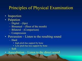
  • 1. Principles of Physical Examination • Inspection • Palpation – Digital – (lips) – Bimanual – (floor of the mouth) – Bilateral – (Comparison) – Compression • Percussion – Listen to the resulting sound – Hard • high pitch has support by bone • Low pitch has less support by bone – Soft • Auscultation – Listen to the direct sound
  • 3. Reading X-ray Films Use a suitable film mount Place in proper position with care Use an illuminator for study Should be mounted that one views them as though he were inside the mouth Clinical examinations are recorded as one views the mouth from in front of the patient After diagnosis, the film is kept in s separate file until treatment is complete and filed with the case record envelope or folder for future references.
  • 4. Proper mounting of the x-rays
  • 5.
  • 6.
  • 7. Interpreting X-ray Films Is the most important work of the dentist in radiology It should be done in a systematic manner Proper consideration given to the age of the patient The history The clinical examination All findings should be recorded on the patient’s chart
  • 8. Patients seen for the first time, and those whose teeth have never been radiographed should; Have a routine examination X-rays of all the teeth includes; Edentulous spaces Edentulous jaws Bitewing are taken when teeth are present
  • 9. If this system is carried out one will often be surprised to find definite diseased conditions on teeth which were not suspected in the clinical examination.
  • 10. When films are mounted it may be found that the examinations is not conclusive in regard to a certain lesion, or that there is a suspicious place which requires further investigations by use of a different angulations or a different method of exposure or clinical reexamination.
  • 11. What a radiograph shows and what it does not; The radiograph does not picture disease; it only records on a photographic film changes in the x- ray density of the tissues brought about by pathologic processes, as well as surgical or medical treatment.
  • 12. An x-ray diagnosis is, therefore made by drawing deductions from the picture, and the dentist must become proficient in associating radiologic signs with disease conditions.
  • 13. Through experience, careful observation, and systematic study of a large number of similar cases, and in comparison of x-ray findings with postoperative findings and the results of pathologic examinations, it will be possible to make radiographic interpretations more accurate
  • 14. Examinations of the following conditions  Dental, periodontal, and periapical tissues 1. Departures from normal, such as abnormality in size, outline, position and number of roots of tooth 2. Pulpless (root-canal filled) teeth, the apices of which are in close relation with the maxillary sinus 3. Indications of attrition, erosion, abrasion, and caries 4. Conditions of the fillings and other restorations 5. Changes in the pulp canal; pulp stones, denticles, and adventitious dentin 6. Secondary evidence of pulp disease 7. Quality of the root canal fillings 8. Evidence of resorption or hypercementosis 9. Radiolucent periapical changes 10. Radioopaque pericapical changes 11. Evidence of extensive involvement from osteitis
  • 15. 12. Other marked deviations from normal bone 13. Condition of the alveolar crest, atrophy, widening of marginal periodontal membrane, and pocket formation in periodontitis (compared to clinical pocket): periodontal bone sclerosis due to stress. 14. Evidence of parietal abscesses, especially between the roots of maxillary molars in periodontitis. 15. Edentulous sections
  • 16. Bone of Maxilla and Mandible • In Orthodontics: the relation of occlusal disturbance to facial abnormalities; disturbance in dentition, presence of cyst, increased or decrease number f deciduous or permanent teeth. • In Traumatic injury: both on the injured side and on the opposite jaw, especially condyle; fracture or luxation of teeth. • Abnormal conditions causing radiolucent areas or osteolytic defects. These may be due to infections, cyst, or tumors. – A. Localized • With osteitic margin • With clear-cut margin (puched-out area) • With cystic margin (cortical bone) • With bone expansion • With perforation of bone • With trabeculated area – B. Diffuse • With generalized involvement • With defined polycystic involvement • With periosteal new formation • Abnormal conditions causing radioaque areas due to abnormal calcified tissues. These may be caused by sequestra, foreign bodies, sclerosed bone, calcified tumors,such as osteoma, and odontoma.
  • 17. Maxillary Sinuses • Conditions causing increased density: exudates, thickened membrane, cyst, tumors. • Foreign bodies: teeth, roots, osteoma. • Cases of increased density due to external, superimposed tumors or inflammatory conditions such as subperiosteal abscesses and edema of side of face.
  • 18. Salivary glands (sialographs) • Infection of gland • Strictures of ducts • Salivary stones • Tumors Temporomanbular Joint • Adhesions with obliterations of joint spaces • Perforation of meniscus • Displacement of meniscus • Position of condyle at rest and in function • Resorption f condyle • Fractures • Ankylosis
  • 19. Terms used in X-ray diagnosis 1. X-ray Density - the property of a given tissue to absorb or prevent the penetration of x-rays. This varies with the quality and intensity of x-rays. 2. Radiolucent - offering slight resistance to roentgen rays. Seen in inflammation, cysts, and central soft tissue. 3. Radiopaque - offering great resistance to roentgen x- rays. Seen in bone sclerosis, and calcified tumors. 4. Osteitic Margin - an indefinite, diffuse margin due to a gradual change from diseased to normal bone. Seen in inflammation and infiltrating tumors. 5. Clear-cut Margin - a sharply demarcated, punched-out, radiolucent area. Seen in perforation of cortex by periapical infections, central tumors, and rarely surface erosions from periosteal tumors.
  • 20. 6. Cystic Margin - formed by dense cortical bone surrounding the area. Seen in crypts around unerupted teeth, cysts, and benign central osteolytic tumors of the jaw. 7. Bone expansion - caused by pressure of cystic content or encapsulated tumor tissue; cortex is resorbed inside and added to outside by the periosteum. Seen in cysts, benign giant cell lesions, fibro-osteoma, ameloblastoma, and most central tumors. 8. Perforation - infiltrating malignant central tumors may perforate the bone. 9. Trabeculated area - the radiolucent area is generally cystic in character, often expands to bone, and is subdivided by septae. Seen in benign giant cell lesions, central myxoma, central angioma (soap bubble effect), and central mixed tumors. 10. Localized involvement - the changes are confined to a definite area in the bone as in most conditions in ostietic margin, clear-cut margin and cystic margin.
  • 21. 11. Diffused generalized involvement - the entire bone or a large part of it is involved, without well-defined limitation. Seen in osteomyelitis, benign giant cell lesionsa, and locally maglignant tumors. 12. Polycystic involvement - a large part of the bone is involved by a tumor growing by extension through new cysts forming at the periphery of the lesion. 13. Periosteal new bone formation - from the periosteum new bone is formed either parallel to the surface or vertical to the periphery. The latter is found in syphilis and as the so-called “sunray effect” in osteogenic sarcoma. 14. Metaplastic bone formation - bone is formed in many tumors as a secondary, not a primary, neoplastic phenomenon. 15. Filling defect - if radiopaque substances are injected and part of a cavity or gland is not filled with it, we speak of this as a filling defect. Filling defects are generally due to the presence of pathologic tissue such as tumors.
  • 22. Laboratory Examinations • Blood Examination – Blood test are used for diagnosis of a great variety of conditions. The test useful as aids in diagnosis of oral conditions are as follows: • Blood count • Hemoglobin estimation • Test for bleeding time and coagulation time • Certain chemical test ( Blood Chemistry) • And similar test (Special Examinations, ex. Kidney, Liver Functions)
  • 23. • Hemoglobin Estimation - this can be made by various methods, the Sahli test by acid hematin method is generally used. The normal hemoglobin value is 14 to 17 gm per 100cc. • The Color Index - the percentage of hemoglobin divided by the percentage of red blood cells gives the color index. The percentage of red blood cells is found by dividing the number of red blood cells present in a cubic millimeter of blood by 5,000,000 (the normal number of cells per cu mm of blood), and multiplying the quotient by 100. Thus, if a person has 2,500,000 red blood cells per cubic millimeter of blood, he has 50 percent of the normal red blood cells; if there are 3,500,000, red blood cell count is 70 percent normal. If the individual’s hemoglobin is 40 percent and the percentage of erythrocytes is 50, the color index is 4 divided by 50, or 0.8. In most anemias the color index is below 1. In pernicious anemia the typical color index is above 1.
  • 24. • The Hematocrit Reading and Volume Index - the hematocrit reading is the percentage of total blood volume composed of erythrocytes. The volume of the red blood cells (hematocrit reading) is determined by rapid centrifuging the blood in a special tube, and then noting the volume of the erythrocyte sediment as compared with the liquid portion of blood. The normal hematocrit value is 42 to 45. The volume index is obtained by dividing the hematocrit (in percentage of normal) by the erythrocyte count (in percentage of normal). A value greater than 1 indicates that the erythrocytes are larger than normal.
  • 25. The Blood Cell Count • Apparatus used to make a blood cell count consist of: – A microscope – A blood lancet – A hemacytometer with a counting chamber with the Neubauer ruling • A drop of blood is obtained by the use of the lancet, the first drop expressed is to be used for the hemoglobin estimation.
  • 26. • The Red Blood Cell Count – Blood is drawn into the pipet, diluted and shaken. A drop is placed in the counting chamber and 100 of the smallest squares are counted. The number of red cells in 1 cu mm of blood is computed. The normal count is 4,000,000 to 5,555,000 per cubic millimeter. A low count suggests anemia. The anemias are usually classified on their etiologic basis i.e. • those due to decreased production of erythrocytes or hemoglobin, • those due to excessive destruction of erythrocytes, • and those due to loss of blood.
  • 27. The size and shape of red blood cells are ascertained by means of a stained smear. • Poikilocytsis - there is irregularity of the shape of the cells (severe anemia and leukemia); sickle cells in large numbers are also found in anemias. • Anisocytosis - there is marked irregularity in the size of the cells • Normoblast - are nucleated red blood cells found in severe anemias and various types of leukemias, and make for an unfavorable prognosis. • Polychromatophilia - applies to cells which do not stain uniformity, but take the basic stain. • Stippling or basophilic degeneration - is a characteristic of poisoning with lead or other metals. • Hypochromasia and Anochromasia are sign of normal degeneration.
  • 28. • The White Blood Cell Count – Blood is drawn into the pipet, diluted and shaken. A drop is placed on the ruled counting chamber and the cells in four of the large squares are counted. The number of cells counted is divided by four to obtain an average and multiplied by 200 (dilution 20 times depth of slide 10). This gives the number of white cells in 1 cu mm of blood. The white cells have the function of protecting the human organism. The number may be decreased through the effects of poisons, increased in case of need. The normal white cell count is between 5,000 and 10,000 cells per cubic millimeter of adult blood.
  • 29. – The high figure occurs in pregnancy, alimentary digestion, and people exposed to the elements. A low count is called “leukopenia”, a high cell count is called “leukocytosis”, occurs in infection, leukemia, and severe cachexia. A mounting cell count means a continuance of infection towards suppuration or spreading of the disease. An increase from leukopenia to normal means improvement. In leukemia an increase is a bad omen
  • 30. • The differential count gives the proportion of cell types present, not necessarily an absolute increase or decrease from normal. The white cell differential count is made by smearing a slide, applying special stains, and counting cell types. Intravital and supravital staining techniques may also be used by the hematologist to drive additional information.
  • 31. Normal Proportions for adults • Polymorphonuclear neutrophils - 50 – 75 percent • Polymorphonuclear eosinophils - 1 – 4 percent • Polymorphonuclear basophils - 0 – 1 percent • Lymphocytes - 20 – 30 percent • Monocytes (large mononuclear lymphocytes, endotheliocytes - 5 – 10 percent
  • 32. • Shift to the left is an expression indicating a change in the types of the polymorphonuclear neutrophils. Schilling divided them into four groups according to their nuclear morphology: – Myelocytes - immature cells with single rounded nucleus (0 percent) – Transitional or young metamyelocytes - with a single round nucleus somewhat indented (0 -2 percent) – Older metamyelocytes - with the nucleus deeply indented but not truly lobulated (3 -5 percent) – Polymorphonuclear cells - (55 -70 percent) • A shift to the left means a relative increase of the first two groups of immature cells; the greater the shift the poorer becomes the prognosis, especially if combined with a leukopenia. The appearance of myelocytes is a grave omen.
  • 33. Blood Cell Sedimentation Test • This is a macroscopic test used to detect serious infection. In blood containing an anticoagulant the cells are allowed to settle, leaving clear plasma. This is called sedimentation, and is more rapid in disease than in health. • Measurement of the time required for the sedimentation (S.T.) is of value in diagnosis. In another test the time is fixed and the quantity and distance of falling are registered (S.R.). Cutler states that the velocity with which the red blood cells settle varies from minute to minute, and that this variation is of extreme importance.
  • 34. • The total sedimentation at the end of 60 minutes expressed in millimeters is called the sedimentation index (normal for men 2 to 8 mm., average 3 to 4 mm., normal for women 2 to 10 mm., average 5 to 6 mm., during menstruation as high as 12 mm.; however, the readings vary greatly with different methods and require careful interpretation). • The sedimentation rate under normal conditions is relatively constant but is higher and varies more in women. It may be accelerated in pregnancy or during menstruation or by pathologic conditions.
  • 35. Bleeding and Coagulation Time • These are important to the dentist • If there is a history of undue bleeding from a former injury or tooth extraction, or if larger surgical operations are planned, the bleeding and clotting time should be ascertained. • These are actually screening tests and more complete studies by a well qualified physician or internist may be indicated when the results are not within normal limits. • It should be remembered, however, that in many instances hemorrhages are due to partly severed blood vessels and other local conditions. The patients should be questioned as to whether any anticoagulant is being used for vascular disease, and generally evaluated clinically.
  • 36. The Bleeding Time • Normal value 2 to 4 minutes • Prolonged bleeding time does not always parallel the clotting time • There is a slight increase in severe anemias • A great increase, 10 to 90 minutes, is found in patients with blood platelets greatly reduced (purpura hemorrhagica, acute leukemia) and in patients with extremely low fibrinogen content of the blood (phosphorus poisoning, destructive liver disease). • The paradox of prolonged bleeding time and normal coagulation time is explained by the fact that in such a disease as thrombocytopenic purpura the number of platelets is too small to plug the defect in the capillaries but large enough to intiate the process of coagulation.
  • 37. The Coagulation Time • Normal value 2 to 8 min • A coagulation time more than thrice the normal is a danger signal. It is not a contraindication to surgery, but when present proper precautions or prophylactic treatment is indicated, preferably after proper medical evaluation. • Prolonged coagulation time occurs in hemophilia, in which it is from one to several hours in anemias, leukemias, in many infectious diseases, and in jaundice. • Prolonged coagulation time slightly above the normal may occur in healthy individuals, and may be cause of prolong oozing from small blood vessels as in patients who have had tonsillectomy or operative procedures in the mouth, including the extraction of teeth.
  • 38. The Clot Retraction Time • If the blood is normal the clot gradually separates from the wall of the tube expressing the serum. The process should be completed after 1 hour. • By other methods, including incubation, this time will vary considerably. The retraction is due to the presence of blood platelets and is independent of the coagulation time. – In thrombocytopenia the coagulation time may be normal but the blood clot will be friable and retracts very little or not at all, even after several days. – In hemophilia the blood coagulates very slowly but the clot when once form has normal retractile power.
  • 39. The Prothrombin Time • Coagulation time does not differentiate among the various factors that disturb the clotting mechanism. The deficiency in prothrombin should be recognized. • Quick’s test is in common use today. The normal for this test is 16 to 17 seconds, but the time varies considerably with the thromboplastin used in the test. • The prothrombin determination is of value to determine the state of prothrombinopenia which may be due – to lack of vitamin K, – because of dietary deficiency or faulty absorption caused by the lack of salts such as occurs in obstructive jaundice, biliary fistula, and sprue, or in persons in whom liver damage has occurred. – Prolonged use of acetylsalicyclic acid (aspirin) may decrease the prothrombin time. • Prothrombinopenia may be corrected in such cases by the administration of vitamin K. Cahn recommends that 1 mg. of vitamin K should be prescribed to every 5 grains of aspirin, if taken constantly.
  • 40. The Tourniquet Test • This is a test for capillary fragility • A square 2 by 2 cm. in size is drawn on the inner aspect of the forearm so that its upper edge is 4 cm. below the crease of the elbow. • The rubber cuff of a sphygmomanometer is then applied to the upper arm and inflated until the pressure is between the systolic and diastolic. After 15 minutes the pressure is released and 5 minutes later the number of petechiae visible in the encircled area are counted. The count of 10 or less petechiae within the square is considered normal. Counts between 10 and 20 are evidence of a pathologic condition. Patient with marked capillary fragility may have counts of 100 or more.
  • 42. Normal Values Normal range values Normal Values Normal range values Prothrombin Time 10 – 13 sec Erythrocytes 4 – 5. 5 x 10⁷ g/L Bleeding Time 1 – 3 minutes Platelets 2 – 4 x 10⁵ per cu. mm. of blood Clotting Time 3 – 6 minutes (Slide) 8 – 15 minutes (Lee/White) Coagulation Time 2 – 8 minutes Platelet count 140 – 440 x 109 Clot retraction time 1 hour Hemoglobin M – 140 – 160 F – 120 - 140 Partial Thromboplastin Time 29 – 34 sec Hematocrit M – 40 – 54 F – 37 - 47 WBC 5 – 10 x 10 g/L Sedimentation Rate Adult 0 – 30mm/hr Children 0 – 145 mm/hr
  • 43. Differentiation of Hemorrhagic Disorders • Coagulation time prolonged, bleeding time and platelet count normal: – Hemophilia – Vitamin K deficiency – Severe liver injury – Idiopathic hypoprothrombinemia – Presence of anticoagulants in the blood – Pseudohemophilia • Coagulation and bleeding time prolonged, platelet count normal: – Same conditions as I – Fibrinogenopenia
  • 44. • Prothrombin time prolonged, Coagulation and bleeding time and platelet count normal: – Hemorrhagic disease of new born – Vitamin K deficiency – Severe liver injury – Idiopathic hypoprothrombinemia • Bleeding time prolonged, coagulation time and platelet count normal: – Hereditary hemorrhagic diathesis • Bleeding time prolonged and presence of thrombocytopenia: – Idiopathic purpura hemorrhagica – Symptomatic thrombocytopenic purpura, due to poisoning by chemical, vegetable and animal agents
  • 45. Blood Chemistry There are a number of blood chemistry test which are of diagnostic interest in certain diseases. The blood for such tests is taken from a vein, sodium citrate solution is added, and the specimen is sent to a laboratory. Generally specific test are indicated for a specific diagnosis.
  • 46. Substance Normal range values Increase Decrease Glucose Substance 4.2 – 6.4 m mol/L Diabetes mellitus Hyperinsulinism, Addison’s disease Urea Nitrogen Substance 3.2 – 6.8 m mol/L Prostatic or intestinal obstruction, shock Severe liver damage Creatinine Substance 44 – 106 u mol/L Nephritis, metallic poisoning, prostatitis Fever, acidosis, diabetes, vomiting, gastrointestinal disturbance Uric Acid Substance 178 – 440 u mol/L Arthritis, nephritis Severe liver damage Cholesterol Substance 3.4 – 6.1 m mol/L Diabetes, nephrosis, xanthomatosis Severe liver damage Alkaline Phosphatase 9 – 35 lu/L Hyperparathyroid, rickets, liver disease, Paget’s osteomalacia LDH - L 109 – 193 lu/L Triglycerides Substance Up to 2.4 m mol/L Calcium Substance 2.1 – 2.5 m mol/L Hyperparathyroid Hypoparathyroid, Uremia
  • 47. Spinal Fluid • Spinal fluid may be examined for syphilis. When reaction to the serologic Wassermann test is negative the spinal fluid may give a positive results, especially in neurosyphilis. Urinalysis • The urine is examined as a routine procedure in all hospitals before administration of an anesthetic. Primarily it gives information regarding renal diseases, but it is also of importance in the study of irregularities or deficiencies in metabolism, which in turn may have a direct relation to oral conditions. The quantity of urine (normal amount 1,000 to 2,000 cc. or 1 to 2 qts. in 24 hours) and frequency of urination should be noted by the clinician; they often give important information.
  • 48. • Albumin - the presence of albumin is seen in fever, pregnancy (toxic), and nephritis, especially if cast are present. Sometimes albumin is present in urine of patients with marked dental infection. • Sugar - Detection may be made by Benedict’s test, simple screening test packets are available. More than 0.2 percent of sugar is found in persons having diabetes mellitus, and also in those with renal diabetes, which is temporary as a rule and frequency due to the excessive use of alcohol, starch, or sugar, or to nervous strain. The presence of excessive sugar in the urine suggest the need for thorough medical study. • Acetone - this is found in the urine of patient’s with acidosis due to the incomplete metabolism of fat. It occurs in association with diabetes mellitus and may indicate cachexia, starvation or coma. • Urea - The quantitative test gives information regarding the body metabolism or the amount of nitrogen excreted, which is eliminated mostly by the kidneys in a form of urea. Urea is usually excreted in amounts of 15 to 40 gms. per 24 hours. It is increased in toxic goiter.