
Recommended
More Related Content
What's hot
What's hot (19)
Viewers also liked
Similar to DVA Cell Proliferation Effects
Similar to DVA Cell Proliferation Effects (20)
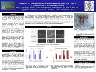
DVA Cell Proliferation Effects
- 1. Methods Abstract The Effects of Commercialized Performance Enhancing Deer Velvet Antler on Cell Proliferation in 3T3 Mouse Fibroblasts Emily Eichelberger, Chelsea Easter, Sydney Shearer, Timothy Shoff, and Irene M. Wolf, PhD Department of Biology, Saint Francis University, Loretto, Pennsylvania 15940 Many athletes seek supplements that accelerate the recovery process, such as insulin-like growth factor 1 (IGF-1). In humans, IGF-1 is stimulated by growth hormones and synthesized primarily by the liver, but also produced by bone and muscle tissue. It is important in skeletal growth by inducing chondrocyte proliferation and has effects on connective tissue proliferation. IGF-1 is an anabolic steroid and banned by the World Anti-Doping Agency (WADA), yet it is still used by many athletes. Commercial companies claim that a product created from Deer Velvet Antler (DVA) mimics IGF-1. According to an article on BornFitness.com, individuals aiming for higher performance and accelerated recovery rates have begun using DVA. However, very little laboratory research has been conducted to support these claims. We hypothesized that IGF-1 and commercialized DVA will enhance cell proliferation in 3T3 Mouse fibroblast cells. Data utilizing scratch assays reveal an increase in cell proliferation when treating the cells with commercialized DVA and DVA plus synthetic IGF-1 in serum. Interestingly, the data indicates that smaller concentrations of DVA induces greater cell proliferation. Conversely, all three drugs show increased cell growth when grown in media without serum. Future studies will be conducted using an MTT assay to confirm the effects of DVA on cell proliferation. 3T3 mouse fibroblast cells were housed in an incubator held at 37°C and 5% CO2. The cells were grown in Dulbecco’s Modified Eagle Medium (DMEM) with high glucose, 10% bovine calf serum (BCS), and 1% penicillin/streptomycin. The cells were plated onto 35 mm petri dishes at 6x10⁴ cells/mL with serum. After 24 hours, the media was replaced with media containing serum (BCS) in trial 1 and without serum in trial 2. The desired concentrations of drugs were added and the cells were then scratched with a pipet tip. A picture was taken with an inverted microscope immediately following the scratch (0 hrs), the scratch was measured and the cells were placed in the incubator for 24 hours. Pictures and measurements were also taken at 24 and 48 hours. The results show an increase in cell proliferation when looking at human and mouse IGF-1, DVA only, and DVA plus synthetic IGF-1. In the scratch assay trials using human IGF-1 and media with serum (BCS), both human IGF-1 and DVA/IGF-1 showed higher cell proliferation rates at higher concentrations. Interestingly, the DVA induced higher growth rates at lower concentrations. In the trials using mouse IGF-1 and media without serum (BCS), all three drugs showed increased cell proliferation rates given at higher concentrations. The results from the MTT Assay are inconclusive and need to be retested in other trials. Overall, more scratch assay trials and MTT Assays need to be performed to further support our hypothesis. An AKT Assay will also be ran to determine if DVA follows the same pathway as IGF-1 to induce cell proliferation. - Department of Biology at Saint Francis University - TriBeta National Biology Honors Society Grant - Saint Francis University School of Sciences Undergraduate Research Grant Methods Approximately 48 hours prior to treatment, the 3T3 cells were cultured in a 96-well plate at 5x10⁴ cells/mL in a 37°C incubator and in 5% CO2. Different concentrations of IGF-1, DVA, and DVA + IGF-1 were used to treat the cells, using DMEM with no serum. Treatments were in triplicate, and repeated three times for n=3. The plate was then incubated for 48 hours. Following the 48 hours, media was refreshed and the drugs were added. The plate was then placed back into the incubator. After 24 hours, an MTT (3-(4,5-Dimethylthiazol-2-yl)-2,5-Diphenyltetrazolium Bromide) assay was performed in order to evaluate and determine cell proliferation activity. 10 microliters of MTT reagent was added to each well plate and was placed back in the incubator to allow the reaction to occur. Contents of the wells were removed and 100 microliters of Crystal Dissolving Solution was added to each well. The final absorbance levels were measured at 570 nm using a spectrophotometer. All results will be subjected to statistical analysis. Figure 2: MTT Assay A) DVA, B) DVA/IGF-1, C) Mouse IGF-1. Darker colors indicate higher levels of cell proliferation. Mouse IGF- 1 show higher growth rates at higher concentrations. The results of this assay were inconclusive. Figure 1: Human IGF-1 Scratch Assay containing Serum. Conclusion Acknowledgements Results Graph 1: Scratch Assay with Serum (Trial 1) Scratch Assay reveals increased cell proliferation at higher concentrations with Human IGF-1 and DVA/IGF-1 plus. Graph 2: Scratch Assay without Serum (Trial 2) Scratch Assay reveals increased cell proliferation at higher concentrations with Mouse IGF-1, DVA, and DVA/IGF-1 Plus. C BA