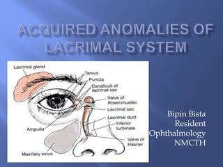
Acquired anomalies of lacrimal system
•Download as PPTX, PDF•
1 like•495 views

simplified slide to learn easily about acquired anomalies of lacrimal system.

Recommended
More Related Content
What's hot
What's hot (20)
Similar to Acquired anomalies of lacrimal system
Similar to Acquired anomalies of lacrimal system (20)
More from Bipin Bista
More from Bipin Bista (20)
Recently uploaded
Recently uploaded (20)
Acquired anomalies of lacrimal system
- 2. h/o : tearing – epiphora or lacrimation. Constant vs intermittent Period of remission vs no remission. Unilateral vs bilateral. h/o lacrimal sac inflammation Subjective ocular discomfort Use of topical medications h/o probing during childhood Prior infections Sinus surgeries/mid- facial trauma Clear tear vs discharge.
- 3. Pseudoepiphora evaluation – tear meniscus, TBUT. Corneal & conjunctival epithelium evaluation : staining, Inferior 1/3rd of cornea is devitalised – more severe malfunction with epithelial loss. Schirmer’s test Corneal irritation Lacrimal outflow evaluation
- 4. Eyelid evaluation Facial nerve dysfunction Carancular hypertrophy & conjunctival chalosis or frank prolapse. ROPAS Routine nasal examination
- 5. This diagnostic test was first outlined by Lester Jones. Dye disappearance test followed by Jones I & II
- 6. Useful for assessing the presence or absence of adequate lacrimal outflow, in unilateral cases. Fluorescein is instilled in fornices, then observe tear film – persistent of significant dye – lacrimal drainage dysfunction. Hard to rule out : allergy, dacryolith or intranasal obstruction.
- 7. Investigate whether lacrimal outflow is under normal condition. Instill fluorescein into conjunctival fornices & recoring it in Inferior meatus by passing a cotton-tipped wire applicator into the region of ostium of NLD at 2 & 5 minutes.
- 8. In this part cannula is placed in the sac & the system is irrigated. If no fluorescein is passed meaning upper ( canalicular ) abnormality. If fluorescein passed meaning lower lacrimal ( sac/duct abnormality.
- 9. Most frequently performed test after DDT. Done to determine the level of occlusion. Difficulty in irrigating, if refluxes from upper canaliculi, complete blockage. Patent : passed freely.
- 10. Direct visualization of lacrimal passage. Helpful in evaluating lacrimal anatomy.
- 11. CONTRAST DACROCYSTOGRAPHY DACROSCINTIGRAPHY Knowing the LDS anatomy by dye injection into LS followed by computerised digital substraction. Physiological evaluation Performs by flowing radionucleotide drops to flow as tear using scintigram.
- 12. Results in epiphora May be too small ( occlusion & stenosis) or too big ( iatrogenic). May be malpositioned or occluded by adjacent structures. Punctal stenosis – treated by dilatation, punctoplasty or stenting. Regular monitoring requires as stenting may cause ‘cheese wiring’.
- 13. May be common, upper or lower. Diagnostic : canalicular probing. SOFT STOP vs HARD STOP. Total Functional Occlusion : weakness of lacrimal pump or inability of tears to pass through even minimal obstruction . Usually it is a physiological condition and can be overcomed by irrigation by creating abnormal hydrostatic pressure.
- 15. 1. Lacrimal plug 2. Medication 3. Infection 4. Inflammatory diseases 5. Trauma 6. Neoplasm
- 16. Punctal & canalicular For dry-eye. Diagnosed by : canalicular probing, high frequency USG Surgical excision of canaliculi & re- anastomosis.
- 17. 2. Medication : systemic chemotherapeutic agents – 5-fluorouracil, docetaxel, idoxuridine. 3. Infection : HSV, Vaccinia virus. 4. Inflammatory disease : pemphigoid, SJS, graft vs host disease. 5. Trauma : permanent damage if not managed at time. 6. If present in medial canthal area, complete excision along with puncta & canaliculi.
- 18. Canalicular stenting : 1st line management. Reconstruction : successful if only few mm is involved. If its proximal then occluded canaliculi is resected and cut ends of canaliculi is anastomosed. If occlusion is distal at common canaliculi, then stenting is required to prevent contracture & for epithelisation. Canaliculodacryocystorhinostomy : If total obstruction. Conjunctivodacryocystorhinostomy : when there’s severe occlusion. Inferior half of caruncle – osteotomy—middle meatus.
- 19. Obstruction of tube with mucus & migration of the tube. Forced inspiration. “MUST DO” Tube foreign body : Pyogenic granuloma formation – Frosted, angled or modified Jones tube. Porous polyethylene-coated tube.
- 20. Usually diagnosed with irrigation. Etiology : Involutional stenosis Dacryolith Sinus disease Trauma Inflammatory disease Lacrimal plugs Radioactive iodine Neoplasm
- 21. Most commonly female than in male. Compression of NLD d/t inflammatory & edematous condition. Management : DCR.
- 22. Dacryolith : cast formation – consists of shed epithelial cells, lipids & amorphous debris with or without calcium deposition. Occassionally occur with Actinomycetes israelli or Candida. Management : DCR without any difficulty.
- 23. Often occurs with or in any instances which may contribute to NL abnormality. H/o – Sinus surgery.
- 24. Naso-orbital fracture. Early treatment with fracture reduction with stenting of NLD. For late : DCR.
- 25. Granulomatous dz Sarcoidosis WG Lethal midline granuloma
- 26. Lacrimal plugs : similar to NLD Obstruction. Radioactive Iodine : Treatment for thyroid disease : Closure of lacrimal apparatus. Neoplasm : bloody punctal discharge, swelling above medial canthal tendon : suspicious h/o malignancy, sinus or NP CT/MRI Biopsy : while surgical intervention Benign : DCR, CDCR Malignancy : certainty of clear margins or freedom from recurrence – DCR or CDCR .
- 27. Intubation & stenting Dacrocystorhinostomy (DCR)
- 28. Treatment of choice Creating an anastomosis between lacrimal sac & nasal cavity through a bony ostium. Types : Internal(endoscopic) & external. Internal DCR : lack of visible scar, shorter recovery period & shorter time . Success rate is more in external (90>70)%. GA .. LA (strong monitoring) Hemostasis
- 30. Fibrosis & occlusion of osteotomy Common canalicular obstruction Inappropriate placement or size of bony ostium
- 31. LACRIMAL GLAND (Dacroadenitis) − Occurs in sterile condition & occurs in consequence of malignancy. − Extremely rare condition. − Gross appearance & abscess formation is quite uncommon. − Emperical therapy
- 32. Caused by variety of bacteria, viruses & mycotic organisms. Most commonly caused by filamentous gram positive rod :Actinomycetes israelli Complaints : persistent weeping, accompanied by follicular conjunctivitis centered in conjunctiva. Punctum : erythematous Milking present. Warm compression, massage & topical antibiotics Canaliculotomy
- 34. Inflammation of lacrimal sac Most commonly due to lacrimal sac obstruction. Chronic tear retention & stasis : secondary. C/F :edema & erythema with distension of lacrimal sac below medial canthal tendon. Complication : Dacryocystocele formation, chronic conjunctivitis, & spread to adjacent structure .
- 36. Irrigation or probing Topical antibiotics : Limited value Oral antibioitics : gram positive are common , gram negative seen in diabetics & immunocompromised. Parenteral Ab : severe cases. Aspiration of lacrimal sac : pyocele, mucocele I & D : Localised abscess . Packing- open Total obstruction : DCR surgery.
- 37. Primary : papilloma, scc Primary tumors of tissue surrounding lacrimal region : eyelid skin (basal & squamous cell), adenoid cystic carcinoma, capillary hemangioma, inverted papilloma, epidermoid carcinoma,osteoma & lymphoma. Metastatic tumors
- 38. Histologically, 45% are benign and 55 % are malignant. Treatment : Dacrocystectomy (DCT) In case of malignancy : DCT with lateral rhinostomy. Exenteration including bone removal in medial canthal area.
- 39. Hypoplasia & agenesis Craniosynostosis Lacrimal gland fistulas Duplication Congenital lacrimal cutaneous fistula Naso-lacrimal duct obstruction .