The skin & integumentary system. ppt
•
8 likes•2,299 views

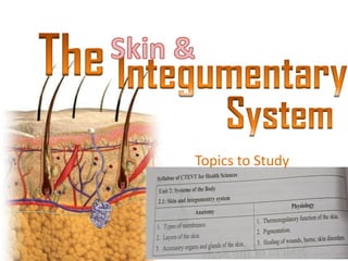
The skin & integumentary system is based on the CTEVT syllabus for diploma in health sciences, Anatomy, and physiology,

Recommended
More Related Content
What's hot
What's hot (20)
Similar to The skin & integumentary system. ppt
Similar to The skin & integumentary system. ppt (20)
More from Bashant Kumar sah
More from Bashant Kumar sah (20)
Recently uploaded
Recently uploaded (20)
The skin & integumentary system. ppt
- 2. The integument as an organ, and is an alternative name for skin. The integumentary system includes the skin and the skin derivatives hair, nails, and glands. The Skin & integument as an organ:
- 3. The Skin & Integument Is the largest system of the body 15-20% of body weight, (4kg ) 1.5 to 2m2 in area, 0.3 to 3mm thickness The integument is made up of two parts: 1. Cutaneous membrane a. Epidermis– Superficial epithelium b. Dermis – underlying Cutaneous tissue with blood supply 2. Accessory structures a. Hair b. Nails c. Exocrine Glands
- 4. Types of Membrane -Thin layers of epithelial tissue -Bound to an underlying layer of connective tissue. -Membranes cover, protect, or separate other structures or tissues in the body. There are four types of Membrane: 1. Cutaneous membranes: -a layer of stratified squamous epithelium (epidermis) firmly attached to a thick layer of dense connective tissue (dermis). -exposed to air and is dry.
- 5. 2.Serous membranes: -consist of simple squamous epithelium (a mesothelium) supported by a layer of connective tissue (areolar). These moist membranes line the closed, internal divisions of the ventral body cavity. The three types of serous membranes are: P3 --The pleura, lining the pleural cavities and covering the lungs; --The peritoneum, lining the peritoneal cavity and covering the abdominal organs; and --The pericardium, lining the pericardial cavity and covering the heart.
- 6. 3. Mucus membranes: -consist of epithelial tissue (usually stratified squamous or simple columnar epithelia) on a layer of loose connective tissue called the lamina propria (from the Latin, meaning “one’s own layer”). -The mucosae line the body cavities that open to the exterior, such as the digestive, respiratory, reproductive, and urinary tracts. -Are kept moist by bodily secretions.
- 7. 4. Synovial Membrane: --are composed of connective tissue. --They surround the cavity of joints, filling the space with the synovial fluid that they make. --The synovial fluid lubricates the ends of the bones allowing them to move freely.
- 8. Protection First line of defense against Bacteria Viruses Protects underlying structures from Ultraviolet (UV) radiation Dehydration Vitamin D production Needed for calcium absorption Sensation Sensory receptors
- 9. Body temperature regulation If too hot Dermal blood vessels dilate Vessels carry more blood to surface so heat can escape If too cold Dermal blood vessels constrict Prevents heat from escaping Excretion Small amounts of waste products are lost through perspiration
- 10. Understanding how the skin can function in these many ways starts with understanding the structure of the 3 layers of skin The Epidermis Epithelial tissue Dermis Dense connective tissue proper – irregular Hypodermis Subcutaneous tissue- loose connective tissue proper and adipose tissue
- 12. Thin Skin Covers most of the body Has four layers of keratinocytes Thick Skin Covers the palms of the hands and soles of the feet Has five layers of keratinocytes
- 13. The Epidermis Is a vascular stratified squamous epithelium Nutrients and oxygen diffuse from capillaries in the dermis Cells of the Epidermis Keratinocytes Contain large amounts of keratin about 90% Strengthens the skins and prevents from excessive water loss.
- 14. Melanocytes: -synthesizes melanin, which barrier against UV radiation. Langerhans’ cells: -class of macrophages, role in immunity acting as antigen cells, produced in bone marrow. Merkel cells: -acts as a touch receptor.
- 15. Thick skin LM 210 Surface Stratum corneum Stratum lucidum Stratum granulosum Stratum spinosum Stratum basale Basement membrane Dermis Papillary layer of dermis E P I D E R M I S
- 16. Stratum Basale Is attached to basement membrane by hemidesmosomes Forms a strong bond between epidermis and dermis Forms epidermal ridges (e.g., fingerprints) Dermal papillae (tiny mounds) Increase the area of basement membrane Strengthen attachment between epidermis and dermis Has many basal cells or germinative cells
- 17. Stratum Spinosum — the ―spiny layer‖ Produced by division of stratum basale Eight to ten layers of keratinocytes bound by desmosomes Cells shrink until cytoskeletons stick out (spiny) Continue to divide, increasing thickness of epithelium Contain dendritic (Langerhans) cells, active in immune response
- 18. Stratum Granulosum — the ― grainy layer ‖ Stops dividing, starts producing Keratin A tough, fibrous protein Makes up hair and nails Keratohyalin Dense granules Cross-link keratin fibers
- 19. Stratum Lucidum — the ―clear layer‖ Found only in thick skin Covers stratum granulosum Stratum Corneum — the ―horn layer ‖ Exposed surface of skin 15 to 30 layers of keratinized cells Water resistant Shed and replaced every 2 weeks
- 20. The Dermis Located between epidermis and subcutaneous layer Anchors epidermal accessory structures (hair follicles, sweat glands) Two components 1. Outer papillary layer 2. Deep reticular layer Dermis
- 21. The Papillary Layer Consists of areolar tissue Contains smaller capillaries, lymphatics, and sensory neurons Has dermal papillae projecting between epidermal ridges The Reticular Layer Consists of dense irregular connective tissue Contains larger blood vessels, lymphatic vessels, and nerve fibers Contains collagen and elastic fibers Contains connective tissue proper
- 22. An inflammation of the papillary layer Caused by infection, radiation, mechanical irritation, or chemicals (e.g., poison ivy) Characterized by itch or pain Characteristics Strong, due to collagen fibers Elastic, due to elastic fibers Flexible
- 24. The Hypodermis (Subcutaneous Layer) Lies below the integument Stabilizes the skin Allows separate movement Made of elastic areolar and adipose tissues Connected to the reticular layer of integument by connective tissue fibers Deposits of Subcutaneous Fat Distribution patterns determined by hormones Reduced by cosmetic liposuction (lipoplasty)
- 26. Accessory organs and glands of the skin: Hair : Nails Sweat glands: Sebaceous glands:
- 27. The Hair Follicle Hair follicles are the organs that form the hairs. Hair follicles are the organs that form the hairs. Located deep in dermis. Produces nonliving hairs. Wrapped in a dense connective tissue sheath. Base is surrounded by sensory nerves (root hair plexus). Control bacteria
- 28. Exposed shaft of hair Sebaceous gland Arrector pili muscle Connective tissue sheath Root hair plexus Accessory Structures of Hair Arrector pili Involuntary smooth muscle Causes hairs to stand up Produces ―goose bumps‖ Sebaceous glands Lubricate the hair
- 29. Regions of the Hair Hair root Lower part of the hair Attached to the integument Hair shaft Upper part of the hair Not attached to the integument Boundary between hair shaft and hair root Arrector pili muscle Hair shaft Sebaceous gland Hair root Connective tissue sheath Hair bulb Hair matrix Hair papilla
- 30. Nails Protect fingers and toes Made of dead cells packed with keratin Metabolic disorders can change nail structure Nail Production Occurs in a deep epidermal fold near the bone called the nail root Free edge of Nail Body of Nail Laternal Nail fold Lunula Eponychium (cuticle)
- 31. Structure of a Nail Nail body The visible portion of the nail Covers the nail bed Lunula The pale crescent at the base of the nail Sides of nails Lie in lateral nail grooves Surrounded by lateral nail folds
- 32. Eponychium Proximal nail fold Nail root Lunula Nail body Epidermis Dermis A longitudinal section Phalanx Hyponychium
- 33. Sweat and Sebaceous glands:
- 34. A. Sweat Glands: Situated in the dermis Excretes urea, salt water and other ions in the form of sweat • Eccrine sweat glands: -Mostly found in palms and soles -Secretion is thin and watery for temperature regulation under control of Automatic Nervous System. • Apocrine sweat glands: -Located in axillary and pubic region -ducts empty in hair follicles -secretion is more viscous( fats and proteins) than eccrine glands -activated by sex hormones and sympathetic autonomic nervous system
- 35. B. Modified sweat glands: Ceruminous glands: -Located in the external ear canal -Secretes wax (cerumin) Mammary glands: -Milk producing and is most dominant in female -secrets milk after parturition C. Sebaceous glands: -Located next to hair follicles and oily secretion called sebum -sebum composed of triglycerides, cholesterol, proteins and electrolytes. -inhibits bacterial growth, lubricates hair shaft and conditions the skin -influenced by sex hormones
- 36. Thermoregulatory function of the skin: Physics of Heat Loss from the Body:
- 37. “Set-Point” for Temperature Control ●Criticalbodycore temperature isabout36.8°C to 37.1°C (98.8°F) ●Drasticchangesoccurin the rates ofboth heatlossandheatproduction. ●Attemperaturesabovethislevel, the rate ofheatlossisgreater thanthat ofheatproduction, sothe bodytemperature fallsandapproaches the 37.1°C level. ●Attemperaturesbelow thislevel, the rate ofheatproduction isgreater thanthat ofheatloss, sothe bodytemperature risesandagain approachesthe37.1°Clevel. ●Thiscrucialtemperature leveliscalledthe “set-point”ofthe temperature control mechanism. ●Thatis, allthe temperature control mechanismscontinuallyattempt to bringthebodytemperature backto thisset-point level.
- 38. Mechanism: -The thalamus is the temperature regulating center responsive to the temperature of the circulating blood. -Controls through autonomic nerve stimulation of the sweat glands and peripheral blood vessels.
- 40. Pigmentation: Skin pigmentation determined by the following pigments: 1. Melanin: dark skinned people produce more melanin although having similar amounts of melanocytes which is stimulated by the exposure to sunlight and protects from UV radiation. 2.Carotene: -gives yellow or orange skin color 3. Bilirubin and biliverdin: -products of degradation haemoglobin and gives yellow skin color 4. Hemoglobin: -gives pinkish color to fair and dark color in black skin.
- 41. Healing of wounds It is defined as a living body response in an attempt to restore the normal structure and function which include regeneration and repair of body parts simultaneously.
- 42. Factors delaying wound healing - Infection - Poor blood supply - Presence of foreign body - Mobilization of wounded part - Age slow in old age - Nutritional deficiency - Diabetes - Bleeding disorders Complications: -Epidermal cyst -Scar formation -Infection may occurs -Neoplasia may develop if untreated for long time
- 43. Burns: The injury which is caused by dry heat is called burns and by moist heat is called scalds. Causes of burns: Dry heat: fire, flame, piece of hot metal Moist heat: boiling water, steam, hot oil Electricity; electric currents and lightning Radiation: burns due to radium and deep X-ray. CLASSIFICATION: Erythema: results from vasodilation due to heat with sign of inflammation. Superficial burns: only superficial layer is lost and very painful due to exposure of nerve ending. Deep burning: whole skin is lost and less painful due to nerve ending burning. Skin grafting is needed.
- 44. Estimation of burns The percentage of body surface burn can be estimated by Wallace rule of NINE
- 45. Skin disorders A. Inflammatory disorders: -viral infections:HPV (human pailoma virus) causes warts herpes virus causes chicken pox, shingles in the skin -Bacterial infections: impetigo and cellulities -Fungal infection: Ringworm and Tinea pedis B. Non- Inflammatory disorders: -Eczema and dermatitis -psoriasis -Malignant tumors: basal cell carcinoma, malignant melanoma, Kaposi’s sarcoma etc.
- 46. THANK YOU