Quaternary structure of protein
•Transferir como PPTX, PDF•
22 gostaram•11,809 visualizações

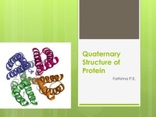
Quaternary structure refers to the arrangement of multiple protein subunits into a single protein complex. Hemoglobin is a common example that is made of two alpha and two beta subunits. The subunits interact through hydrophobic interactions, hydrogen bonding, and other bonds. Globular proteins tend to have quaternary structure that clusters the subunits into a spherical shape, while fibrous proteins form long coils or sheets through interactions between subunits. Quaternary structure allows proteins to take on specialized functions beyond what individual subunits could achieve alone.

Recomendados
Recomendados
Mais conteúdo relacionado
Mais procurados
Mais procurados (20)
Semelhante a Quaternary structure of protein
Semelhante a Quaternary structure of protein (20)
Mais de Arjun K Gopi
Mais de Arjun K Gopi (20)
Último
Último (20)
Quaternary structure of protein
- 2. Protiens: Proteins were first described by the Dutch chemist Gerhardus Johannes Mulder and named by the Swedish chemist Jöns Jakob Berzelius in 1838. Early nutritional scientists such as the German Carl von Voit believed that protein was the most important nutrient for maintaining the structure of the body, because it was generally believed that "flesh makes flesh."
- 3. The amino acids in a polypeptide chain are linked by peptide bonds. Once linked in the protein chain, an individual amino acid is called a residue, and the linked series of carbon, nitrogen, and oxygen atoms are known as the main chain or protein backbone. The peptide bond has two resonance forms that contribute some double-bond character and inhibit rotation around its axis, so that the alpha carbons are roughly coplanar. The other two dihedral angles in the peptide bond determine the local shape assumed by the protein backbone. The end of the protein with a free carboxyl group is known as the C-terminus or carboxy terminus, whereas the end with a free amino group is known as the N- terminus or amino terminus.
- 4. Levels of Proteins: Four levels of structure are frequently cited in discussions of protein architecture. Primary Structure Secondary Structure Tertiary Structure Quaternary Structure
- 5. Quaternary Protein - Structure The quaternary protein structure involves the clustering of several individual peptide or protein chains into a final specific shape. A variety of bonding interactions including hydrogen bonding, salt bridges, and disulfide bonds hold the various chains into a particular geometry. There are two major categories of proteins with quaternary structure - fibrous and globular.
- 6. Fibrous Proteins: Actually, the final beta-pleated sheet structure of silk is the result of the interaction of many individual protein chains. Specifically, hydrogen bonding on amide groups on different chains is the basis of beta-pleated sheet in silk proteins. SILK
- 7. Other fibrous proteins such as the keratins in wool and hair are composed of coiled alpha helical protein chains with other various coils analogous to those found in a rope. Other keratins are found in skin, fur, hair, wool, claws, nails, hooves, horns, scales, beaks, feathers, actin and mysin in muscle tissues and fibrinogen needed for blood clots.
- 8. Globular Proteins: On the other hand, globular proteins may have a combination of the above types of structures and are mostly clumped into a shape of a ball. Major examples include insulin, haemoglobin, and most enzymes. INSULIN
- 9. The most common example used to illustrate quaternary structure is the hemoglobin protein. Hemoglobin's quaternary structure is the package of its monomeric subunits. Hemoglobin is composed of four monomers. There are two α-chains, each with 141 amino acids, and two β-chains, each with 146 amino acids. Because there are two different subunits, hemoglobin exhibits heteroquaternary structure. If all of the monomers in a protein are identical, there is homoquaternary structure
- 10. Quaternary structure refers to the association of multiple individual protein chains into a single protein with multiple subunits. The arrangement of the subunits gives rise to a stable structure, which can usually be dissociated in the laboratory, but is very strongly bound in vivo. The subunits may be identical or different When they are different, each subunit tends to have a different function STRUCTURAL DISCRIPTION
- 11. A common shorthand for describing such proteins is to use Greek letters for each type of subunit, and subscript numeral to specify numbers of units. A protein designated a2bg consists of two a units and one each of b and g. The subunits usually are held together by hydrophobic interactions, the clustering serving to reduce exposure of hydrophobic side chains to the solvent. Occasionally, ionic interactions between carboxylate and amino side chains may contribute.
- 12. Biological Function: Enzymes are generally globular proteins and range from just 62 amino acid residues in size, for the monomer of 4- oxalocrotonate tautomerase, to over 2,500 residues in the animal fatty acid synthase. A small number of RNA-based biological catalysts exist, with the most common being the ribosome; these are referred to as either RNA-enzymes or ribozymes. The activities of enzymes are determined by their three- dimensional structure. However, although structure does determine function, predicting a novel enzyme's activity just from its structure is a very difficult problem that has not yet been solved.
- 13. Several proteins are actually assemblies of more than one polypeptide chain, which in the context of the larger assemblage are known as protein subunits. In addition to the tertiary structure of the subunits, multiple-subunit proteins possess a quaternary structure, which is the arrangement into which the subunits assemble. Enzymes composed of subunits with diverse functions are sometimes called holoenzymes, in which some parts may be known as regulatory subunits and the functional core is known as the catalytic subunit. Examples of proteins with quaternary structure include haemoglobin, DNA polymerase, and ion channels. Other assemblies referred to instead as multiprotein complexes also possess quaternary structure. Examples include nucleosomes and microtubules.
- 14. Classification of enzymes: Oxidoreductases: catalyze oxidation/reduction reactions Transferases: transfer a functional group (e.g. a methyl or phosphate group) Hydrolases: catalyze the hydrolysis of various bonds Lyases: cleave various bonds by means other than hydrolysis and oxidation Isomerases: catalyze isomerization changes within a single molecule Ligases: join two molecules with covalent bonds.
- 15. Protein structure determination • X-Ray crystallography • NMR spectroscopy • Mass spectroscopy
- 16. Types of protiens: Conjugated protein Lipoproteins Glycoproteins Phosphoproteins Metalloprotein etc.
- 17. THANK YOU