SlideShare a Scribd company logo
1 of 29
FLUID MOSAIC MODEL OF CELL MEMBRANE
TIGHT JUNCTION
CYTOSKELETON
BY – ARUSHI PAL
*
*The fluid mosaic model was first proposed by S. J
SINGER and GARTH L. NICLSON in 1972 to explain
the structure of the plasma membrane
.
*WHAT IS FLUID MOSAIC MODEL ?
The fluid mosaic model describe the cell membrane as
a tapestry of several types of molecules ( phospholipid ,
cholesterol , and protein) that are constantly moving .
This movement helps the cell membrane maintain its role
as a barrier between the inside and outside of cell
membrane.
*Points regarding on FMM :
*The study of fluid mosaic model is clearly
studied after electron microscope .
*It composed of lipid bilayer, lipid are fatty
acid
Hydrophobic tail – present inside
Hydrophilic head – present outside , because
to prevent hydrophobic tail from aqueous
enviroment
lipid component mainly consist of
phosphoglyceride
they also contain protein and
carbohydrate.
RBC 52% protein and 40% lipids
there are two types of protein
*Integral protein
*Peripheral protein
*Integral protein – protein are on the buried
area.
*Peripheral protein – protein are on the surface
area.
*
*TRANSPORT
PASSIVE OSMOSIS
ACTIVE
1- PASSIVE- If any molecule from this lipid
bilayer, transport or travel without any energy
called passive transport. Means no ATP is
required.
*2 – ACTIVE
if any molecules from this lipid bilayer transport
or travel with energy called active transport
means ATP is required.
Polar molecules like Na and K
3 – OSMOSIS
Movement of water from high concentration to
low concentration
*
*Phospholipid molecules shows two types of
movement :
Transition Flip flop
movement movement
1- Transition movement : movement of
phospholipid molecules in same direction .
2 - Flip flop movement : movement of phospholipid
movement between two layers.
TIGHTJUNCTION
*
*Tight junction are areas where the membrane of two
adjacent cell join together to form a barrier .
* The cell membrane are connected by strands of
transmembrane protein such as claudins and
occludin.
*Tight junction binds cell together , prevent molecules
from passing in between cell and also helps to
maintain the polarity of cells. They are only found in
vertebrate , animals with a backbone and skeleton
*
*
*To help cells from a barrier that prevent
molecules from getting through and to stop
protein in the cell membrane from moving around
.
*Tight junction are often found at epithelial cells (
which are cells that line the surface of the body
and line body cavities .)
* not only epithelial cell do separate the body from
the surrounding environment , they also separate
surface with in the body , therefore it is very
important that the permeability of molecules
through layer of epithelial cell is tightly controlled.
*if molecules are blocked by tight junction and physically
unable to pass through the space in between cells , they
must enter through other methods that involve entering
the cells themselves.
*They could pass through special protein in the celll
membrane or be engulfed by the cell through
endocytosis.
*Using this method , the cell has greater control over what
material it take in and allow to pass through epithelial cell
, certain protein must be kept on certain sides of the cells.
the apical or outside layer of the sheets of cells contains
protein that only let certain substances pass through .
The basal layer or inside layer is where cells let molecules
pass through them by expelling them from their membrane
in a process called exocytosis .
the tight junction keep the correct proteins on the correct
sides of the cells in order to function this also help
maintain the polarity of cell.
Another function is simply to hold cell together . The
branching protein strands of tight junction link adjacent cell
together tightly so that they from a sheet.
*
*Tight junction are a branching network of
protein strands on the surface of a cell that link
with each other through out the surface of the
membrane . The strands formed by trans
membrane protein on the surface of the cell
membrane that are adjacent to each other.
* there are about 40 different protein at tight these
protein can be grouped into four main types.
* 1 – trans membrane protein – are wedged in the
middle of the cell membrane and are responsible
for adhesion and permeability
*2 – scaffolding proteins – organize transmembrane protein
*3- signaling proteins – are responsible for forming the tight
junction and regulating the barrier
*4 – regulation proteins – regulate what protein are brought to
the cell membrane in vesicle.
*Claudins and occludin – are the two main types of protein
present at tight junction and they are both transmembrane
protein
*
* claudins – are important in forming tight junction
*Occludin – play more of a role in keeping the tight junction
stable and maintaining the barrier between cell that’s keeps
unwanted molecules out .
*Other cell junction :
*Other cell junction are gap junction and anchoring junction
*1 – gap junction – are channels in cells that let adjacent cell
communicate with one another without having to send
molecules through ECF surrounding the cell.
*2 - anchoring junction – hold cell together with anchoring
protein such as catenins and cadherins . The cell cytoskeleton is
tethered to proteins that link adjacent cell.
*
* a type of cell junction in which cells are connected by a mass
of protein ( anchoring junction )
* a types of cell junction that allows adjacent cell to exchange
molecule ( gap junction )
*
*The cytoskeleton is responsible for cell shape ,
motility ( movement ) of the cell as a whole and
motility of organelles with in a cells .
*There are the three types of filaments in the
cytoplasm of most vertebrate cells:
* 1- microfilaments
* 2 – microtubules
* 3 – intermediate filaments
*
*The thickest are the microtubules ( 20 nm in a
diameter ) which consist primarily of tubulin protein
.
*Each tubulin subunit is a made of one alpha and one
beta tubulin that are attached to each other , so
technically tubulin is a heterodimer not a monomer .
*Since it looks like tube , it is named as microtubule
*It is pipe like structure .
*In a microtubules structure tubulin monomers are
linked both at their ends along their sides ( laterally )
. This means that microtubules have distinct ends ,
called the positive and negative ends
*
*Transportation of water , ions or small molecules
.
*Cytoplasmic streaming .
*Formation of fibers or aster of the mitotic or
meiotic spindle during cell division .
*Formation of structural unit of the centrioles
basal granules , cilia and flagella
*
*
*Tough and flexible
*The thinnest are the microfilaments ( 7nm in a diameter ) which
are solid and are principally made of two strands of a globular
proteins called actin .
*For this reason , microfilaments are known as actin filaments.
*Actin is powered by ATP to assemble filaments from , which
serves as a track for movements of a motor protein called
myosin .
*FUNCTION:
1- they maintain the shape of the cell .
2- From contractile component of cell mainly of the muscles
cell.
3- WBC can move to the site of an infection and engulf the
pathogen due to microfilaments.
*
*The of the middle order are called intermediate
filaments 10nm in a diameter .
* they have been classified according to their
constitute proteins such as desmin filaments ,
keratin filaments , neurofilaments and glial
filaments .
*FUNCTION :
* it contribute to cellular structural elements and
are often crucial in holding together tissue like
skin .
*
*The cytoskeleton is responsible for lots of
important cellular function :
* providing cell shape .
*It allows cells to move .
*Engulf particles .
*Transport vesicles through cytosol.
*Separate chromosome during cell division.
*It allow our muscles to contract.

More Related Content

What's hot

Cell Structure & Cell Division
Cell Structure & Cell DivisionCell Structure & Cell Division
Cell Structure & Cell DivisionShahin Hameed
 
Structure of a human cell
Structure of a human cellStructure of a human cell
Structure of a human cellManishKharel6
 
Cell Physiology 1-dr. roomi
Cell Physiology 1-dr. roomiCell Physiology 1-dr. roomi
Cell Physiology 1-dr. roomicapellea
 
Muskuloskeletal system for 1st year bsc nursing
Muskuloskeletal system for 1st year bsc nursingMuskuloskeletal system for 1st year bsc nursing
Muskuloskeletal system for 1st year bsc nursingRGCN
 
CELL ANATOMY AND PHYSIOLOGY SLIDESHARE
CELL ANATOMY AND PHYSIOLOGY SLIDESHARE CELL ANATOMY AND PHYSIOLOGY SLIDESHARE
CELL ANATOMY AND PHYSIOLOGY SLIDESHARE Jitendra Bhargav
 
Cell membrane (Plasma Membrane) & Cellular Junctions
Cell membrane (Plasma Membrane) & Cellular JunctionsCell membrane (Plasma Membrane) & Cellular Junctions
Cell membrane (Plasma Membrane) & Cellular JunctionsPradeep Singh Narwat
 
Connective Tissue
Connective TissueConnective Tissue
Connective TissueReiss Perez
 
cartilage structure and function
cartilage structure and functioncartilage structure and function
cartilage structure and functionNickoNivash
 
Lecture 3. (synovial joints)
Lecture 3. (synovial joints)Lecture 3. (synovial joints)
Lecture 3. (synovial joints)Ayub Abdi
 
HUMAN CELL ANATOMY
HUMAN CELL ANATOMYHUMAN CELL ANATOMY
HUMAN CELL ANATOMYCHANDANA K
 

What's hot (20)

Plasma membrane
Plasma membranePlasma membrane
Plasma membrane
 
Cell Structure & Cell Division
Cell Structure & Cell DivisionCell Structure & Cell Division
Cell Structure & Cell Division
 
Structure of a human cell
Structure of a human cellStructure of a human cell
Structure of a human cell
 
Muscle Tissue
Muscle TissueMuscle Tissue
Muscle Tissue
 
Plasma Membrane
Plasma MembranePlasma Membrane
Plasma Membrane
 
Connective tissue.pptx
Connective tissue.pptxConnective tissue.pptx
Connective tissue.pptx
 
Cell Physiology 1-dr. roomi
Cell Physiology 1-dr. roomiCell Physiology 1-dr. roomi
Cell Physiology 1-dr. roomi
 
Bone ppt
Bone pptBone ppt
Bone ppt
 
Muskuloskeletal system for 1st year bsc nursing
Muskuloskeletal system for 1st year bsc nursingMuskuloskeletal system for 1st year bsc nursing
Muskuloskeletal system for 1st year bsc nursing
 
CELL ANATOMY AND PHYSIOLOGY SLIDESHARE
CELL ANATOMY AND PHYSIOLOGY SLIDESHARE CELL ANATOMY AND PHYSIOLOGY SLIDESHARE
CELL ANATOMY AND PHYSIOLOGY SLIDESHARE
 
formations of tissues of body
formations of tissues of  body formations of tissues of  body
formations of tissues of body
 
Cell membrane (Plasma Membrane) & Cellular Junctions
Cell membrane (Plasma Membrane) & Cellular JunctionsCell membrane (Plasma Membrane) & Cellular Junctions
Cell membrane (Plasma Membrane) & Cellular Junctions
 
Connective tissue
Connective tissueConnective tissue
Connective tissue
 
Connective Tissue
Connective TissueConnective Tissue
Connective Tissue
 
cartilage structure and function
cartilage structure and functioncartilage structure and function
cartilage structure and function
 
connective tissue
connective tissueconnective tissue
connective tissue
 
Epithelium ppt...
Epithelium ppt...Epithelium ppt...
Epithelium ppt...
 
Lecture 3. (synovial joints)
Lecture 3. (synovial joints)Lecture 3. (synovial joints)
Lecture 3. (synovial joints)
 
Epithelial tissue 1
Epithelial tissue 1Epithelial tissue 1
Epithelial tissue 1
 
HUMAN CELL ANATOMY
HUMAN CELL ANATOMYHUMAN CELL ANATOMY
HUMAN CELL ANATOMY
 

Similar to Nursing biochemistry unit 3

Introduction to Anatomy (Lec 4) 2021-2022.pdf
Introduction to Anatomy (Lec 4) 2021-2022.pdfIntroduction to Anatomy (Lec 4) 2021-2022.pdf
Introduction to Anatomy (Lec 4) 2021-2022.pdfM SHAKEEL
 
INTRODUCTION AND CELLULAR LEVEL OF ORGANIZATION.pptx
INTRODUCTION AND CELLULAR LEVEL OF ORGANIZATION.pptxINTRODUCTION AND CELLULAR LEVEL OF ORGANIZATION.pptx
INTRODUCTION AND CELLULAR LEVEL OF ORGANIZATION.pptxprajkatakamble201720
 
Cell & its organelles, inter cellular conections
Cell & its organelles, inter cellular conectionsCell & its organelles, inter cellular conections
Cell & its organelles, inter cellular conectionsDr K Ambareesha Goud PhD
 
cell organisation .pptx
cell organisation .pptxcell organisation .pptx
cell organisation .pptxchetanadakhare
 
CYTOSKELETON STRUCTURE.ppt
CYTOSKELETON STRUCTURE.pptCYTOSKELETON STRUCTURE.ppt
CYTOSKELETON STRUCTURE.pptEstherShobhaR
 
The Cytoskeleton- An overview
The Cytoskeleton- An overviewThe Cytoskeleton- An overview
The Cytoskeleton- An overviewDariyus Kabraji
 
Cytoskeleton structure. Microfilaments, microtubules and intermediate filaments.
Cytoskeleton structure. Microfilaments, microtubules and intermediate filaments.Cytoskeleton structure. Microfilaments, microtubules and intermediate filaments.
Cytoskeleton structure. Microfilaments, microtubules and intermediate filaments.GURUNath18
 
Cellular components
Cellular componentsCellular components
Cellular componentsFathiah
 
1. Lecture 2-The cell.pdf
1. Lecture 2-The cell.pdf1. Lecture 2-The cell.pdf
1. Lecture 2-The cell.pdfHussein Ali
 
Cytoskeleton - microtubules ,microfilaments and intermediate filaments
Cytoskeleton - microtubules ,microfilaments and intermediate filamentsCytoskeleton - microtubules ,microfilaments and intermediate filaments
Cytoskeleton - microtubules ,microfilaments and intermediate filamentsBIOTECH SIMPLIFIED
 
THE CYTOSKELETON
THE CYTOSKELETONTHE CYTOSKELETON
THE CYTOSKELETONMariya Raju
 

Similar to Nursing biochemistry unit 3 (20)

Cytoskeleton
CytoskeletonCytoskeleton
Cytoskeleton
 
Mdsc 1001 pbl problem 1
Mdsc 1001 pbl problem 1Mdsc 1001 pbl problem 1
Mdsc 1001 pbl problem 1
 
CELL PHYSIOLOGY
CELL PHYSIOLOGYCELL PHYSIOLOGY
CELL PHYSIOLOGY
 
CMB_L28_2008s.ppt
CMB_L28_2008s.pptCMB_L28_2008s.ppt
CMB_L28_2008s.ppt
 
Introduction to Anatomy (Lec 4) 2021-2022.pdf
Introduction to Anatomy (Lec 4) 2021-2022.pdfIntroduction to Anatomy (Lec 4) 2021-2022.pdf
Introduction to Anatomy (Lec 4) 2021-2022.pdf
 
INTRODUCTION AND CELLULAR LEVEL OF ORGANIZATION.pptx
INTRODUCTION AND CELLULAR LEVEL OF ORGANIZATION.pptxINTRODUCTION AND CELLULAR LEVEL OF ORGANIZATION.pptx
INTRODUCTION AND CELLULAR LEVEL OF ORGANIZATION.pptx
 
Cell & its organelles, inter cellular conections
Cell & its organelles, inter cellular conectionsCell & its organelles, inter cellular conections
Cell & its organelles, inter cellular conections
 
cell organisation .pptx
cell organisation .pptxcell organisation .pptx
cell organisation .pptx
 
Basic cytology
Basic cytologyBasic cytology
Basic cytology
 
Plant cell; Overview.pptx
Plant cell; Overview.pptxPlant cell; Overview.pptx
Plant cell; Overview.pptx
 
CYTOSKELETON STRUCTURE.ppt
CYTOSKELETON STRUCTURE.pptCYTOSKELETON STRUCTURE.ppt
CYTOSKELETON STRUCTURE.ppt
 
The Cytoskeleton- An overview
The Cytoskeleton- An overviewThe Cytoskeleton- An overview
The Cytoskeleton- An overview
 
Cytoskeleton structure. Microfilaments, microtubules and intermediate filaments.
Cytoskeleton structure. Microfilaments, microtubules and intermediate filaments.Cytoskeleton structure. Microfilaments, microtubules and intermediate filaments.
Cytoskeleton structure. Microfilaments, microtubules and intermediate filaments.
 
Cytoskeleton
CytoskeletonCytoskeleton
Cytoskeleton
 
Cellular components
Cellular componentsCellular components
Cellular components
 
Ocular Biology
Ocular BiologyOcular Biology
Ocular Biology
 
1. Lecture 2-The cell.pdf
1. Lecture 2-The cell.pdf1. Lecture 2-The cell.pdf
1. Lecture 2-The cell.pdf
 
Cytoskeleton - microtubules ,microfilaments and intermediate filaments
Cytoskeleton - microtubules ,microfilaments and intermediate filamentsCytoskeleton - microtubules ,microfilaments and intermediate filaments
Cytoskeleton - microtubules ,microfilaments and intermediate filaments
 
Cell movement
Cell movementCell movement
Cell movement
 
THE CYTOSKELETON
THE CYTOSKELETONTHE CYTOSKELETON
THE CYTOSKELETON
 

Recently uploaded

SKILL OF INTRODUCING THE LESSON MICRO SKILLS.pptx
SKILL OF INTRODUCING THE LESSON MICRO SKILLS.pptxSKILL OF INTRODUCING THE LESSON MICRO SKILLS.pptx
SKILL OF INTRODUCING THE LESSON MICRO SKILLS.pptxAmanpreet Kaur
 
General Principles of Intellectual Property: Concepts of Intellectual Proper...
General Principles of Intellectual Property: Concepts of Intellectual  Proper...General Principles of Intellectual Property: Concepts of Intellectual  Proper...
General Principles of Intellectual Property: Concepts of Intellectual Proper...Poonam Aher Patil
 
Towards a code of practice for AI in AT.pptx
Towards a code of practice for AI in AT.pptxTowards a code of practice for AI in AT.pptx
Towards a code of practice for AI in AT.pptxJisc
 
How to Give a Domain for a Field in Odoo 17
How to Give a Domain for a Field in Odoo 17How to Give a Domain for a Field in Odoo 17
How to Give a Domain for a Field in Odoo 17Celine George
 
Spellings Wk 3 English CAPS CARES Please Practise
Spellings Wk 3 English CAPS CARES Please PractiseSpellings Wk 3 English CAPS CARES Please Practise
Spellings Wk 3 English CAPS CARES Please PractiseAnaAcapella
 
Making communications land - Are they received and understood as intended? we...
Making communications land - Are they received and understood as intended? we...Making communications land - Are they received and understood as intended? we...
Making communications land - Are they received and understood as intended? we...Association for Project Management
 
Holdier Curriculum Vitae (April 2024).pdf
Holdier Curriculum Vitae (April 2024).pdfHoldier Curriculum Vitae (April 2024).pdf
Holdier Curriculum Vitae (April 2024).pdfagholdier
 
Google Gemini An AI Revolution in Education.pptx
Google Gemini An AI Revolution in Education.pptxGoogle Gemini An AI Revolution in Education.pptx
Google Gemini An AI Revolution in Education.pptxDr. Sarita Anand
 
Fostering Friendships - Enhancing Social Bonds in the Classroom
Fostering Friendships - Enhancing Social Bonds  in the ClassroomFostering Friendships - Enhancing Social Bonds  in the Classroom
Fostering Friendships - Enhancing Social Bonds in the ClassroomPooky Knightsmith
 
Wellbeing inclusion and digital dystopias.pptx
Wellbeing inclusion and digital dystopias.pptxWellbeing inclusion and digital dystopias.pptx
Wellbeing inclusion and digital dystopias.pptxJisc
 
Interdisciplinary_Insights_Data_Collection_Methods.pptx
Interdisciplinary_Insights_Data_Collection_Methods.pptxInterdisciplinary_Insights_Data_Collection_Methods.pptx
Interdisciplinary_Insights_Data_Collection_Methods.pptxPooja Bhuva
 
Sociology 101 Demonstration of Learning Exhibit
Sociology 101 Demonstration of Learning ExhibitSociology 101 Demonstration of Learning Exhibit
Sociology 101 Demonstration of Learning Exhibitjbellavia9
 
Python Notes for mca i year students osmania university.docx
Python Notes for mca i year students osmania university.docxPython Notes for mca i year students osmania university.docx
Python Notes for mca i year students osmania university.docxRamakrishna Reddy Bijjam
 
Food safety_Challenges food safety laboratories_.pdf
Food safety_Challenges food safety laboratories_.pdfFood safety_Challenges food safety laboratories_.pdf
Food safety_Challenges food safety laboratories_.pdfSherif Taha
 
Jamworks pilot and AI at Jisc (20/03/2024)
Jamworks pilot and AI at Jisc (20/03/2024)Jamworks pilot and AI at Jisc (20/03/2024)
Jamworks pilot and AI at Jisc (20/03/2024)Jisc
 
How to setup Pycharm environment for Odoo 17.pptx
How to setup Pycharm environment for Odoo 17.pptxHow to setup Pycharm environment for Odoo 17.pptx
How to setup Pycharm environment for Odoo 17.pptxCeline George
 
Key note speaker Neum_Admir Softic_ENG.pdf
Key note speaker Neum_Admir Softic_ENG.pdfKey note speaker Neum_Admir Softic_ENG.pdf
Key note speaker Neum_Admir Softic_ENG.pdfAdmir Softic
 
2024-NATIONAL-LEARNING-CAMP-AND-OTHER.pptx
2024-NATIONAL-LEARNING-CAMP-AND-OTHER.pptx2024-NATIONAL-LEARNING-CAMP-AND-OTHER.pptx
2024-NATIONAL-LEARNING-CAMP-AND-OTHER.pptxMaritesTamaniVerdade
 
REMIFENTANIL: An Ultra short acting opioid.pptx
REMIFENTANIL: An Ultra short acting opioid.pptxREMIFENTANIL: An Ultra short acting opioid.pptx
REMIFENTANIL: An Ultra short acting opioid.pptxDr. Ravikiran H M Gowda
 
How to Create and Manage Wizard in Odoo 17
How to Create and Manage Wizard in Odoo 17How to Create and Manage Wizard in Odoo 17
How to Create and Manage Wizard in Odoo 17Celine George
 

Recently uploaded (20)

SKILL OF INTRODUCING THE LESSON MICRO SKILLS.pptx
SKILL OF INTRODUCING THE LESSON MICRO SKILLS.pptxSKILL OF INTRODUCING THE LESSON MICRO SKILLS.pptx
SKILL OF INTRODUCING THE LESSON MICRO SKILLS.pptx
 
General Principles of Intellectual Property: Concepts of Intellectual Proper...
General Principles of Intellectual Property: Concepts of Intellectual  Proper...General Principles of Intellectual Property: Concepts of Intellectual  Proper...
General Principles of Intellectual Property: Concepts of Intellectual Proper...
 
Towards a code of practice for AI in AT.pptx
Towards a code of practice for AI in AT.pptxTowards a code of practice for AI in AT.pptx
Towards a code of practice for AI in AT.pptx
 
How to Give a Domain for a Field in Odoo 17
How to Give a Domain for a Field in Odoo 17How to Give a Domain for a Field in Odoo 17
How to Give a Domain for a Field in Odoo 17
 
Spellings Wk 3 English CAPS CARES Please Practise
Spellings Wk 3 English CAPS CARES Please PractiseSpellings Wk 3 English CAPS CARES Please Practise
Spellings Wk 3 English CAPS CARES Please Practise
 
Making communications land - Are they received and understood as intended? we...
Making communications land - Are they received and understood as intended? we...Making communications land - Are they received and understood as intended? we...
Making communications land - Are they received and understood as intended? we...
 
Holdier Curriculum Vitae (April 2024).pdf
Holdier Curriculum Vitae (April 2024).pdfHoldier Curriculum Vitae (April 2024).pdf
Holdier Curriculum Vitae (April 2024).pdf
 
Google Gemini An AI Revolution in Education.pptx
Google Gemini An AI Revolution in Education.pptxGoogle Gemini An AI Revolution in Education.pptx
Google Gemini An AI Revolution in Education.pptx
 
Fostering Friendships - Enhancing Social Bonds in the Classroom
Fostering Friendships - Enhancing Social Bonds  in the ClassroomFostering Friendships - Enhancing Social Bonds  in the Classroom
Fostering Friendships - Enhancing Social Bonds in the Classroom
 
Wellbeing inclusion and digital dystopias.pptx
Wellbeing inclusion and digital dystopias.pptxWellbeing inclusion and digital dystopias.pptx
Wellbeing inclusion and digital dystopias.pptx
 
Interdisciplinary_Insights_Data_Collection_Methods.pptx
Interdisciplinary_Insights_Data_Collection_Methods.pptxInterdisciplinary_Insights_Data_Collection_Methods.pptx
Interdisciplinary_Insights_Data_Collection_Methods.pptx
 
Sociology 101 Demonstration of Learning Exhibit
Sociology 101 Demonstration of Learning ExhibitSociology 101 Demonstration of Learning Exhibit
Sociology 101 Demonstration of Learning Exhibit
 
Python Notes for mca i year students osmania university.docx
Python Notes for mca i year students osmania university.docxPython Notes for mca i year students osmania university.docx
Python Notes for mca i year students osmania university.docx
 
Food safety_Challenges food safety laboratories_.pdf
Food safety_Challenges food safety laboratories_.pdfFood safety_Challenges food safety laboratories_.pdf
Food safety_Challenges food safety laboratories_.pdf
 
Jamworks pilot and AI at Jisc (20/03/2024)
Jamworks pilot and AI at Jisc (20/03/2024)Jamworks pilot and AI at Jisc (20/03/2024)
Jamworks pilot and AI at Jisc (20/03/2024)
 
How to setup Pycharm environment for Odoo 17.pptx
How to setup Pycharm environment for Odoo 17.pptxHow to setup Pycharm environment for Odoo 17.pptx
How to setup Pycharm environment for Odoo 17.pptx
 
Key note speaker Neum_Admir Softic_ENG.pdf
Key note speaker Neum_Admir Softic_ENG.pdfKey note speaker Neum_Admir Softic_ENG.pdf
Key note speaker Neum_Admir Softic_ENG.pdf
 
2024-NATIONAL-LEARNING-CAMP-AND-OTHER.pptx
2024-NATIONAL-LEARNING-CAMP-AND-OTHER.pptx2024-NATIONAL-LEARNING-CAMP-AND-OTHER.pptx
2024-NATIONAL-LEARNING-CAMP-AND-OTHER.pptx
 
REMIFENTANIL: An Ultra short acting opioid.pptx
REMIFENTANIL: An Ultra short acting opioid.pptxREMIFENTANIL: An Ultra short acting opioid.pptx
REMIFENTANIL: An Ultra short acting opioid.pptx
 
How to Create and Manage Wizard in Odoo 17
How to Create and Manage Wizard in Odoo 17How to Create and Manage Wizard in Odoo 17
How to Create and Manage Wizard in Odoo 17
 

Nursing biochemistry unit 3

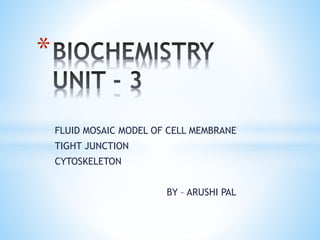
  • 1. FLUID MOSAIC MODEL OF CELL MEMBRANE TIGHT JUNCTION CYTOSKELETON BY – ARUSHI PAL *
  • 2. *The fluid mosaic model was first proposed by S. J SINGER and GARTH L. NICLSON in 1972 to explain the structure of the plasma membrane . *WHAT IS FLUID MOSAIC MODEL ? The fluid mosaic model describe the cell membrane as a tapestry of several types of molecules ( phospholipid , cholesterol , and protein) that are constantly moving . This movement helps the cell membrane maintain its role as a barrier between the inside and outside of cell membrane.
  • 3. *Points regarding on FMM : *The study of fluid mosaic model is clearly studied after electron microscope . *It composed of lipid bilayer, lipid are fatty acid
  • 4. Hydrophobic tail – present inside Hydrophilic head – present outside , because to prevent hydrophobic tail from aqueous enviroment lipid component mainly consist of phosphoglyceride they also contain protein and carbohydrate. RBC 52% protein and 40% lipids there are two types of protein
  • 5. *Integral protein *Peripheral protein *Integral protein – protein are on the buried area. *Peripheral protein – protein are on the surface area.
  • 6.
  • 7. * *TRANSPORT PASSIVE OSMOSIS ACTIVE 1- PASSIVE- If any molecule from this lipid bilayer, transport or travel without any energy called passive transport. Means no ATP is required.
  • 8. *2 – ACTIVE if any molecules from this lipid bilayer transport or travel with energy called active transport means ATP is required. Polar molecules like Na and K 3 – OSMOSIS Movement of water from high concentration to low concentration
  • 9. * *Phospholipid molecules shows two types of movement : Transition Flip flop movement movement 1- Transition movement : movement of phospholipid molecules in same direction . 2 - Flip flop movement : movement of phospholipid movement between two layers.
  • 11. * *Tight junction are areas where the membrane of two adjacent cell join together to form a barrier . * The cell membrane are connected by strands of transmembrane protein such as claudins and occludin. *Tight junction binds cell together , prevent molecules from passing in between cell and also helps to maintain the polarity of cells. They are only found in vertebrate , animals with a backbone and skeleton
  • 12. *
  • 13. * *To help cells from a barrier that prevent molecules from getting through and to stop protein in the cell membrane from moving around . *Tight junction are often found at epithelial cells ( which are cells that line the surface of the body and line body cavities .) * not only epithelial cell do separate the body from the surrounding environment , they also separate surface with in the body , therefore it is very important that the permeability of molecules through layer of epithelial cell is tightly controlled.
  • 14. *if molecules are blocked by tight junction and physically unable to pass through the space in between cells , they must enter through other methods that involve entering the cells themselves. *They could pass through special protein in the celll membrane or be engulfed by the cell through endocytosis. *Using this method , the cell has greater control over what material it take in and allow to pass through epithelial cell , certain protein must be kept on certain sides of the cells.
  • 15. the apical or outside layer of the sheets of cells contains protein that only let certain substances pass through . The basal layer or inside layer is where cells let molecules pass through them by expelling them from their membrane in a process called exocytosis . the tight junction keep the correct proteins on the correct sides of the cells in order to function this also help maintain the polarity of cell. Another function is simply to hold cell together . The branching protein strands of tight junction link adjacent cell together tightly so that they from a sheet.
  • 16. * *Tight junction are a branching network of protein strands on the surface of a cell that link with each other through out the surface of the membrane . The strands formed by trans membrane protein on the surface of the cell membrane that are adjacent to each other. * there are about 40 different protein at tight these protein can be grouped into four main types. * 1 – trans membrane protein – are wedged in the middle of the cell membrane and are responsible for adhesion and permeability
  • 17. *2 – scaffolding proteins – organize transmembrane protein *3- signaling proteins – are responsible for forming the tight junction and regulating the barrier *4 – regulation proteins – regulate what protein are brought to the cell membrane in vesicle. *Claudins and occludin – are the two main types of protein present at tight junction and they are both transmembrane protein * * claudins – are important in forming tight junction *Occludin – play more of a role in keeping the tight junction stable and maintaining the barrier between cell that’s keeps unwanted molecules out .
  • 18. *Other cell junction : *Other cell junction are gap junction and anchoring junction *1 – gap junction – are channels in cells that let adjacent cell communicate with one another without having to send molecules through ECF surrounding the cell. *2 - anchoring junction – hold cell together with anchoring protein such as catenins and cadherins . The cell cytoskeleton is tethered to proteins that link adjacent cell. * * a type of cell junction in which cells are connected by a mass of protein ( anchoring junction ) * a types of cell junction that allows adjacent cell to exchange molecule ( gap junction )
  • 19.
  • 20. * *The cytoskeleton is responsible for cell shape , motility ( movement ) of the cell as a whole and motility of organelles with in a cells . *There are the three types of filaments in the cytoplasm of most vertebrate cells: * 1- microfilaments * 2 – microtubules * 3 – intermediate filaments
  • 21. * *The thickest are the microtubules ( 20 nm in a diameter ) which consist primarily of tubulin protein . *Each tubulin subunit is a made of one alpha and one beta tubulin that are attached to each other , so technically tubulin is a heterodimer not a monomer . *Since it looks like tube , it is named as microtubule *It is pipe like structure . *In a microtubules structure tubulin monomers are linked both at their ends along their sides ( laterally ) . This means that microtubules have distinct ends , called the positive and negative ends
  • 22.
  • 23. * *Transportation of water , ions or small molecules . *Cytoplasmic streaming . *Formation of fibers or aster of the mitotic or meiotic spindle during cell division . *Formation of structural unit of the centrioles basal granules , cilia and flagella *
  • 24. * *Tough and flexible *The thinnest are the microfilaments ( 7nm in a diameter ) which are solid and are principally made of two strands of a globular proteins called actin . *For this reason , microfilaments are known as actin filaments.
  • 25.
  • 26. *Actin is powered by ATP to assemble filaments from , which serves as a track for movements of a motor protein called myosin . *FUNCTION: 1- they maintain the shape of the cell . 2- From contractile component of cell mainly of the muscles cell. 3- WBC can move to the site of an infection and engulf the pathogen due to microfilaments.
  • 27. * *The of the middle order are called intermediate filaments 10nm in a diameter . * they have been classified according to their constitute proteins such as desmin filaments , keratin filaments , neurofilaments and glial filaments . *FUNCTION : * it contribute to cellular structural elements and are often crucial in holding together tissue like skin .
  • 28.
  • 29. * *The cytoskeleton is responsible for lots of important cellular function : * providing cell shape . *It allows cells to move . *Engulf particles . *Transport vesicles through cytosol. *Separate chromosome during cell division. *It allow our muscles to contract.