SlideShare uma empresa Scribd logo
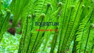
EQUISETUM
Structure and Life Cycle
Habitat of Equisetum:
• The plant body of Equisetum has an aerial part and an underground
rhizome part. The rhizome is perennial, horizontal, branched and
creeping in nature. The aerial part is herbaceous and usually annual.
Majority of the species are small with a size range in between 15 and
60 cm in height and 2.0 cm in diameter.
• Equisetums generally grow in wet or damp habitats and are
particularly common along the banks of streams or irrigation canals
(E. debile, E. palustre). However, some species are adapted to xeric
condition (e.g., Equisetum arvense).
Some species of Equisetum are indicators of the mineral content of the
soil in which they grow. Some species accumulate gold (about 4.5
ounce per ton of dry wt.), thus they are considered as ‘gold indicator
plants.
Structure of Equisetum:
The Sporophyte:
The sporophytic plant body of Equisetum is differentiated into
 Stem
 Roots
 Leaves
 Stem:
The stem of Equisetum has two parts: perennial, underground, much-branched
rhizome and an erect, usually annual aerial shoot. The branching is monopodial,
shoots are differentiated into nodes and internodes.
• Some shoots are profusely branched, green (chlorophyllous) and purely vegetative. The
others are fertile, unbranched, brownish in colour (achlorophyllous) and have terminal
strobili.
• The underground rhizome and the aerial axis appear to be articulated or jointed due to the
presence of distinct nodes and internodes. Externally, the internodes have longitudinal ridges
and furrows and, internally, they are hollow, tube-like structures. The ridges of the successive
internodes alternate with each other and the leaves are normally of the same number as the
ridges on the stem.
Internal Features of Stem:
 In T.S., the stem of Equisetum appears wavy in outline with ridges and furrows.
The epidermal cell walls are thick, cuticularised and have a deposition of
siliceous material.
 Stomata are distributed only in the furrows between the ridges. A hypodermal
sclerenchymatous zone is present below each ridge which may extend up to
stele in E. giganteum. The cortex is differentiated into outer and inner regions.
 The outer cortex is chlorenchymatous, while the inner cortex is made up of
thin-walled parenchymatous cells. There is a large air cavity in the inner
cortex corresponding to each furrow and alternating with the ridges, known
as vallecular canal.
 New leaves and branches of Equisetum are produced by the apical
meristem, however, most of the length of the stem are due to the activity of
intercalary meristem located just above each node. The activity of
intercalary meristem causes rapid elongation of the inter- nodal region.
 The vascular bundles are arranged in a ring which lies opposite to the ridges in
position and alternate with the vallecular canals of the cortex. Vascular bundles
are conjoint, collateral and closed. In the mature vascular bundle, protoxylum is
disorganised to form a carinal cavity which lies opposite to the ridges.
 The vascular bundles remain unbranched until they reach the level of node. At
the nodal region, each vascular bandle trifurcates (divided into three parts).
The xerophytic features are:
(i) Ridges and furrows in the stem,
(ii) Deposition of silica in the epidermal cells,
(iii) Sunken stomata,
(iv) Sclerenchymatous hypodermis,
(v) Reduced and scaly leaves, and
(vi) photosynthetic tissue in the stem.
The hydrophytic characteristics on the other hand are:
(i) Well-developed aerating system like carinal
canal, vallecular canal and central pith cavity.
(ii) Reduced vascular elements.
Root:
 The primary root is ephemeral. The slender adventitious roots arise endogenously at the
nodes of the stems. In T.S., the root shows epidermis, cortex and stele from periphery to the
centre. The epidermis consists of elongated cells, with or without root hairs.
 The cortex is extensive; cells of the outer cortex often have thick walls (sclerenchymatous)
and those of the inner cortex are thinner parenchymatous.
A large metaxylem element is present in the centre of the stele
and the protoxylem strands lie around it. The space between the
protoxylem groups is filled with phloem. There is no pith.
Leaves:
The leaves of Equisetum are small, simple, scale-like and isophyllous; they are
attached at each node, united at least for a part of the length and thus form a
sheath around the stem. The sheath has free and pointed teeth-like tips.
The number of leaves per node varies according to the species. The species with
narrow stems have few leaves (e.g., 2-3 leaves in E. scirpoides) and those with
thick stem have many leaves (e.g., up to 40 leaves in E. schaffneri).
The number of leaves at a node corresponds to the number of ridges
on the internode below. The leaves do not perform any photosynthetic
function and their main function is to provide protection to young
buds at the node.
Life cycle:
• Sex Organs of Equisetum:
• i. Antheridium:
• In monoecious species, antheridia develop later than archegonia. They are of
two types — projecting type and embedded type. Antheridia first appear on the
lobes of the gametophyte. The periclinal division of the superficial antheridial
initial gives rise to jacket initial and an androgonial cell.
The jacket initial divides anticlinally to form a single-layered jacket.
The repeated divisions of androgonial cells form numerous cells
which, on metamorphosis, produce spermatids/antherozoids. The
antherozoids escape through a pore created by the separation of the
apical jacket cell. The apical part of the antherozoid is spirally coiled,
whereas the lower part is, to some extent, expanded.
ii. Archegonium:
Any superficial cell in the marginal meristem acts as an archegonial initial which undergoes
periclinal division to form a primary cover cell and an inner central cell. The cover cell, by
two vertical divisions at right angle to each other, forms a neck. The central cell divides
transversely to form a primary neck canal cell and a venter cell.
Two neck canal cells are produced from the primary neck canal cell. While,
the venter cell, by a transverse division, forms the ventral canal cell and an
egg.
At maturity, an archegonium has a projecting neck comprising of three to
four tiers of neck cells arranged in four rows, two neck canal cells of
unequal size, a ventral canal cell, and an egg at the base of the embedded
venter.
Fertilization:
Water is essential for fertilization. The gametophyte must be covered with a thin layer of
water in which the motile antherozoides swim to the archegonia. The neck canal cells
and ventral canal cell of the archegonia disintegrate to form a passage for the entry of
antherozoids.
Many antherozoids pass through the canal of the archegonium but only one of them
fuses with the egg. Thus diploid zygote is formed. Generally more than one archegonia
are fertilized in a prothallus.
Embryo (The New Sporophyte):
The embryo is the mother cell of the next sporophytic generation. Unlike most
pteridophytes, several sporophytes develop on the same prothallus. The first
division of the zygote is transverse. This results in an upper epibasal cell and
lower hypobasal cell. The embryo is therefore exoscopic (where the apical cell is
duacted outward. No suspensor is formed in Equisetum.
The epibasal and hypobasal cells then divide at right angles to the
oogonial wall, and as a result a tour-celled quadrant stage is established.
All the four cells of the quadrant are of different size and shape.
The four-celled embryo undergoes subsequent divisions and the future
shoot apex originate from the largest cell and leaf initials from the remai-
ning cells of one quadrant of the epibasal hemisphere.
Later the root grows directly downward and penetrate the gametophytic tissue
to reach the soil or substratum. A number of such sporophytes may develop
from a large mature gametophyte if more than one egg is fertilized
"Equisetum" Structural development Reproduction

Mais conteúdo relacionado

Mais procurados

Mais procurados (20)

ginkoales.pptx
ginkoales.pptxginkoales.pptx
ginkoales.pptx
 
Psilotum
PsilotumPsilotum
Psilotum
 
Anthoceros
AnthocerosAnthoceros
Anthoceros
 
Anthoceros
AnthocerosAnthoceros
Anthoceros
 
Chlorophyta
ChlorophytaChlorophyta
Chlorophyta
 
Marchantia ppt
Marchantia pptMarchantia ppt
Marchantia ppt
 
Riccia ppt
Riccia ppt Riccia ppt
Riccia ppt
 
Notothylas by p.n
Notothylas by p.nNotothylas by p.n
Notothylas by p.n
 
Pteris ppt
Pteris pptPteris ppt
Pteris ppt
 
5 2 -pinus
5   2 -pinus5   2 -pinus
5 2 -pinus
 
Structure, reproduction, life history and systematic position of Lycopodium
Structure, reproduction, life history and systematic position of LycopodiumStructure, reproduction, life history and systematic position of Lycopodium
Structure, reproduction, life history and systematic position of Lycopodium
 
Marchantia
MarchantiaMarchantia
Marchantia
 
Anthoceros ppt
Anthoceros pptAnthoceros ppt
Anthoceros ppt
 
Oedogonium
OedogoniumOedogonium
Oedogonium
 
Selaginella ppt
Selaginella pptSelaginella ppt
Selaginella ppt
 
APOGAMY & APOSPORY.pptx
APOGAMY & APOSPORY.pptxAPOGAMY & APOSPORY.pptx
APOGAMY & APOSPORY.pptx
 
Anthoceros.ppt
Anthoceros.pptAnthoceros.ppt
Anthoceros.ppt
 
Morphological, anatomical and reproductive characters of Marchantia
Morphological, anatomical and reproductive characters of MarchantiaMorphological, anatomical and reproductive characters of Marchantia
Morphological, anatomical and reproductive characters of Marchantia
 
Psilotum
PsilotumPsilotum
Psilotum
 
Life cycle of pinus
Life cycle of pinusLife cycle of pinus
Life cycle of pinus
 

Semelhante a "Equisetum" Structural development Reproduction

Pteris : features, anatomy, morphology and lifecycle
Pteris : features, anatomy, morphology and lifecyclePteris : features, anatomy, morphology and lifecycle
Pteris : features, anatomy, morphology and lifecycle
Cherry
 
MARSILEA notes in detail for II year Botany.ppt
MARSILEA  notes in detail for II year Botany.pptMARSILEA  notes in detail for II year Botany.ppt
MARSILEA notes in detail for II year Botany.ppt
aigil2
 

Semelhante a "Equisetum" Structural development Reproduction (20)

Pteris : features, anatomy, morphology and lifecycle
Pteris : features, anatomy, morphology and lifecyclePteris : features, anatomy, morphology and lifecycle
Pteris : features, anatomy, morphology and lifecycle
 
Sphenopsida details
Sphenopsida detailsSphenopsida details
Sphenopsida details
 
Vegetative characters of equisetum
Vegetative characters of equisetumVegetative characters of equisetum
Vegetative characters of equisetum
 
Life-cycle-of-Sphagnum.pdf
Life-cycle-of-Sphagnum.pdfLife-cycle-of-Sphagnum.pdf
Life-cycle-of-Sphagnum.pdf
 
Pteridophytes
PteridophytesPteridophytes
Pteridophytes
 
Gametofite and sporofite in bryopsida
Gametofite and sporofite in bryopsidaGametofite and sporofite in bryopsida
Gametofite and sporofite in bryopsida
 
Type polytrichum
Type polytrichumType polytrichum
Type polytrichum
 
2. anthocerospp
2. anthocerospp2. anthocerospp
2. anthocerospp
 
Funaria
FunariaFunaria
Funaria
 
Marchantia jb
Marchantia jbMarchantia jb
Marchantia jb
 
Marchantia, Dr.V.Vijaya, Assistant Professor of Botany, E.M.G. Yadava Women's...
Marchantia, Dr.V.Vijaya, Assistant Professor of Botany, E.M.G. Yadava Women's...Marchantia, Dr.V.Vijaya, Assistant Professor of Botany, E.M.G. Yadava Women's...
Marchantia, Dr.V.Vijaya, Assistant Professor of Botany, E.M.G. Yadava Women's...
 
Reproduction In Sphenopsida
Reproduction In SphenopsidaReproduction In Sphenopsida
Reproduction In Sphenopsida
 
Bryopsida – funaria
Bryopsida – funariaBryopsida – funaria
Bryopsida – funaria
 
Ginko
GinkoGinko
Ginko
 
Riccia
RicciaRiccia
Riccia
 
Riccia
RicciaRiccia
Riccia
 
Riccia Bryophytes presentation
Riccia             Bryophytes         presentationRiccia             Bryophytes         presentation
Riccia Bryophytes presentation
 
Marsilea details
Marsilea detailsMarsilea details
Marsilea details
 
Anatomy Of Coniferales.pptx
Anatomy Of Coniferales.pptxAnatomy Of Coniferales.pptx
Anatomy Of Coniferales.pptx
 
MARSILEA notes in detail for II year Botany.ppt
MARSILEA  notes in detail for II year Botany.pptMARSILEA  notes in detail for II year Botany.ppt
MARSILEA notes in detail for II year Botany.ppt
 

Último

Gliese 12 b: A Temperate Earth-sized Planet at 12 pc Ideal for Atmospheric Tr...
Gliese 12 b: A Temperate Earth-sized Planet at 12 pc Ideal for Atmospheric Tr...Gliese 12 b: A Temperate Earth-sized Planet at 12 pc Ideal for Atmospheric Tr...
Gliese 12 b: A Temperate Earth-sized Planet at 12 pc Ideal for Atmospheric Tr...
Sérgio Sacani
 
Detectability of Solar Panels as a Technosignature
Detectability of Solar Panels as a TechnosignatureDetectability of Solar Panels as a Technosignature
Detectability of Solar Panels as a Technosignature
Sérgio Sacani
 
Pests of sugarcane_Binomics_IPM_Dr.UPR.pdf
Pests of sugarcane_Binomics_IPM_Dr.UPR.pdfPests of sugarcane_Binomics_IPM_Dr.UPR.pdf
Pests of sugarcane_Binomics_IPM_Dr.UPR.pdf
PirithiRaju
 
Pests of Green Manures_Bionomics_IPM_Dr.UPR.pdf
Pests of Green Manures_Bionomics_IPM_Dr.UPR.pdfPests of Green Manures_Bionomics_IPM_Dr.UPR.pdf
Pests of Green Manures_Bionomics_IPM_Dr.UPR.pdf
PirithiRaju
 
The solar dynamo begins near the surface
The solar dynamo begins near the surfaceThe solar dynamo begins near the surface
The solar dynamo begins near the surface
Sérgio Sacani
 
Aerodynamics. flippatterncn5tm5ttnj6nmnynyppt
Aerodynamics. flippatterncn5tm5ttnj6nmnynypptAerodynamics. flippatterncn5tm5ttnj6nmnynyppt
Aerodynamics. flippatterncn5tm5ttnj6nmnynyppt
sreddyrahul
 
Climate extremes likely to drive land mammal extinction during next supercont...
Climate extremes likely to drive land mammal extinction during next supercont...Climate extremes likely to drive land mammal extinction during next supercont...
Climate extremes likely to drive land mammal extinction during next supercont...
Sérgio Sacani
 
The importance of continents, oceans and plate tectonics for the evolution of...
The importance of continents, oceans and plate tectonics for the evolution of...The importance of continents, oceans and plate tectonics for the evolution of...
The importance of continents, oceans and plate tectonics for the evolution of...
Sérgio Sacani
 

Último (20)

Ostiguy & Panizza & Moffitt (eds.) - Populism in Global Perspective. A Perfor...
Ostiguy & Panizza & Moffitt (eds.) - Populism in Global Perspective. A Perfor...Ostiguy & Panizza & Moffitt (eds.) - Populism in Global Perspective. A Perfor...
Ostiguy & Panizza & Moffitt (eds.) - Populism in Global Perspective. A Perfor...
 
Tissue engineering......................pptx
Tissue engineering......................pptxTissue engineering......................pptx
Tissue engineering......................pptx
 
National Biodiversity protection initiatives and Convention on Biological Di...
National Biodiversity protection initiatives and  Convention on Biological Di...National Biodiversity protection initiatives and  Convention on Biological Di...
National Biodiversity protection initiatives and Convention on Biological Di...
 
Gliese 12 b: A Temperate Earth-sized Planet at 12 pc Ideal for Atmospheric Tr...
Gliese 12 b: A Temperate Earth-sized Planet at 12 pc Ideal for Atmospheric Tr...Gliese 12 b: A Temperate Earth-sized Planet at 12 pc Ideal for Atmospheric Tr...
Gliese 12 b: A Temperate Earth-sized Planet at 12 pc Ideal for Atmospheric Tr...
 
METHODS OF TRANSCRIPTOME ANALYSIS....pptx
METHODS OF TRANSCRIPTOME ANALYSIS....pptxMETHODS OF TRANSCRIPTOME ANALYSIS....pptx
METHODS OF TRANSCRIPTOME ANALYSIS....pptx
 
NuGOweek 2024 full programme - hosted by Ghent University
NuGOweek 2024 full programme - hosted by Ghent UniversityNuGOweek 2024 full programme - hosted by Ghent University
NuGOweek 2024 full programme - hosted by Ghent University
 
Detectability of Solar Panels as a Technosignature
Detectability of Solar Panels as a TechnosignatureDetectability of Solar Panels as a Technosignature
Detectability of Solar Panels as a Technosignature
 
GBSN - Microbiology (Lab 1) Microbiology Lab Safety Procedures
GBSN -  Microbiology (Lab  1) Microbiology Lab Safety ProceduresGBSN -  Microbiology (Lab  1) Microbiology Lab Safety Procedures
GBSN - Microbiology (Lab 1) Microbiology Lab Safety Procedures
 
electrochemical gas sensors and their uses.pptx
electrochemical gas sensors and their uses.pptxelectrochemical gas sensors and their uses.pptx
electrochemical gas sensors and their uses.pptx
 
Pests of sugarcane_Binomics_IPM_Dr.UPR.pdf
Pests of sugarcane_Binomics_IPM_Dr.UPR.pdfPests of sugarcane_Binomics_IPM_Dr.UPR.pdf
Pests of sugarcane_Binomics_IPM_Dr.UPR.pdf
 
Pests of Green Manures_Bionomics_IPM_Dr.UPR.pdf
Pests of Green Manures_Bionomics_IPM_Dr.UPR.pdfPests of Green Manures_Bionomics_IPM_Dr.UPR.pdf
Pests of Green Manures_Bionomics_IPM_Dr.UPR.pdf
 
GBSN - Microbiology (Lab 2) Compound Microscope
GBSN - Microbiology (Lab 2) Compound MicroscopeGBSN - Microbiology (Lab 2) Compound Microscope
GBSN - Microbiology (Lab 2) Compound Microscope
 
The solar dynamo begins near the surface
The solar dynamo begins near the surfaceThe solar dynamo begins near the surface
The solar dynamo begins near the surface
 
Aerodynamics. flippatterncn5tm5ttnj6nmnynyppt
Aerodynamics. flippatterncn5tm5ttnj6nmnynypptAerodynamics. flippatterncn5tm5ttnj6nmnynyppt
Aerodynamics. flippatterncn5tm5ttnj6nmnynyppt
 
Hemoglobin metabolism: C Kalyan & E. Muralinath
Hemoglobin metabolism: C Kalyan & E. MuralinathHemoglobin metabolism: C Kalyan & E. Muralinath
Hemoglobin metabolism: C Kalyan & E. Muralinath
 
word2vec, node2vec, graph2vec, X2vec: Towards a Theory of Vector Embeddings o...
word2vec, node2vec, graph2vec, X2vec: Towards a Theory of Vector Embeddings o...word2vec, node2vec, graph2vec, X2vec: Towards a Theory of Vector Embeddings o...
word2vec, node2vec, graph2vec, X2vec: Towards a Theory of Vector Embeddings o...
 
Climate extremes likely to drive land mammal extinction during next supercont...
Climate extremes likely to drive land mammal extinction during next supercont...Climate extremes likely to drive land mammal extinction during next supercont...
Climate extremes likely to drive land mammal extinction during next supercont...
 
Structural annotation................pptx
Structural annotation................pptxStructural annotation................pptx
Structural annotation................pptx
 
Topography and sediments of the floor of the Bay of Bengal
Topography and sediments of the floor of the Bay of BengalTopography and sediments of the floor of the Bay of Bengal
Topography and sediments of the floor of the Bay of Bengal
 
The importance of continents, oceans and plate tectonics for the evolution of...
The importance of continents, oceans and plate tectonics for the evolution of...The importance of continents, oceans and plate tectonics for the evolution of...
The importance of continents, oceans and plate tectonics for the evolution of...
 

"Equisetum" Structural development Reproduction

  • 2. Habitat of Equisetum: • The plant body of Equisetum has an aerial part and an underground rhizome part. The rhizome is perennial, horizontal, branched and creeping in nature. The aerial part is herbaceous and usually annual. Majority of the species are small with a size range in between 15 and 60 cm in height and 2.0 cm in diameter. • Equisetums generally grow in wet or damp habitats and are particularly common along the banks of streams or irrigation canals (E. debile, E. palustre). However, some species are adapted to xeric condition (e.g., Equisetum arvense).
  • 3.
  • 4. Some species of Equisetum are indicators of the mineral content of the soil in which they grow. Some species accumulate gold (about 4.5 ounce per ton of dry wt.), thus they are considered as ‘gold indicator plants.
  • 5. Structure of Equisetum: The Sporophyte: The sporophytic plant body of Equisetum is differentiated into  Stem  Roots  Leaves  Stem: The stem of Equisetum has two parts: perennial, underground, much-branched rhizome and an erect, usually annual aerial shoot. The branching is monopodial, shoots are differentiated into nodes and internodes.
  • 6. • Some shoots are profusely branched, green (chlorophyllous) and purely vegetative. The others are fertile, unbranched, brownish in colour (achlorophyllous) and have terminal strobili. • The underground rhizome and the aerial axis appear to be articulated or jointed due to the presence of distinct nodes and internodes. Externally, the internodes have longitudinal ridges and furrows and, internally, they are hollow, tube-like structures. The ridges of the successive internodes alternate with each other and the leaves are normally of the same number as the ridges on the stem.
  • 7.
  • 8.
  • 9. Internal Features of Stem:  In T.S., the stem of Equisetum appears wavy in outline with ridges and furrows. The epidermal cell walls are thick, cuticularised and have a deposition of siliceous material.  Stomata are distributed only in the furrows between the ridges. A hypodermal sclerenchymatous zone is present below each ridge which may extend up to stele in E. giganteum. The cortex is differentiated into outer and inner regions.
  • 10.  The outer cortex is chlorenchymatous, while the inner cortex is made up of thin-walled parenchymatous cells. There is a large air cavity in the inner cortex corresponding to each furrow and alternating with the ridges, known as vallecular canal.  New leaves and branches of Equisetum are produced by the apical meristem, however, most of the length of the stem are due to the activity of intercalary meristem located just above each node. The activity of intercalary meristem causes rapid elongation of the inter- nodal region.
  • 11.  The vascular bundles are arranged in a ring which lies opposite to the ridges in position and alternate with the vallecular canals of the cortex. Vascular bundles are conjoint, collateral and closed. In the mature vascular bundle, protoxylum is disorganised to form a carinal cavity which lies opposite to the ridges.  The vascular bundles remain unbranched until they reach the level of node. At the nodal region, each vascular bandle trifurcates (divided into three parts).
  • 12.
  • 13. The xerophytic features are: (i) Ridges and furrows in the stem, (ii) Deposition of silica in the epidermal cells, (iii) Sunken stomata, (iv) Sclerenchymatous hypodermis, (v) Reduced and scaly leaves, and (vi) photosynthetic tissue in the stem.
  • 14. The hydrophytic characteristics on the other hand are: (i) Well-developed aerating system like carinal canal, vallecular canal and central pith cavity. (ii) Reduced vascular elements.
  • 15. Root:  The primary root is ephemeral. The slender adventitious roots arise endogenously at the nodes of the stems. In T.S., the root shows epidermis, cortex and stele from periphery to the centre. The epidermis consists of elongated cells, with or without root hairs.  The cortex is extensive; cells of the outer cortex often have thick walls (sclerenchymatous) and those of the inner cortex are thinner parenchymatous.
  • 16. A large metaxylem element is present in the centre of the stele and the protoxylem strands lie around it. The space between the protoxylem groups is filled with phloem. There is no pith.
  • 17.
  • 18.
  • 19. Leaves: The leaves of Equisetum are small, simple, scale-like and isophyllous; they are attached at each node, united at least for a part of the length and thus form a sheath around the stem. The sheath has free and pointed teeth-like tips. The number of leaves per node varies according to the species. The species with narrow stems have few leaves (e.g., 2-3 leaves in E. scirpoides) and those with thick stem have many leaves (e.g., up to 40 leaves in E. schaffneri).
  • 20. The number of leaves at a node corresponds to the number of ridges on the internode below. The leaves do not perform any photosynthetic function and their main function is to provide protection to young buds at the node.
  • 21.
  • 22. Life cycle: • Sex Organs of Equisetum: • i. Antheridium: • In monoecious species, antheridia develop later than archegonia. They are of two types — projecting type and embedded type. Antheridia first appear on the lobes of the gametophyte. The periclinal division of the superficial antheridial initial gives rise to jacket initial and an androgonial cell.
  • 23. The jacket initial divides anticlinally to form a single-layered jacket. The repeated divisions of androgonial cells form numerous cells which, on metamorphosis, produce spermatids/antherozoids. The antherozoids escape through a pore created by the separation of the apical jacket cell. The apical part of the antherozoid is spirally coiled, whereas the lower part is, to some extent, expanded.
  • 24. ii. Archegonium: Any superficial cell in the marginal meristem acts as an archegonial initial which undergoes periclinal division to form a primary cover cell and an inner central cell. The cover cell, by two vertical divisions at right angle to each other, forms a neck. The central cell divides transversely to form a primary neck canal cell and a venter cell.
  • 25. Two neck canal cells are produced from the primary neck canal cell. While, the venter cell, by a transverse division, forms the ventral canal cell and an egg. At maturity, an archegonium has a projecting neck comprising of three to four tiers of neck cells arranged in four rows, two neck canal cells of unequal size, a ventral canal cell, and an egg at the base of the embedded venter.
  • 26.
  • 27.
  • 28. Fertilization: Water is essential for fertilization. The gametophyte must be covered with a thin layer of water in which the motile antherozoides swim to the archegonia. The neck canal cells and ventral canal cell of the archegonia disintegrate to form a passage for the entry of antherozoids. Many antherozoids pass through the canal of the archegonium but only one of them fuses with the egg. Thus diploid zygote is formed. Generally more than one archegonia are fertilized in a prothallus.
  • 29. Embryo (The New Sporophyte): The embryo is the mother cell of the next sporophytic generation. Unlike most pteridophytes, several sporophytes develop on the same prothallus. The first division of the zygote is transverse. This results in an upper epibasal cell and lower hypobasal cell. The embryo is therefore exoscopic (where the apical cell is duacted outward. No suspensor is formed in Equisetum.
  • 30. The epibasal and hypobasal cells then divide at right angles to the oogonial wall, and as a result a tour-celled quadrant stage is established. All the four cells of the quadrant are of different size and shape. The four-celled embryo undergoes subsequent divisions and the future shoot apex originate from the largest cell and leaf initials from the remai- ning cells of one quadrant of the epibasal hemisphere.
  • 31.
  • 32. Later the root grows directly downward and penetrate the gametophytic tissue to reach the soil or substratum. A number of such sporophytes may develop from a large mature gametophyte if more than one egg is fertilized