Inner body tube of nematodes
•Transferir como PPTX, PDF•
2 gostaram•3,196 visualizações

for agriculture students

Recomendados
Mais conteúdo relacionado
Mais procurados
Mais procurados (20)
Semelhante a Inner body tube of nematodes
Semelhante a Inner body tube of nematodes (20)
Mais de AnurAg Kerketta
Mais de AnurAg Kerketta (20)
Último
Último (20)
Inner body tube of nematodes
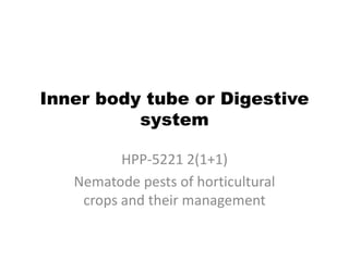
- 1. Inner body tube or Digestive system HPP-5221 2(1+1) Nematode pests of horticultural crops and their management
- 2. Magnify view of anatomy of nematode
- 3. Inner body tube • The digestive system of nematodes includes the stoma, oesophagus, intestine and posterior gut. The inner body tube is divided into 3 main regions. 1. Stomodeum : which constitute the stoma, oesophagus and cardia. 2. Mesenteron : which constitute the intestine. 3. Proctodeum : which is the posterior –most region comprising rectum and anal opening.
- 4. 1. Stomodeum • Stoma is the portion of the inner body tube lying between the oral opening and oesophagus. It includes: mouth and lips, the stoma and the oesophagus • The stomatal opening is small and slit like and is surrounded by six lips
- 5. Mouth and lips • The and lips are also associated with the feeding activity. • Generally there are 6 lips which surround the mouth. • In some cases they may be reduced by partial fusion of 3 or by complete fusion to form a united ring around the mouth.
- 6. Stoma or buccal cavity • Mouth cavity or buccal cavity forms the feeding apparatus and lie between the mouth and oesophagus. • Plant parasitic nematodes are armed with a protrusible stylet which is usually hollow and functions like a hypodermic needle. • In, Secernentea, the stylet is thought to be derived from fusion of the stomatal lining and therefore called as stomatostylet.
- 7. Stylet of plant parasitic nematode
- 8. • The buccal spear is also found in other parasitic as well as predatory nematodes. • The stomatostylet is developed from the fusion of the walls of the buccal cavity. • In order dorylaimida the stylet arises in two pieces, a buccal portion, embedded in the oesophageal wall (odontostyle extension or odontophore) and a replaceable stylet odontostyle or onchiostyle. • The replaceable stylet formed within the oesophagus. At each moult, extension of the cell moves the stylet forward to displace the old stylet from its basal extension and a new stylet is formed.
- 9. Oesophagus or pharynx • It is the largest part of the Stomodeum and found between stoma and intestine. The pharynx is mainly a food transporter pumping food from the low pressure stoma to the high pressure intestine. • Internally the pharynx is lined with cuticle and externally by a membrane (basal lamella). It contains radial muscle, oesophageal glands and valves, which prevent the regurgitation of food.
- 12. • The oesophago-intestinal valve or cardia is a part of stomodeum, which lies at the junction of oesophagus and intestine. • It controls the passage of food from oesophagus to intestines in uni-directional flow.
- 13. 2. Mesenteron or intestine • The intestine is hollow generally a straight tube formed from single layer of epithelial cells backed by a well developed basal lamina. • In some nematode three distinct region could be distinguished which are the anterior or ventricular region, the mid intestinal region and the posterior or prerectal region. • The plasma membrane which lines the lumen of the intestine is thrown into fine finger like projection known as microvilli. They increase the surface area of the intestine and are both secretary and absorptive in function.
- 15. Microvilli seen on the inner lining • The intestinal cell is surrounded by a plasma membrane the whole intestine is separated from the pseudocoelom by a basement membrane. • These cells are rich in mitochondria golgicomplex, ER, ribosome, glycogen, lipid, fatty acid etc. • The food moves in intestine by the ingestion of more food and also by the locomotory activity of the nematode
- 16. 3. Protodeum • Proctodeum comprises rectum and anus. • The intestinal tube is connected with a narrow small tube at the posterior end, through a valve known as rectum. • It regulates the flow of undigested food material which is to be passed outside the nematode body through a ventrally located aperture known as anus. Anal opening
- 17. • In male nematode, the rectum joins with the hind part of the testis forming a common opening known as cloaca. In female, there is a separate opening. Glands • Oesophageal and rectal glands are present in nematodes. The oesophageal gland enter the stomodeum and rectal gland enter proctodeum.
- 18. • Three uninucleated oesophageal glands. One gland on dorsal and other tow ventro lateral or sub ventral in position. These gland connect with the lumen of the oesophagus by means of ducts, often by means of a terminal swelling or ampulla. • The oesophageal glands have important role in hatching host penetration and also establishment of host parasitic relationship. Rectal glands • Are responsible for the copious production of gelatinous mucopolysaccharides matrix in which eggs are deposited as a mass. It protects the eggs from adverse environmental conditions.