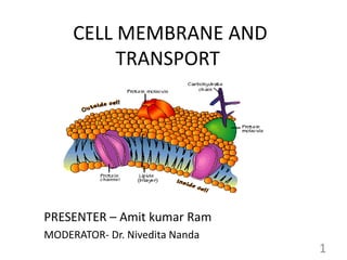
2. Contents :
Introduction
Cell membrane structure
Classification of Transport system
Associated disorders
Summary
2
3. Cell Membrane
“Possibly the decisive step [in the
origin of life] was the formation of
the first cell, in which chain
molecules were enclosed by a semi-
permeable membrane which kept
them together but let their food in.”
James Danielli
(1911-1984)
3
4. Cell Membrane
Defined as a biological membrane or an outer membrane
of a cell.
Divide the internal space of eukaryotic cells into discrete
compartments to segregate processes and components
4
5. Cell Membrane functions
• Mechanical structure – maintain the physical integrity of
cell and hold the cytoskeleton in place.
• Selective permeability – Gases, hydrophibic and small non
polar molecules can easily pass through it.
• Transport – certain molecules pass through passively
other need various transporters.
• Markers and signalling – some surface proteins act as cell
marker and helps in cell signalling.
5
7. Fluid Mosaic model
• Proposed by Singer and Nicolson in 1972
• Composed of a phospholipid bilayer with a collage of
many different proteins, lipids and carbohydrates.
• A membrane is a mosaic
– Proteins and other molecules are embedded in a
framework of phospholipids.
• A membrane is fluid
– Most protein and phospholipid molecules can move
laterally.
– Flip-Flop movement is restricted and is catalysed by an
enzyme “Flippase”- important during membrane lipid
synthesis
7
9. Lipid composition varies across the two leaflets of
the same membrane.
9
Changes in distribution
have biological
consequences
Platelet is able to play
its role in clot
formation only when
phosphatidylserine
moves to outer leaflet.
Phosphatidylserine
exposure also act as
marker for programmed
cell death
10. Membrane proteins
• Classified depending on
type of interaction with
bilayer.
i. Integral membrane
proteins- pass through
bilayer
ii. Peripheral membrane
proteins- associate with
bilayer by non-covalent
interactions
iii. Lipid-anchored proteins.
10
11. Peripheral membrane proteins
Attached to membrane
through electrostatic
interaction and H-bond.
Extracted by treating
membrane with high salt or
alkaline pH
Serve as regulator for
membrane bound enzymes,
also may limit the mobility of
integral proteins.
Examples – Ankyrin, spectrin
11
12. Integral membrane proteins
• Held in the membrane by
hydrophobic interaction with
lipids
• Need detergent to remove.
• Types –
a) Single Transmembrane
-Glycophorin
a) Multiple Transmembrane
- Bacteriorhodopsin
12
13. Certain Integral Proteins Mediate Cell-Cell
Interactions and Adhesion
13
• Intigrin serve as binding
site for extracellular
proteins like Collagen and
Fibronectin
• It also regulate platelet
aggregation at the site of
wound.
• Mutation in intigrin gene
encoding CD18 cause
Leukocyte adhesion
deficiency in humans
14. Lipid-anchored membrane proteins
• Covalently linked to
membrane by short
oligosachharide linked to a
molecule of GPI embedded on
outer leaflet.
• Example –Scrapie protein PrPc
• Some linked to inner leaflet
like Ras and Src proteins have
been implicated in
transformation of normal cell
to malignant cell.
14
15. Lipid-anchored membrane proteins
• One of protein is responsible for
sleeping sickness.
• Protozoan parasite carried by
tsetse flies survives in blood by
virtue of dense cell surface coat
made of a GPI anchored
glycoprotein.(Eg- Transamidase
complex)
• Several hundreds of glycoprotein
variants to invade host immune
system.
15
Trypanosome brucie
16. Atomic Force
Microscopy (AFM) to
Visualize Membrane
Proteins
16
Purified E.coli
aquaporin
F0-chloroplast
ATP synthase
17. Six major functions of membrane proteins
Transport Enzymatic activity Signal transduction
Cell-cell recognition Intercellular joining Attachment to the
cytoskeleton ECM
17
19. Transport through cell membrane
Classification based on function
Membrane transport
Active Via mainly by
ATP-driven transporters
(pumps)
Passive
Simple
diffusion
Facilitated
Via various
transporters Via Ion channels
19
Primary Active
transport
Secondary
active transport
20. Transporters Can Be Grouped into Super
families Based on Their Structures
A. α Helix type channels
1. Voltage gated ion channel VIC superfamily
- Voltage-gated k+ channels
2. Major intrinsic protein family
- Aquaporins
3. Ligand gated ion channel
- Acetylcholine receptor
B. β barrel porins
- General bacterial porin (GBP) family
20
21. Transporters Can Be Grouped into Super
families Based on Their Structures
C. Pore forming toxins
- Diptheria toxin family
D. Porters : Uniports, symports, and antiporters
1. Sugar porter family
-GLUT1 glucose transporter of erythrocyte
2. Solute-Na+ transporter
- Na+ –glucose symporter in epithelial cells
3. HCO - transporters
- HCO - –Cl - antiporter 21
22. Transporters Can Be Grouped into Super
families Based on Their Structures
E. Non Ribosomal Synthesized porters
-Valinomycin carrier family
F. Diphosphate bond hydrolysis-driven transporters (use
PPi not ATP)
• ATP binding cassette superfamily
• A type ATPase superfamily
• P-type ATPase superfamily
22
24. Facilitated diffusion
• Carrier mediated and involves
transporter protein.
• A transporter protein reduces the
∆G€ for transmembrane diffusion
of the solute.
• It does this by forming non-
covalent interactions with the
dehydrated solute to replace the
H-bonding with water and by
providing hydrophilic
transmembrane passageway.
• Example – Glucose transporters.
24
26. The Glucose Transporter of Erythrocytes
Mediates Passive Transport
Proposed structure of GLUT1 – Type III Transmembrane
protein
• Contains 12 transmembrane α-helices of which 9 contains three or
more polar or charged amino acid residues often separated by
several hydrophobic residues.
26
27. Proposed structure of GLUT1
Fig : A helical wheel- distribution
of polar and non polar residues on
surface of helical segment
Fig: Side by side association of five or six
amphipathic helices, each with its polar
face oriented towards central cavity, can
produce a transmembrane channel lined
with polar and charged residues.
27
28. Model of glucose transport into erythrocytes by
GLUT1
The transporter exist in two conformation T1, with the
glucose-binding site exposed on the outer surface of the
plasma membrane, and T2, with its binding site exposed
on the inner surface.
28
29. Regulation by insulin of glucose transport by
GLUT4 into a myocyte
29
Type I
(juvenile
onset)
diabetes
mellitus
30. Active Transport
• Transport against electrochemical gradient.
• Thermodynamically unfavorable – coupled with
exergonic process
• ATP hydrolysis occurs.
• Two types- primary and secondary
30
31. Primary Active transport
• Directly utilizes metabolic energy for the transport
process.
• Includes ion pumps
• Na + K + ATPase
- maintenance of intracellular cations and thus cell volume
- Protein synthesis by maintaining high concn. of k +
- Maintains resting membrane potential
- Mediates action of hormones like thyroxine,
aldosterone, and insulin
31
32. Primary Active transport
• Ca + + ATPase
- Maintain high concn. Of Ca+ in ECF
- helps in storage of Ca+ in SR needed for instant muscle contraction
• H + K + ATPase – pumps H+ ion for HCL secretion
- Secrets H + ion into tubular fluid – urine acidification
Types Examples Location in cell
P-Type
(phosphorylation
type)
Na+K+ ATPase
Ca++ ATPase
Plasmamembrane
Sarcoplasmic
reticulum
V-type(Vacuolar type H+K+ ATPase Plasma membrane
F-type(Energy
coupling factor)
ATP synthase Inner Mitochondrial
membrane
ABC transporter CFTR proteins
MDR-1 protein
Plasma membrane
Plasma membrane 32
Classification of ATPase
33. Postulated mechanism
of Na+ and K+ transport
by the Na+K+ ATPase
Component of Digitalis
used to treat
congestive Heart
Failure
33
34. ABC Transporters
Pumps Amino-acids, metal ion
many hydrophobic compounds
including drugs, out of cells
against concentration gradients.
Multi-drug transporters(MDR1)
• responsible for resistance of
certain tumors to some
generally effective antitumor
drugs.
• Has broad substrate specificity
including chemotherapeutic
drugs adriamycin, doxorubicin,
and vinablastin.
ABC transporter
34
• ABCB4- Familial intrahepatic
cholestasis type3
• ABCC2- Dublin-Jhonson’s
Syndrome
• ABCD1- Adenoleukodystophy
• ATP7A- Menkes disease
• ATP7B- Wilson’s disease
35. Secondary active Transport
• Transport of one solute
against its concentration
gradient by using the
energy generated by
gradient of another
solute transport.
• Example – reabsorption
of glucose from kidney
tubule or intestine.
35
37. Ion channels
Salient features
• They are transmembrane proteins and may exist in α-
helical or β-barrel structure.
• Selective for one particular ion
• Different channels are available for Na+ k+, Ca++and Cl-
• Well regulated by presence of “gates”
• Two main types – Ligand-gated and Voltage-gated
• Activities are affected by certain drugs.
• Mutation of genes encoding them cause specific
disease.
37
38. Ligand-gated channels
• Specific molecule binds to receptor and opens the
channel.
• Example – Acetylcholine receptor in post synaptic
membrane. It is a complex of five subunits having
binding site for acetylcholine.
Structure of Acetylcholine receptor 38
39. Voltage gated channels
• Opens or close in response to
change in membrane potential
39
Voltage-gated Na+ channel of
neurons-
The voltage-sensing
mechanism involves
movement of helix 4
perpendicular to the plane of
the membrane in response to
a change in potential
40. Many naturally occuring toxins act as Ion Channels
Produced by Puffer fish, an ingredient of
Japanese delicacy Fugu act by binding to the
voltage gated Na+ channels of neurons and
preventing normal action potential.
Concentrated in shellfish which become
highly poisonous to organism higher up the
food chain
The active component of crurae
block the acetylcholine receptor
or k+ channels
40
41. Ionophores
• They are membrane shuttle for
specific ions.
• Produced by microorganisms and
used as antibiotics.
• Increase the permeability of
membranes by acting channel
formers, thus ion gradient is
dissipated.
• Two types – Mobile ion
carriers(Valinomycin) and channel
formers(Gramicidin)
Valinomycin, a
peptide ionophore
that binds K+
41
42. Water channel (Aquaporin)
• Family of Integral membrane proteins.
• Provide channels for rapid movement of water molecules
across all plasma membrane.
• Ten aquaporins are known in humans.
• RBC contain 2x10⁵ copies of AQP-1 per cell.
• Plasma membrane of PCT cells contain
five different
aquaporin types.
Structure of Aquaporin 42
44. Gap junction
• Structure that permits direct transfer of small
molecule(upto 1200Da).
• Composed of family of protein called connexins.
• Mutations in genes encoding connexins are associated
with cardiovascular diseases and X-linked form of charcot-
Marrie-tooth disease.
44
49. Receptor Mediated Endocytosis
• Phosphatidylinositol 4.5 bisphosphate(PIP2) and the protein
dynamin are necessary for the pinching off clathrin-coated
vesicles from the cell surface.
• Low density lipoprotein(LDL) molecule and its receptor are
internalized by means of coated pits containing the LDL
receptors.
• There is dark side to receptor-mediated endocytosis in that
viruses which cause such disease as hepatitis, poliomyelitis,
and AIDS initiate their damage by this mechanism.
• Iron toxicity also begins with excessive uptake due to
endocytosis. 49
53. Cystic Fibrosis
• Common in Caucasian population and are carriers.
• Obstruction of GIT & Respiratory tract leading to bacterial
infection of airway & death due to respiratory insufficiency
before the age of 30.
53
54. Myasthenia Gravis
• Auto immune disease
• Decrease in Ach receptors on motor end
plate.
• The anti-AchR antibodies compete with Ach
to bind to AchR, producing receptor
blockade.
54
55. Myasthenia Gravis
• Womens are more affected.
• The extraocular muscles and eyelids
are often involved in early course of
disease.
• Weakness increases during
prolonged use of muscle and
improves after rest or sleep.
• Treated by AchE inhibitors
- Pyridostigmine bromide
- Neostigmine bromide
55
56. Summary
• Membranes are complex structure composed of
phospholipid bilayer, proteins and carbohydrates.
• The Fluid –mosaic models forms a useful basis for
thinking about membrane structure.
• Integral proteins firmly embed the bilayer while
peripheral proteins are attached to the inner or outer
surface.
• Certain hydrophobic molecules freely diffuse across
membranes but the movement of other is restricted
because of their size or charge. 56
57. Summary
• Passive and active (usually ATP-dependent) mechanisms
maintain gradient of various ions across membrane.
• Glucose enter cells by facilitated diffusion via different
transporters.
• Na+ K+ ATPase is the key enzyme in regulating intracellular
concentration of Na+ and K+.
• Ligand or voltage gated ion channels transport charged
molecules(Na +, K+, Ca+ + etc.)
57
58. Summary
• Ionophores carries specific ions dissipating the energy of
electrochemical ion gradients.
• Water moves across membranes through aquaporins.
Defective aquaporins leads to certain disease like
Nephrogenic DI.
• Large molecules enter or leave cells through endocytosis
or exocytosis.
• Mutation that affect to structure of membrane
proteins(receptor, transporter, ion channels, enzymes, &
structural proteins) may cause disease.
• Examples include cystic fibrosis, Myesthenia Gravis.
58
59. References
• Cox. MM, Nelson. DL; Lehninger Principle of
Biochemistry; 5th edition.
• G.M. Cooper; The cell-A molecular approach; 5th edition.
• Gerald Karp; Cell and Molecular Biology 6th edition.
• Murray RK, Bender DA, Botham KM, Kennelly PJ et al;
Harper Illustrated Biochemistry; 28th edition.
• Harrison’s Principles of Internal Medicine 17th edition.
• Lodish H; Molecular Biology of Cell; 6th edition.
• Yeagle PL; The structure of Biological Membranes 2nd
edition.
• Clapham DE; Symmetry, selectivity and the 2003 Nobel
prize cell 2003; 115:641
59