Lameness diagnoses in equines
•Download as PPTX, PDF•
0 likes•97 views

Lameness diagnoses in equines

Recommended
More Related Content
What's hot
What's hot (20)
Similar to Lameness diagnoses in equines
Similar to Lameness diagnoses in equines (20)
More from DR AMEER HAMZA
More from DR AMEER HAMZA (20)
Recently uploaded
Recently uploaded (20)
Lameness diagnoses in equines
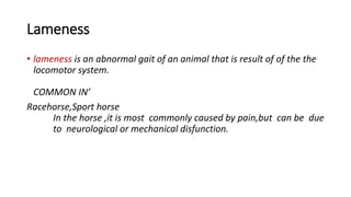
- 1. Lameness • lameness is an abnormal gait of an animal that is result of of the the locomotor system. COMMON IN’ Racehorse,Sport horse In the horse ,it is most commonly caused by pain,but can be due to neurological or mechanical disfunction.
- 2. CAUSES; • DUE TO ; • Pain • Orthropedic caus circulatory causes infectious causes mechanical and neurological lamness is also one of the cause of lameness
- 3. Mechanical lamness: • It is causes by physical abnormality such as scar tissue tht prevents normal motion of limb . • Eg;Upward fixation of patella and fibrotic myopathy ,but any type of adhesion or fibrosis can cause mechanical lameness.
- 4. Neurological lamness; • Due to result of infection ,trauma,toxicities,or congenital disease.signs are more commonly associated neurologic cause include unilateral muscle atrophy,peresis,paralysis,or dysmetria. • Stringhalt • Shivers • Wobbler disease • neoplasic
- 6. LAMNESS DIAGNOSIS IN EQUINES • Nuclear scintography nuclear scintography or bone scan involves injecting a radioactive substance often technectium into the horse and then measuring uptake ,which is strongest in the areas of rapid bone remodling . The bone scan is often useful for lameness that cant be easily localized to one area ,that effects multiple limbs,or lamness that is thought to originate in areas not easily imaged by other means such as vertebral colomn . It is relatively non invasive requiring an initial injection of the radioisotope , and the sedation through out the procedure .
- 7. Scintigraphy
- 8. Blood or synovial fluid testing • Blood and synovial fluid may be tested for pathogens in the case of infected synovial structures. Both cytology and bacterial culture can be used to help identify the cause of infection . In adult horses ,septic arthritis or tenosynovitis are most commonly secondary to joint injection, penetrating injury , or following surgery ,and are often from staphylococcus infection . Foals often develop septic arthritis secondary to systemic infection and hematogenous to the joints .
- 10. Where do bone scan • The bone scan allows imaging of pelvis ,vertebral colomn and upper limbs ,which are areas that are usually imaged by radiograph on the adult horse ,due to their size .it allows some evaluation of soft tissues which is generally not imaged well by radiographs.
- 11. Computed tomography CT • Computed tomography CT is an imaging that produces a b 3dimensional radiograph . A series of plain radiograph are taken .In spiral around site of interest ,and the individual 2D radiograph are converted into 3D image by a computer .The image may be manipulated to view in different planes ,such as cross section ,making it possible to see an injury from multiple propectives and improving diagnostic capabilities when compared to plain radiograph .
- 13. Magnetic resonance imaging MRI • MRI produces 3 dimensional image that allows for exceptional evaluation of soft tissue structures as well as detection of bony change and presence of excessive fluid accumulation associated with inflammation .MRI takes a significat amount of time acquire an image ,which translates to long anesthesia times and therefore reduces the size of the area that may be imaged in a single session . The area thought to be associated with lameness must be placed in MRI. MRI is therefore inappropriate for any lamness that can not be localized to a specific region of the limb .
- 15. Thermography • Thermography or thermal imaging ,measaures the heat gradient of skin by detection of infrared radiation. Because heat is cardinal sign of inflammation , thermal imaging can be used to detect inflammation that ,may be the cause of lameness , and at times discover a subclinical injury . When used, horses must be placed In an area free of sunlight exposure , drafts ,or other sources of outside heat , and hair length should be uniform in the area imaged . Benefits include non –invasiveness and the potential for early identification of injury , and detection of early contralateral limb injury in the case of orthopedic patients .
- 17. Arthroscopy • Arthroscooy involves placing a small camera through a hole into a joint or other synovial structure .It requires general anesthesia ,but allows through visualization of the synovial membrane and articular cartilage .Treatment may often be performed at the same time . Arthroscopy is most commonly used for chip fractures of the knee and fetlock joint , osteochondritis dissecans ,and proliferative synovitis .
- 19. Body mounted inertial sensor system • Inertial system systems (ISS) generally refers to wireless inertial sensor (acceleromotors And gyroscopes) transmitting precision movement data back to a computer. Asymmetry of motion can be measured using ISS attached to the horse body . A computer application then quantifies lameness by measuring the asymmetry of movement between left and right sides of the body . Some more sophisticated computer applications are able to determine the limb or limbs involved and the point in the stride cycle that the horse is differentialy unloading the limb .
- 20. Treatment • Appopriate treatment for lameness depends on the condition diagnosed, but a minimum it usually includes rest or decreased activity and anti –inflammatory medications . Other treatment options , such as corrective shoeing , joint injections ,and regenerative therapies , and the financial limits of the owner . Consultation with a veterinarian is generally recommended , even for mild cases , as some types of lameness may worsen if not properly diagnosed and treated.
- 21. References • Adams Stephan ‘lamness in equines ‘ THE MARK VETERINARY MANUAL ONLINE . Mark publishing group retrieved 21 December 2014 • Maxie , MG , Physick sheared ,PW.’Aortic –lilac thrombosis in horse ‘’ vet pathol .