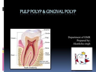
1. PULP POLYP & GINGIVAL POLYP
Department of OMR
Prepared by:
Akanksha singh
2. Whatispulppolyp?
Pulp polyp is also known as chronic
hyperplastic pulpitis.
It is inflammation of the pulp due to extensive
carious exposure of the young pulp.
It is a result of irreversible pulpitis.
3. • Pulp polyp arte mainly seen in the molars.
• It is a development of the granulation tissue.
Histology
• The surface of the pulp polyp is covered by
the stratified squamous epithelium.
•This epithelium is derived from the gingiva or
from the epithelial cells of the mucosa and
tongue.
Stratified sq.
Epithelium covering the
Pulp polyp
4. Causes
1. Slow progressive exposure of the pulp due to the
carious lesions.
2. Mechanical irritation of the pulp due to chewing.
3. Bacterial infection.
Symptoms
Pulp polyp is usually symptomless, except during
mastication, when pressure of the food bolus may
cause discomfort.
Clinical features
1. Asymptomatic.
2. Mainly seen in the deciduous molars as well as the
permanent 1st molars.
3. If the mass is large then it interferes with the closure
of the mouth.
4. Discomfort during mastication of food.
5. Bleeding.
5. Diagnosis
The appearance of the polypoid tissue is clinically characteristic, a
fleshy reddish pulpal mass fills most of the pulp chamber or
cavity. It also interferes with the closure of the mouth.
In early stages it is less sensitive but when they grow the become
more vascular and hence the bleeding is more common.
One must not confuse it with gingival enlargement, as in the later
stage it appears similar to it.
They can be differentiated by the use of the probe and the thermal
test.
Differential diagnosis
The disorder must be distinguished from the proliferating gingival
tissue.
Treatment
1. Extirpation of the pulp.
2. Extraction of the tooth.
Extirpation
of pulp
6. Gingivalpolyp
Gingival polyp is an localized increase in the size of the
gingiva.
It relates to the term epulis, denoting a localized tumor
or the lump on the gingiva.
Synonym: gingival enlargement, hypertrophic gingivitis
or gingival hyperplasia.
Classification
(According to etiologic factors)
1. Inflammatory enlargement
2. Drug induced enlargement
3. Enlargement due to systemic diseases
4. Neoplastic enlargement
5. False enlargement
10. Symptoms
One of the more common characteristics of this condition is having red,
bleeding gums.
Other symptoms associated with gum overgrowth include:
•Tender gums
•Inflammation
•Pain
•Bad breath
•Plaque buildup on teeth
In more severe cases, the gums can completely cover the teeth,
affecting hygiene and teeth alignment.
Cause
There are mainly 3 causes for the gingival hyperplasia:
• Inflammatory gum enlargement
• Systemic cause
• HGF (hereditary gingival fibromatous)
11. Diagnosis
The diagnosis depends on the underlying cause of the enlargement of the
gingiva.
For the diagnosis of different enlargements one must see the area that is
affected, which is described below:
1. Inflammatory enlargement : interdental papilla and marginal
papilla
2. Drug induced enlargement: interdental papilla and facial and
lingual gingival margins
3. Systemic enlargement: mostly marginal gingiva & sometimes IDP
4. Neoplastic enlargement: marginal gingiva along with the masses in
the interproximal spaces
5. False enlargement: labial gingiva- bulbous marginal distortion
13. Differencebetweenpulppolypandgingivalpolyp
Pulp polyp
1. soft edematous more
reddish in appearance
2. Friable
3. On passing the probe we
can trace its origin with
the tooth
4. Endodontic therapy or
extraction in extreme
cases
gingival polyp
1. Comparatively firm
with color looks similar
to the adjacent gingiva
2. Non friable
3. On passing the probe we
can trace the origin
around the tooth
4. Removal of the cause
e.g. removal of the
calculus.