STUDY ON ENHANCEMENT OF OSSEOINTEGRATION OF THE BIO-ACTIVE TITANIUM IMPLANT B...
BCUR Poster
1. Aidan Seeley & M. H. Helfrich
aidan.seeley.11@aberdeen.ac.uk & m.helfrich@abdn.ac.uk; see http://www.abdn.ac.uk/ims/research/musculoskeletal/
The Musculoskeletal Research Programme, The Institute of Medical Sciences,
The University of Aberdeen, Foresterhill, Aberdeen AB25 2ZD
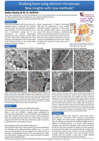
Transmission Electron Microscopy (TEM) of Osteocytes
Tibia and femur from a three month old rat where high pressure
frozen in the Leica EMPACT2. Samples where dehydrated by
automatic freeze substitution in the Leica EM AFS2 and embedded in
epon resin. Ultrathin sections where cut using a diatome diamond
knife and stained with lead citrate and uranyl acetate and viewed
using the Jeol 1400 Plus TEM. This method was used to study the
pericellular matrix in the osteocyte lacuna.
Scanning Electron Microscopy (SEM) of Osteocytes
Tibia and fibula taken from a three month old rat where dehydrated
and infiltrated with methymethacrylate (MMA) resin in a tissue
processor. 9% phosphoric acid was applied to the block surface to
dissolve the hydroxyapatite mineral of the bone leaving the MMA
resin filled spaces. Samples were then coated with gold and viewed
using the Zeiss EVO MA10 SEM.
Scanning Electron Microscopy (SEM) of Osteocytes
Our studies illustrate that osteocytes reside within lacuna
surrounded by a pericellular matrix containing unmineralized
type I collagen and matrix vesicles, suggesting osteocytes may
have the capacity to synthesize and mineralize collagen matrix in
addition to releasing calcium in a process called osteocytic
osteolysis. We show that osteocytes make contact with blood
vessels and that their processes are longer and more numerous
than previously described. This study illustrates the complexity of
the osteocyte network in vivo and why it is a major simplification
to study individual osteocytes in vitro. We conclude that the
methods used here provide a useful tool to obtain detailed
anatomical knowledge about the osteocyte network.
There are three types of cells in bone tissue; the
osteoblast, which is responsible for formation
of bone matrix, the osteoclast, responsible for
bone-resorption, and the osteocyte that has a
role in mechanical sensing and in the
maintenance of mineral homeostasis.
Osteocytes are found encased in cavities within
the bone called lacuna. Osteocytes have many
projections emanating from the cell body which
travel through the bone in the tunnels called
canaliculi as shown in Figure 1.
E F
G H
Image E shows an overview of resin etched cortical bone illustrating the
interacting between the osteocyte canalicular system (orange arrow) and an
osteonic canal (red arrow). Note how it appears that direct interactions
between the two exist with canaliculi containing osteocyte processes touching
the osteonic canal containing endothelial cells suggesting direct contact of
osteocyte processes with endothelial cells. Image F shows the pericellular
space (red arrow) which we known to contain collagen fibers, see images A-D.
in which an osteocyte resides (orange arrow) with canaliculi eminating from
this space. These contain the osteocyte processes. The osteocyte canaliculi
contain the osteocyte processes can be clearly seen by the blue arrow. Image
G shows the connections between two osteocytes. Image H shows the
connections between osteocytes and the endosteal (END) surface.
This project was funded by Medical Research Scotland and the Musculoskeletal Research Programme at the University of Aberdeen. We acknowledge technical support from Mr J. Greenwood, Miss L. Wight, Mr K. MacKenzie and Mrs G. Milne
from the Microscopy and Histology Core Facility at the Institute of Medical Sciences, the University of Aberdeen. See http://www.abdn.ac.uk/ims/facilities/microscopy-histology.php
Transmission Electron Microscopy (TEM) of Osteocytes
A B
DC
Image A and B show TEM micrographs of an osteocyte located in the femural
cortex. The pericellular matrix (PCM) is filled with type I collagen fibers. Red arrows
show transverse osteocyte processes and blue arrows shown longitudinal osteocyte
processes running in their canaliculi, the spaces within the bone. Images C and D
show TEM micrographs of the pericelullar matrix (PCM) surrounding an osteocyte.
Red arrows show longitudinal fibers, blue arrows show transverse fibers and orange
arrows show matrix vesicles, the start sites of mineralisation.
Recent advancements in Electron Microscopy
(EM) offer the opportunity for more in-depth
anatomical studies of osteocytes. In this project
two new methods where used to study bone
tissue in the electron microscope: a method to
freeze tissue under high pressure and then study
very thin sections of that tissue to look inside the
bone cells and a method to study the lacuna and
canaliculi by embedding bone with resin, cutting
the block and etching the surface with acid to
create casts. Figure 1. Structure of compact bone. Taken from:
Sheir, Butler & Lewis Hole’s Human Anatomy. 10th
Edition, McGraw Hill, Boston 2004.