Lung cancer
•
19 likes•8,604 views

Lung cancer is a type of cancer that begins in the lungs. Your lungs are two spongy organs in your chest that take in oxygen when you inhale and release carbon dioxide when you exhale. Lung cancer is the leading cause of cancer deaths in the United States, among both men and women

Recommended
More Related Content
What's hot
Similar to Lung cancer
Similar to Lung cancer (20)
More from Abhay Rajpoot
More from Abhay Rajpoot (20)
Recently uploaded
Recently uploaded (20)
Lung cancer
- 1. Lung cancer PRESENTED BY: ABHAY RAJPOOT
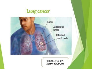
- 2. INTRODUCTION Lung cancer is a type of cancer that begins in the lungs. Your lungs are two spongy organs in your chest that take in oxygen when you inhale and release carbon dioxide when you exhale. Lung cancer is the leading cause of cancer deaths in the United States, among both men and women.
- 4. DEFINITION Cancer arising in the air passage (bronchial Cancer) which is characterized by uncontrolled growth in tissues of lungs
- 5. INCIDENCE It is the leading cause of death among all racial groups in US. Accounting for 31% of all ca deaths in men & 27% of deaths in women. In 2005 more than 1,68,000 people died from lung ca in US; an estimated 1,84000 new cases were diagnosed in the same year.
- 6. RISK FACTORS: It increases with age, occurring most commonly in clients over age 50. 80% of lung ca are caused due to smoking. There is dose response relationship between the smoking & lung ca. Exposure to ionizing radiation & inhaled irritants, asbestos. Exposure to radon (a radioactive gas).
- 7. ETIOLOGY: Cigarette smoking which contain 43 known chemical carcinogens & ca promoters is most significant cause of ca. Genetic abnormality chromosome 3 with loss of genetic material. Alteration of tumor suppressor gene.
- 8. LUNG CA STAGING: Stages Primary tumor(T stage) Regional lymph node (N) Distant metastasis (M) Stage-0 To-no evidence of primary tumor . Tx- malignant cells in the bronchopulmonary secretions, but no tumor visualized. Mx- presence of distant metastasis cannot be assessed. Stage -1 Tis-carcinoma in situ T1- tumor that is 3 cm in diameter or less, with no evidence of invasion No- no regional lymph node metastasis. Mo- no distant metastasis. Stage-2 T2- tumor that is greater than 3 cm in diameter or invades visceral pleura or has associated atelectesis or pneumonia. N1- metastasis or direct extension to peribronchial or ipsilateral hilar node Stage -3 T3- tumor with direct extension into an adjacent structure or any tumor with associated pleural effusion or ipsilateral hilar nodes N2- metastasis to ipsilateral mediastinal nodes Stage-4 T4 – tumor that invades mediastinum or involve the heart , great vessels, trachea, esophagus, vertebral body or carina; presence of malignant plural effusion. N3- metastasis to contralateral M1- distant metastasis
- 9. PATHOPHYSIOLOGY: Due to etiological factors Damage of bronchial epithelial cells Mutation of bronchial epithelial cells Epithelial cells become neoplastic
- 10. TYPES OF LUNG CA Acc. To cell type tumor can divide into : s. no. Cell type & prevelence Presentation & associated manifestation spread 1 Small-cell carcinoma (20-25%) Central lesion with hilar mass common, early meditational involvement,, no cavitations, SIADH, Cushing syndrome Aggressive tumor, more than 40% of clients have distant metastasis at time of presentation 2 Adenocarcinoma (20-40%) Peripheral mass involving; few local symptoms Early metastasis to CNS skeleton, & adrenal glands 3 Squamus cell carcinoma (30-32%) Central lesion located in large bronchi; clients presents with cough, dyspnea, atelectasis & wheezing Spread by local invasion 4 Large cell carcinoma (10-15%) Usually peripheral lesion that is larger than that associated with adenocarcinoma & tends to cavitate; gynecomastia Early metastasis
- 11. CONTI… For clinical purpose the three cell type are frequently classified as – Non-small cell carcinoma: it accounts for 75% of lung ca. each cell type differs in incidence, presentation & manner of spread. Small cell lung ca:accounts for app. 25% of lung ca grow rapidly & spread early. These tumors have paraneoplstic properties, such as ACTH. ADH, parathrome like hormone. Bronchogenic ca: tumors begin as mucosal lesions that grow to form masses that obstruct the bronchi. Frequently spread via lymph system to nodes & other organs such as brain, bones & liver.
- 12. CLINICAL MANIFESTATION: Respiratory: Cough Hemoptysis Wheezing & dyspnea Chest pain dull or pleuritic Hoarseness & dysphagia Pleural effusion
- 13. DIAGNOSTIC EVALUATION: Chest x-ray: usually provide the Ist evidence of lung cancer. It may be used as a screening tool for lung ca. Sputum specimen: is sent for cytologic examination. The sputum sample is collected on arising in the morning. If malignant cells are found in the sputum more invasive examinations are required. Bronchoscopy: done to visualize & obtain tissue for biopsy from the tumor. CT-scan: it is used to evaluate & localize tumors in the lung parenchyma & pleura. CT scanning can also detect distant tumor metastasis & evaluate tumor response to treatment. Cytologic examination: cells or tissues for cytologic examination & biopsy may be obtained by aspirating fluid from a pleural effusion, percutaneous needle biopsy & lymph node biopsy.
- 14. CONTI… CBC, liver function test & serum electrolytes: Including ca are obtained to evaluate for evidence of metastatic disease or paraneoplstic syndromes. Tuberculin test is performed to rule out TB as a cause of symptoms. Pulmonary function test: may be performed prior to the initiation of treatment if the client has manifestations of respiratory insufficiency (e.g. dyspnea, low oxygen saturation level).
- 15. MANAGEMENT: Chemotherapy: Used in combination, chemotherapeutic drugs to be attached at different parts of the cell cycle & in different ways, increasing the effectiveness of therapy. Chemotherapy drugs that commonly used are- Vance Alkaloids (Vinblastine), Doxorubicin, Taxanes (Docetaxel), Plantin analogus (Cisplantin, & Carboplantin).
- 16. CONTI… Radiation therapy: It is used alone or in combination with surgery & chemotherapy. Goals- Treatment- prior to surgery, R/T may be used to debulk tumors. Palliative- (symptom relief) it may also be used to relieve manifestation such as cough, hemoptysis & dyspnea from bronchial obstruction. R/T may be delivered by external beam radiation to the primary tumor site or by intraluminal radiation or brachytherapy.
- 17. SURGICAL MANAGEMENT The goal of surgery is to remove all involved tissue while preserving as much as functional lung as possible. s. no. procedure description Used for 1 Laser bronchoscopy Bronchoscopy guided laser used to resect tumor Tumors localized in a main bronchus 2 mediastinoscopy Visualization of mediastinum using an endoscope passed through a suprasternal incision Evaluation & biopsy of a meditational tumors & lymph nodes 3 thoracotomy Incision into the chest wall Access the lung & thoracic cavity for surgery
- 18. 4 Wedge resection Removal of an individual bronchovascular segment of a lobe Peripheral lung tumor with no evidence of extension to chest wall or metastasis 5 Segmental resection Resection of a section of a major bronchus with reconstruction of remaining normal bronchus Small lesion of major bronchus 6 Sleeve resection Resection of a section of a major bronchus with reconstruction of remaining normal bronchus Small lesion of a major bronchus 7 lobectomy Removal of a single lung lobe Tumor confined to a single lobe 8 pneumonecto my Removal of an entire lung Tumor widespread throughout the lung, involving the main bronchus or fixed to the hilum
- 19. Nursing management: Ineffective breathing pattern r/t tumor & treatment of tumor. Activity intolerance r/t resectional lung surgery & inoperable lung ca. Acute pain r/t surgical procedure or terminal stage of ca. Anticipatory grieving r/t advanced diagnosis of lung ca.
- 20. THANK YOU