
Recommended
More Related Content
What's hot
What's hot (20)
Viewers also liked
Viewers also liked (15)
Similar to Spinal tb
Similar to Spinal tb (20)
More from sayf aldeen hussam
Recently uploaded
Recently uploaded (20)
Spinal tb
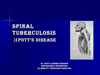
- 1. SPINALSPINAL TUBERCULOSISTUBERCULOSIS (POTT(POTT‘S DISEASE‘S DISEASE(( Dr. SAYF ALDEEN HUSSAMDr. SAYF ALDEEN HUSSAM ORTHOPEDIC DEPARTENTORTHOPEDIC DEPARTENT AL-WASITY TEACHING HOSPITALAL-WASITY TEACHING HOSPITAL
- 2. INTRODUCTIONINTRODUCTION Tuberculous spondylitis is one ofTuberculous spondylitis is one of the oldest demonstrated diseasesthe oldest demonstrated diseases of humankindof humankind It has been documented in ancientIt has been documented in ancient mummies from Egyptmummies from Egypt According to WHO(2006), about one thirdAccording to WHO(2006), about one third of the world’s population is infected byof the world’s population is infected by Mycobacterium TB, and 9 millionMycobacterium TB, and 9 million
- 3. INTRODUCTIONINTRODUCTION Three percent are suffering fromThree percent are suffering from skeletal TB.skeletal TB. Vertebral TB is the most commonVertebral TB is the most common form of skeletal TB and accounts forform of skeletal TB and accounts for 50% of all cases of skeletal TB.50% of all cases of skeletal TB. Almost 50% are from pediatric groupAlmost 50% are from pediatric group Neurological complications are the mostNeurological complications are the most cripplingcrippling complications of spinal TB ( Incidence :complications of spinal TB ( Incidence : 10 to 43%10 to 43%(.(.
- 5. PATHOANATOMYPATHOANATOMY early infection begins in the metaphysis of the vertebral body spreads under the anterior longitudinal ligament and leads to: - contiguous multilevel involvement - skip lesion or noncontiguous segments (15%( - paraspinal abscess formation (50%( usually anterior and can be quite large (much more common in TB than pyogenic infections( initially does not involve the disc space (distinguishes from pyogenic osteomyelitis, but can be misdiagnosed as a neoplastic lesion(
- 6. PATHOANATOMYPATHOANATOMY (cont(cont.(.( chronic infection leads to: severe kyphosis :because the infection is often diagnosed late, there is often much more severe kyphosis in granulomatous spinal infections compared to pyogenic infections sinus formation Pott's paraplegia
- 8. Recently, two distinct patternsRecently, two distinct patterns of spinal TB can be identified,of spinal TB can be identified, - the classic form, called- the classic form, called spondylodiscitis (SPD)spondylodiscitis (SPD) - atypical form characterized by- atypical form characterized by spondylitis without diskspondylitis without disk involvement (SPwD). whichinvolvement (SPwD). which seems to be the most commonseems to be the most common pattern of spinal TBpattern of spinal TB
- 9. CLINICAL FEATURESCLINICAL FEATURES Constitutional symptomsConstitutional symptoms:: 1-Malaise1-Malaise 2-Loss of weight2-Loss of weight // appetiteappetite 3-Night sweats3-Night sweats 4-Evening rise of temperature4-Evening rise of temperature
- 10. CliniCal FeaturesCliniCal Features Specific SymptomsSpecific Symptoms:: 1-Pain1-Pain//Night criesNight cries 2-Stiffness2-Stiffness 3- Deformity3- Deformity 4- Restricted ROM4- Restricted ROM 5-Enlarged lymph nodes5-Enlarged lymph nodes 6-Abscess6-Abscess 7-Neurodeficit7-Neurodeficit
- 11. POtt’s ParaPleGia Paraplegia is the most feared complication of spinal tuberculosis. Divided into: 1-Early-onset paresis: (usually within 2 years of disease onset) is due to pressure by inflammatory oedema, an abscess, caseous material, granulation tissue or sequestra. The patient presents with lower limb weakness, upper motor neuron signs, sensory dysfunction and incontinence. In these cases the prognosis for neurological recovery following surgery is good 2-Late onset paresis:may develop two to three decades after active infection is due to direct cord compression from increasing
- 12. PhysiCal examinatiOnPhysiCal examinatiOn The physical examination inThe physical examination in Potts disease should include thePotts disease should include the followingfollowing:: 1-Careful assessment of spinal1-Careful assessment of spinal alignmentalignment 2-Inspection of skin, with attention2-Inspection of skin, with attention to detection of sinusesto detection of sinuses 3-Abdominal evaluation for3-Abdominal evaluation for subcutaneous flank masssubcutaneous flank mass
- 13. PhysiCalPhysiCal examinatiOnexaminatiOn (COnt(COnt.(.( Large, cold abscesses ofLarge, cold abscesses of paraspinal tissues or psoasparaspinal tissues or psoas muscle may protrude under themuscle may protrude under the inguinal ligament and may erodeinguinal ligament and may erode into the perineum or glutealinto the perineum or gluteal areaarea.. Pott disease that involves thePott disease that involves the upper cervical spine can causeupper cervical spine can cause rapidly progressive symptomsrapidly progressive symptoms.. Retropharyngeal abscesses occurRetropharyngeal abscesses occur
- 14. tyPes OF vertebraltyPes OF vertebral lesiOnslesiOns 11.. ParadiscalParadiscal(commonest(commonest)) 22.. CentralCentral 33.. AnteriorAnterior 44.. AppendicularAppendicular 55.. ArticularArticular
- 15. DiaGnOstiC stuDiesDiaGnOstiC stuDies 1. Laboratory studies1. Laboratory studies 2. Imaging2. Imaging 3. Cultures3. Cultures
- 16. labOratOry stuDieslabOratOry stuDies The WBC count, ESR, and CRP levelThe WBC count, ESR, and CRP level may be elevated in these patients.may be elevated in these patients. These values are generallyThese values are generally nonspecificnonspecific Patients with active tuberculosis orPatients with active tuberculosis or previous exposure toprevious exposure to MycobacteriumMycobacterium will normally exhibit a positivewill normally exhibit a positive tuberculin purified protein derivativetuberculin purified protein derivative skin test, although false-negativeskin test, although false-negative results may occur.results may occur.
- 17. imaGinGimaGinG A-Plain radiograph signsA-Plain radiograph signs :: 1- Reduced disc space1- Reduced disc space 2-Blurred paradiscal margins2-Blurred paradiscal margins 3-Destruction of bodies3-Destruction of bodies 4-Loss of trabecular pattern4-Loss of trabecular pattern 5-Increased prevertebral soft tissue5-Increased prevertebral soft tissue shadowshadow 6-Subluxation6-Subluxation //dislocationdislocation 7-Decreased lordosis7-Decreased lordosis//KyphosisKyphosis
- 18. Reduced disc spaceReduced disc space
- 19. Blurred paradiscal marginsBlurred paradiscal margins
- 20. Increased prevertebral soft tissue shadowIncreased prevertebral soft tissue shadow
- 21. Destruction of vertebral bodiesDestruction of vertebral bodies
- 22. KyphosisKyphosis
- 23. ImagIngImagIng (cont(cont.(.( B-Computed tomographyB-Computed tomography ((CTCT)):: CT demonstrates abnormalities earlier thanCT demonstrates abnormalities earlier than plain radiography .plain radiography . •• CT is of great value in the demonstration ofCT is of great value in the demonstration of any calcification within the cold abscessany calcification within the cold abscess (pathognomic for Tb)(pathognomic for Tb) •• Regions which are difficult to visualize onRegions which are difficult to visualize on plain films like :plain films like : 1. Cranio-vertebral junction (CVJ)1. Cranio-vertebral junction (CVJ) 2. Cervico-dorsal region,2. Cervico-dorsal region, 3. Sacrum3. Sacrum 4. Sacro-iliac joints.4. Sacro-iliac joints. 5. Posterior spinal tuberculosis because
- 25. ImagIngImagIng (cont(cont.(.( C-MRI:C-MRI: •• Detect marrowDetect marrow infiltration ininfiltration in vertebral bodies,vertebral bodies, leading to earlyleading to early diagnosisdiagnosis.. ••Detect Changes ofDetect Changes of diskitisdiskitis •• Assessment ofAssessment of extraduralextradural abscesses /
- 26. ImagIngImagIng (cont(cont.(.( d-USGd-USG :: -to find out primary TB in abdomen-to find out primary TB in abdomen -Detect cold abscess-Detect cold abscess -Guided aspiration -Guided aspiration
- 27. culturescultures CT guided biopsy with cultures and staining effective at obtaining diagnosis should be tested for acid- fast bacilli (AFB) mycobacteria (acid-fast bacilli) may take 10 weeks to grow in culture PCR allows for faster identification (95% sensitivity and 93% accuracy)
- 28. TreaTmenTTreaTmenT Nonsurgical: 1●pharmacotherapy: isoniazid, rifampin, and pyrazanamide therapy indications drugs are the mainstay of treatment in most cases technique treated with isoniazid, rifampin, and pyrazanamide for 9 to 18 months ethambutol and streptomycin added for part of treatment 2● spinal orthosis indications may be used for pain control and prevention of deformity
- 29. TreaTmenT (conTTreaTmenT (conT.(.( Operative: indications: neurologic deficit spinal instability or progressive kyphosis Advanced disease with caseation, fibrosis, and avascularity that limits antibiotic penetration failure of nonoperative treatment after 3 to 6 months
- 30. Surgical TreaTmenTSurgical TreaTmenT SurgerySurgery indicationindication 11 DecompressionDecompression(+/-(+/- fusionfusion(( Too advanced disease, Failure toToo advanced disease, Failure to respond to conservative therapyrespond to conservative therapy 22 DebridementDebridement +/-+/- decompressiondecompression +/-+/- fusionfusion Recurrence of disease or ofRecurrence of disease or of neural complicationsneural complications 33 Anterior transposition ofAnterior transposition of cordcord ((ExtrapleuralExtrapleural anterolateralanterolateral approachapproach(( Sever KyphosisSever Kyphosis ((>60 degree>60 degree) + /) + / neural deficitneural deficit 44 LaminectomyLaminectomy Extradural granulomaExtradural granuloma// OldOld healed disease presenting ashealed disease presenting as secondary canal stenosissecondary canal stenosis // Posterior spinal diseasePosterior spinal disease
- 31. complicaTionScomplicaTionS ParaplegiaParaplegia Cold abscessCold abscess Spinal deformitySpinal deformity SinusesSinuses Secondary infectionSecondary infection Amyloid diseaseAmyloid disease FatalityFatality
- 32. Follow upFollow up Patient evaluated at 3 months interval upto 2 yearsPatient evaluated at 3 months interval upto 2 years Clinical EvaluationClinical Evaluation :: Weight gainWeight gain Pain reliefPain relief Free ROMFree ROM Resolution of abscessesResolution of abscesses Neurological recoveryNeurological recovery RadiologicalRadiological EvaluationEvaluation :: Decreased soft tissue shadowDecreased soft tissue shadow Disappearance of erosionsDisappearance of erosions Return of mineralization GraftReturn of mineralization Graft incorporationincorporation Bony ankylosisBony ankylosis