
Recomendados
Mais conteúdo relacionado
Mais procurados
Mais procurados (20)
Destaque
Destaque (20)
Semelhante a Body Tissues: The 4 Primary Types
Semelhante a Body Tissues: The 4 Primary Types (20)
Mais de zernwoman
Mais de zernwoman (20)
Último
Último (20)
Body Tissues: The 4 Primary Types
- 1. Body Tissues • Tissues – Groups of cells with similar structure and function – Four primary types • Epithelial tissue (epithelium) • Connective tissue • Muscle tissue • Nervous tissue
- 2. Epithelial Tissues • Locations – Body coverings – Body linings – Glandular tissue • Functions – Protection – Absorption – Filtration / Excretion – Secretion
- 3. Epithelium Characteristics • Cells fit closely together and often form sheets – Tight junctions • The apical surface is the free surface of the tissue • The lower surface of the epithelium rests on a basement membrane (acellular) • Avascular (no blood supply) • Regenerate easily if well nourished – Through what process?
- 4. Classification of Epithelia • Number of cell layers – Simple—one layer – Stratified—more than one layer Apical surface Basal Simple surface Apical surface Basal surface Stratified (a) Classification based on number of cell layers Figure 3.17a
- 5. Classification of Epithelia • Shape of cells – Squamous • flattened – Cuboidal • cube-shaped – Columnar • column-like
- 6. Simple Epithelia • Simple squamous – Single layer of flat cells – Location - usually forms membranes • Lines body cavities • Lines lungs and capillaries – Functions in diffusion, filtration, or secretion in membranes – g http://www .youtube.co m/watch?v =d- f3RL0KiUg
- 7. Simple Epithelia • Simple cuboidal – Single layer of cube-like cells – Locations • Common in glands and their ducts (ex: salavary glands) • Forms walls of kidney tubules • Covers the ovaries - Functions – secretion & absorption Nucleus of Simple simple cuboidal cuboidal epithelial epithelial cells cell Basement Basement membrane membrane Connective tissue Photomicrograph: Simple cuboidal (b) Diagram: Simple cuboidal epithelium in kidney tubules (250×).
- 8. Simple Epithelia http://www.youtube.c • Simple columnar om/watch?v=u7yGj6i5 lBA – Single layer of tall cells Video shows brush border of small – Often includes mucus-producing goblet cells intestine – Location - lines digestive tract – Functions in secretion and absorption; those located in intestines contain microvilli to increase the surface area for more absorption. http://www.youtube.com/watch?v=tYCLligRoMY Simple columnar Nucleus of simple epithelial columnar epithelial cell cell Goblet cell Basement membrane Connective Basement tissue membrane Photomicrograph: Simple columnar epithelium of the small intestine (430×). (c) Diagram: Simple columnar
- 9. Simple Epithelia • Pseudostratified columnar – Single layer, but some cells are shorter than others – Often looks like a double layer of cells but all cells rest on the basement membrane – Location - respiratory tract, where it is ciliated – Functions in absorption or secretion; contain goblet cells for secretion of mucus http://www.youtube.com/watch?v=FQwqhblxz3I http://w ww.youtu Cilia be.com/w Pseudo- atch?v=m stratified iEEluVlem epithelial Pseudo- Q layer stratified Video epithelial shows layer epithelial Basement tissue membrane lining Basement trachea membrane Connective tissue Photomicrograph: Pseudostratified ciliated columnar epithelium lining (d) Diagram: Pseudostratified (ciliated) the human trachea (430×). columnar
- 10. Stratified Epithelia • Stratified squamous – Cells at the apical surface are flattened – Functions as a protective covering where friction is common – Locations - lining of the: • Skin • Mouth • Esophagus Nuclei Stratified squamous Stratified epithelium squamous epithelium Basement Basement membrane membrane Connective Photomicrograph: Stratified tissue squamous epithelium lining of (e) Diagram: Stratified squamous the esophagus (140×).
- 11. Stratified Epithelia • Stratified cuboidal—two layers of cuboidal cells; functions in protection • Stratified columnar —surface cells are columnar, cells underneath vary in size and shape; functions in protection Both rare in humans
- 12. Stratified Epithelia • Transitional epithelium – Composed of modified stratified squamous epithelium – Functions in stretching and the ability to return to normal shape – Location - lines organs of the urinary system
- 15. Glandular Epithelium • Gland – One or more cells responsible for secreting a particular product – Secretions contain protein molecules in an aqueous (water-based) fluid
- 16. Glandular Epithelium • Two major gland types – Endocrine gland • Ductless since secretions diffuse into blood vessels • All secretions are hormones – Exocrine gland • Secretions empty through ducts to the epithelial surface • Include sweat and oil glands
- 17. Connective Tissue • Found everywhere in the body • Functions – Binds body tissues together – Supports the body – Provides protection
- 18. Connective Tissue Characteristics • Most are vascular (except tendons, ligaments & cartilage) • The cells of this tissue have many different types of fibers attached to the cell membrane called Extracellular Matrix (ECM) which can vary from very solid (bone) to liquid (blood) – Non-living material that surrounds living cells
- 19. Extracellular Matrix Fibers There are different types but we will focus on collagen fibers which are very strong. There are also elastin fibers which provide elasticity & laminin fibers which help bind tissues (keep them together).
- 20. Connective Tissue Types – classified based on the types of cells & the matrix (fibers) surrounding the cells Types Include: • Bone (osseous tissue) • Cartilage • Dense (Fibrous)Connective tissue (ligaments, tendons) • Loose Connective tissue (areolar & adipose) • Blood
- 21. Connective Tissue Types • Bone – Composed of • Bone cells in lacunae (cavities) • Hard matrix of calcium salts • Large numbers of collagen fibers – Functions to protect and support the body
- 22. Connective Tissue Types • Cartilage – Composed of • Rubbery matrix b/t cells – Locations • Larynx • Entire fetal skeleton prior to birth • External ear • Joints – Functions as a more flexible skeletal element than bone
- 23. Connective Tissue Types • Dense connective tissue – Contains Fibroblasts - cells that make fibers such as collagen – Locations • Tendons—attach skeletal muscle to bone • Ligaments—attach bone to bone at joints • Dermis—lower layers of the skin
- 26. Connective Tissue Types • Loose connective tissue types – Areolar tissue • Under epithelial tissue • Soft, pliable tissue • Can soak up excess fluid (causes edema)
- 27. Connective Tissue Types • Loose connective tissue types – Adipose tissue • Many cells contain large lipid deposits • Functions – Insulates the body – Protects some organs – Serves as a site of fuel storage
- 28. Connective Tissue Types • Blood (vascular tissue) – Blood cells surrounded by fluid matrix called blood plasma – Fibers are visible during clotting – Functions as the transport vehicle for materials
- 29. Connective Tissue Connective Cell type & or Functions Location in the Extra Notes Tissue Type matrix body composition Bone Cartilage Dense Connective Tissue Loose Connective Tissue - Areolar Loose Connective Tissue - Addipose Blood
- 30. Muscle Tissue • Function is to produce movement • Three types – Skeletal muscle – Cardiac muscle – Smooth muscle
- 31. Muscle Tissue Types • Skeletal muscle – Under voluntary control – Contracts to pull on bones or skin – Produces gross body movements or facial expressions – Characteristics of skeletal muscle cells • Striated (stripes) • Multinucleate (more than one nucleus) • Long, cylindrical cells
- 32. Muscle Tissue Types • Cardiac muscle – Under involuntary control – Found only in the heart – Function is to pump blood – Characteristics of cardiac muscle cells • Striated • One nucleus per cell • Cells are attached to other cardiac muscle cells at intercalated disks
- 33. Muscle Tissue Types • Smooth muscle – Under involuntary muscle – Found in walls of hollow organs such as stomach, uterus, and blood vessels – Characteristics of smooth muscle cells • No visible striations • One nucleus per cell • Spindle-shaped cells
- 34. Nervous Tissue • Composed of neurons and nerve support cells • Function is to send impulses to other areas of the body – Irritability – Conductivity • Support cells called neuroglia insulate, protect, and support neurons • Location – brain, spinal cord, throughout body tissues
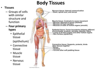
- 35. Nervous tissue: Internal communication • Brain, spinal cord, and nerves Muscle tissue: Contracts to cause movement • Muscles attached to bones (skeletal) • Muscles of heart (cardiac) • Muscles of walls of hollow organs (smooth) Epithelial tissue: Forms boundaries between different environments, protects, secretes, absorbs, filters • Lining of GI tract organs and other hollow organs • Skin surface (epidermis) Connective tissue: Supports, protects, binds other tissues together • Bones • Tendons • Fat and other soft padding tissue Figure 3.22
- 36. Tissue Repair (Wound Healing) • Regeneration – Replacement of destroyed tissue by the same kind of cells • Fibrosis – Repair by dense (fibrous) connective tissue (scar tissue) • Whether regeneration or fibrosis occurs depends on: – Type of tissue damaged – Severity of the injury
- 37. Events in Tissue Repair • Inflammation – Capillaries become very permeable (leaky) – Clotting proteins migrate into the area from the blood stream – A clot walls off the injured area • Granulation tissue forms – Growth of new capillaries – Rebuild collagen fibers • Regeneration of surface epithelium – Scab detaches
- 38. Regeneration of Tissues • Tissues that regenerate easily – Epithelial tissue (skin and mucous membranes) – Fibrous connective tissues and bone • Tissues that regenerate poorly – Skeletal muscle • Tissues that are replaced largely with scar tissue – Cardiac muscle – Nervous tissue within the brain and spinal cord