Short case...Vertebral hemangioma and retromedullary lipoma
•
1 gostou•888 visualizações

Short case...Vertebral hemangioma and retromedullary lipoma

Recomendados
Mais conteúdo relacionado
Mais de Professor Yasser Metwally
Mais de Professor Yasser Metwally (20)
Último
Último (20)
Short case...Vertebral hemangioma and retromedullary lipoma
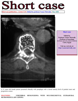
- 1. Short case publication... version 1.24 | Edited by professor Yasser Metwally | May 2008 Short case Edited by Professor Yasser Metwally Professor of neurology Ain Shams university school of medicine Cairo, Egypt Visit my web site at: http://yassermetwally.com A 51 years old female patient presented clinically with paraplegia with a dorsal sensory level of gradual onset and progressive course. DIAGNOSIS: VERTEBRAL HEMANGIOMA WITH MULTISEGMENTAL EXTRADURAL RETROMEDULLARY LIPOMA
- 2. Figure 1. Dorsal vertebral hemangioma with epidural extension and fat overgrowth. Postcontrast CT scan of D4 vertebra showing the characteristic appearance of vertebral hemangioma of alternating coarse trabeculae and low- density cystic areas. Notice the dotted appearance of the vertebral, mostly due to destruction of transverse trabeculae and preservation and thickening of the longitudinal trabeculae. Such haemangiomas are usually of the cavernous type. There is also resorption of bone, with replacement by sinusoids, and thickening of remaining trabeculae. The cortical margin is intact in most cases and may bulge at the posterior aspect. Histologically, many abnormal vascular channels of varying calibre are seen interspersed within a fatty matrix and thickening of vertical trabiculations. The coarse, vertical, and thickened trabecular pattern, with osseous reinforcement (trabecular thickening) adjacent to the vascular channels cause bone resorption and reactive fat overgrowth in between the thickened trabeculae. Reactive fat overgrowth in between the thickened trabeculae is responsible for the radiolucency that is observed between the hyperdense thickened trabeculae. This appearance (the dot appearance.... trabecular thickening) on radiographs represents a response to stress and has been likened to corduroy. Vertebral fractures at the site of these hemangiomas are unusual because of this trabecular reinforcement and thickening. Figure 2. Post intravenous contrast CT scan image (A) and CT myelography (B,C). Notice the epidural extension of the hemangioma that showed contrast enhancement (A). The remnant of dye in the subarachnoid spaces (B,C) showed that the spinal cord is actually pushed anteriorly by an epidural mass of fat density (epidural lipoma)
- 3. Figure 3. CT myelography showing the vertebral hemangioma and the multisegmental retromedullary lipoma pushing the spinal cord anteriorly. The retromedullary lipoma is probably reactive to the epidural hemangioma rather than a true neoplasm. Operative finding, however, revealed an epidural angiolipoma. Addendum A new version of short case is uploaded in my web site every week (every Saturday and remains available till Friday.) To download the current version follow the link quot;http://pdf.yassermetwally.com/short.pdfquot;. You can download the long case version of this short case during the same week from: http://pdf.yassermetwally.com/case.pdf or visit web site: http://pdf.yassermetwally.com To download the software version of the publication (crow.exe) follow the link: http://neurology.yassermetwally.com/crow.zip At the end of each year, all the publications are compiled on a single CD-ROM, please contact the author to know more details. Screen resolution is better set at 1024*768 pixel screen area for optimum display For an archive of the previously reported cases go to www.yassermetwally.net, then under pages in the right panel, scroll down and click on the text entry quot;downloadable short cases in PDF formatquot; References 1. Metwally, MYM: Textbook of neurimaging, A CD-ROM publication, (Metwally, MYM editor) WEB-CD agency for electronic publishing, version 9.1a January 2008