Short case...Focal midbrain glioma
•
1 like•325 views

Short case...Focal midbrain glioma http://yassermetwally.com http://yassermetwally.net

Recommended
More Related Content
What's hot
Viewers also liked
Viewers also liked (20)
Similar to Short case...Focal midbrain glioma
Similar to Short case...Focal midbrain glioma (20)
More from Professor Yasser Metwally
More from Professor Yasser Metwally (20)
Recently uploaded
Recently uploaded (20)
Short case...Focal midbrain glioma
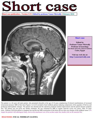
- 1. Short case publication... Version 3.13 | Edited by professor Yasser Metwally | February 2010 Short case Edited by Professor Yasser Metwally Professor of neurology Ain Shams university school of medicine Cairo, Egypt Visit my web site at: http://yassermetwally.com The patient is a 44 years old male patient, who presented clinically at the age of 10 years complaining of clinical manifestations of increased intracranial pressure. MRI at that time (Figures 1,2,3,4,5) revealed a focal midbrain glioma inducing compression of the aqueduct of Sylvius and producing hydrocephalic changes. The patient was shunted and the operation produced marked improvement and the patient became symptom free. The patient was not given any further treatment. He was examined by MRI at regular intervals (every two years). After 34 years (now...February 2010) the patient is symptom free and the last MRI examination of the brain did not show any changes of the midbrain tumor. (To inspect the patient's full radiological study, click on the attachment icon of the acrobat reader then double click on the attached file) DIAGNOSIS: FOCAL MIDBRAIN GLIOMA
- 2. Figure 1. Precontrast CT scan showing midbrain calcification. Mild midbrain hypodensity is also probably present. The midbrain glioma could not be appreciated by CT scan and the patient was misdiagnosis as congenital aqueduct stenosis Figure 2. Pre (A) and postcontrast (B) MRI T1 images showing enlargement of the midbrain, involving mainly the posterior part with a large irregular hypointensity on the precontrast image (A) involving the middle line area and extending from the interpeduncular area anteriorly to the periaqueductal area posteriorly, the tumor is apparently involving the medial parts of the midbrain. The aqueduct of Sylvius is compressed, pushed posteriorly. Dense patchy enhancement is observed on postcontrast image (B) and involved the linear hypointense zone observed on the precontrast image. The tumor is apparently sparing the crus cerebri and the lateral parts of the midbrain and its main bulk is located in the tectal plate posteriorly.
- 3. Figure 3. Pre (A) and postcontrast (B) MRI images showing enlargement of the midbrain, involving mainly the posterior part. The aqueduct of Sylvius is compressed, pushed posteriorly and elongated (C). Dense patchy enhancement is observed on postcontrast image (B). Moderate hydrocephalic changes are also observed. Figure 4. MRI T2 image (A) and FLAIR image (B). The tumor is hyperintense on the MRI T2, FLAIR images and involves the medial parts of the midbrain with selective sparing of the crus cerebri bilaterally and the lateral zones of the midbrain. The mainly part of the tumor is located posteriorly in the tectal plate and the tumor extends from the interpeduncular area anteriorly to the periaqueductal area posteriorly.
- 4. Figure 5. The midbrain glioma References 1. Metwally, MYM: Textbook of neurimaging, A CD-ROM publication, (Metwally, MYM editor) WEB-CD agency for electronic publishing, version 11.1a December 2010 Addendum A new version of short case is uploaded in my web site every week (every Saturday and remains available till Friday.) To download the current version follow the link "http://pdf.yassermetwally.com/short.pdf". You can download the long case version of this short case during the same week from: http://pdf.yassermetwally.com/case.pdf or visit web site: http://pdf.yassermetwally.com To download the software version of the publication (crow.exe) follow the link: http://neurology.yassermetwally.com/crow.zip At the end of each year, all the publications are compiled on a single CD-ROM, please contact the author to know more details. Also to view a list of the previously published case records follow the following link (http://wordpress.com/tag/case- record/) or click on it if it appears as a link in your PDF reader To inspect the patient's full radiological study, click on the attachment icon of the acrobat reader then double click on the attached file