Case record...Intramedullary ependymoma
•
1 gostou•1,180 visualizações

Case record...Intramedullary ependymoma

Recomendados
Mais conteúdo relacionado
Mais procurados
Mais procurados (20)
Semelhante a Case record...Intramedullary ependymoma
Semelhante a Case record...Intramedullary ependymoma (20)
Mais de Professor Yasser Metwally
Mais de Professor Yasser Metwally (20)
Último
Último (20)
Case record...Intramedullary ependymoma
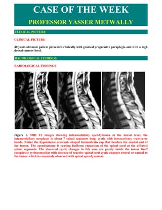
- 1. CASE OF THE WEEK PROFESSOR YASSER METWALLY CLINICAL PICTURE CLINICAL PICTURE 40 years old male patient presented clinically with gradual progressive paraplegia and with a high dorsal sensory level. RADIOLOGICAL FINDINGS RADIOLOGICAL FINDINGS Figure 1. MRI T2 images showing intramedullary ependymoma at the dorsal level, the intramedullary neoplasm is about 7 spinal segments long, cystic with intracavitary transverse bands. Notice the hypointense crescent- shaped hemosiderin cap that borders the caudal end of the tumor. The ependymoma is causing fusiform expansion of the spinal cord at the affected spinal segments. The observed cystic changes in this case are purely inside the tumor itself (neoplastic syringomyelia) with absence of reactive spinal cord cystic changes rostral or caudal to the tumor which is commonly observed with spinal ependymomas.
- 2. Figure 2. MRI T1 postcontrast images showing the cystic intramedullary multisegmental ependymoma, with intracavitary transverse bands. Notice the hypointense hemosiderin cap at the caudal end of the tumor. Very faint contrast enhancement is observed at (B). The tumor is more or less well defined. The signal intensity of the intracavitary content is different from that of CSF. The observed cystic changes in this case are purely inside the tumor itself (neoplastic syringomyelia) with absence of reactive spinal cord cystic changes rostral or caudal to the tumor which is commonly observed with spinal ependymomas. The ependymoma is isointense relative to the spinal cord on T1-weighted MR images Figure 3. Cross sections at the tumor level showing the asymmetric location of the cystic ependymoma and the intracavitary transverse bands. The signal intensity of the intracavitary content is different from that of CSF. The ependymoma is isointense relative to the spinal cord on T1-weighted MR images
- 3. Table 1. In general the intramedullary ependymoma had the following radiological characteristics 1. The existence of the hemosiderin cap at the extreme ends of the tumor 2. The cystic nature of the tumor with intracavitary transverse bands 3. The tumor is more or less well defined DIAGNOSIS: DIAGNOSIS: SPINAL INTRAMEDULLARY EPENDYMOMA DISCUSSION DISCUSSION Spinal ependymoma Prevalence Ependymoma is the most common intramedullary spinal neoplasm in adults, accounting for up to 60% of all glial spinal cord tumors (19). From the four largest studies of patients with spinal cord ependymomas reported in the literature (5,19,24,25), the following demographic information emerges: These lesions tend to manifest in young adulthood, with a mean age at presentation of 38.8 years and are more common in male patients (57.4%). Cord ependymomas occur most commonly in the cervical region, with 44% involving the cervical cord alone and an additional 23% extending into the upper thoracic region. About 26% are located in the thoracic cord alone. Only 6.5% involve either the distal thoracic cord or the conus medullaris (5,19,24,25). Myxopapillary ependymoma, a variant type, is, in rare cases, found in the subcutaneous tissue of the sacrococcygeal region, usually without any connection with the spinal canal (26). It is believed that these arise from either heterotopic ependymal cell rests or vestigial remnants of the distal neural tube during canalization and retrogressive differentiation (26,27). Clinical Presentation As with most primary intramedullary tumors, there is frequently a long antecedent history before the diagnosis of an intramedullary ependymoma is established. The mean duration of symptoms was 36.5 months for the 183 patients from the four largest studies (5,19,24,25). A large majority of patients with spinal cord ependymomas have relatively mild clinical symptoms. Most of the 183 patients (81%) could walk without assistance at presentation (5,24,25). In general, the less preoperative neurologic deficit existing at presentation, the better the postoperative outcome (5,25). Typically, patients initially present with mild symptoms, and there is no objective evidence of neurologic deficits, which often leads to a delay in diagnosis (5,19,24,25). Some ependymomas may even be a source of subarachnoid hemorrhage (28,29). At diagnosis, patients with spinal cord ependymomas typically have back or neck pain (67%), sensory deficits (52%), motor weakness (46%), or bowel or bladder dysfunction (15%)
- 4. (5,19,24,25). The predominance of sensory symptoms (85% of patients with pain and other sensory deficits combined) may be directly related to the more central location of these tumors (5). Spinal cord ependymomas are believed to arise from ependymal cells that line the central canal. Theoretically, this central location makes it likely that the crossing spinothalamic tracts will be compressed or interrupted. Dominant motor symptoms are commonly associated with very large ependymomas and a poorer postoperative outcome secondary to the increased surgical risk associated with resection of these larger lesions (5). Hoshimaru et al (25) found that patients with a shorter duration of symptoms tended to have a better postoperative outcome. Lesions of the thoracic cord are associated with poorer surgical outcomes, perhaps because of its relatively tenuous vascular supply, compared with lesions of the cervical spinal cord. This was especially true in patients with evidence of arachnoid scarring or cord atrophy at surgery (4). Table 2. Prognostic factors in spinal ependymomas Theoretically, this central location makes it likely that the crossing spinothalamic tracts will be compressed or interrupted. Dominant motor symptoms are commonly associated with very large ependymomas and a poorer postoperative outcome secondary to the increased surgical risk associated with resection of these larger lesions (5) Patients with a shorter duration of symptoms tended to have a better postoperative outcome. Lesions of the thoracic cord are associated with poorer surgical outcomes, perhaps because of its relatively tenuous vascular supply, compared with lesions of the cervical spinal cord. Patients with evidence of arachnoid scarring or cord atrophy at surgery (4). The 5-year survival rate for patients with spinal cord ependymomas is approximately 82%, regardless of how severe the preoperative neurologic deficits. The 20-year survival rate, however, is much worse for patients who present with a major neurologic dysfunction (50%) than for patients with a minor neurologic impairment (33%) (1) Intramedullary ependymomas are characterized by slow growth and tend to compress adjacent spinal cord tissue rather than infiltrate it. Accordingly, there is almost always a cleavage plane, which facilitates microsurgical resection, the treatment of choice (5,19,24). Patients frequently have worsened symptoms in the immediate postoperative period secondary to edema and possibly transient interference with spinal cord blood flow (5,25,30). Postoperative radiation therapy is reserved for recurrent disease, which is much more commonly seen in cases of subtotal resection (5,25,30). The patient's preoperative neurologic status is the most important predictor of outcome (5,19,24). Earlier surgical resection is associated with fewer and less severe neurologic deficits. Recurrence is substantially reduced when a complete gross total resection can be performed (5,19,30). The 5-year survival rate for patients with spinal cord ependymomas is approximately 82%, regardless of how severe the preoperative neurologic deficits. The 20-year survival rate, however, is much worse for patients who present with a major neurologic dysfunction (50%) than for patients with a minor neurologic impairment (33%) (1). The lungs, retroperitoneum, and lymph nodes are the most common extraspinal sites of metastatic spread (31). Pathologic Characteristics Most ependymomas displace rather then infiltrate adjacent neural tissue. Because ependymomas
- 5. are believed to arise from ependymal cells of the central canal within the spinal cord, symmetric cord expansion is the rule. These soft, friable, well-marginated lesions are frequently gray and have associated syringomyelia (32). Polar cysts are a common finding (62% in the study by Brotchi and Fischer [24]). True tumoral cysts are less common (22% of cases) (24). Small feeding vessels at the ventral surface are commonly noted at surgical resection (5,25). The myxopapillary variant is virtually always located along the filum terminale with occasional extension to the conus medullaris and may appear as a soft, tannish quot;bagquot; of tissue (32). Uniform, moderately hyperchromatic nuclei are typical findings at histologic examination (32). Six histologic types are recognized: cellular (the classic and most common type), papillary, clear cell, tanycytic, myxopapillary, and melanotic (the least common type) (32). Perivascular pseudorosettes are virtually required to establish the diagnosis of ependymoma but may be less conspicuous in less cellular types of ependymomas. With use of the World Health Organization (WHO) classification (24), almost all spinal cord ependymomas can be classified as either grade I or grade II. Malignant types are rare (32). Cystic degeneration is seen in 50% of cases, and hemorrhage is common (especially at the superior and inferior margins of the tumor). In contrast to intracranial ependymomas, calcification is uncommon. Cellular type The tumours are highly cellular and composed of polygonal cells and little supporting stroma. Two architectural features are found Cellular Ependymal tubules (ependymal rosettes) :- Composed of concentric arrangement of cilia ted ependymal cells around a genuine cavity Cellular Perivascular pseudorosettes :- Perivascular arrangement of ependymal cells Cellular forming pseud rosettes Papillary type (common spinally) Papillary The papillary ependymomas:- The ependymal cells rests upon glial fibrillary stromas Papillary Myxopapillary ependymomas:- The connective tissue stroma is the seat Papillary myxomatous degeneration Rarely, a cord ependymoma may extend exophytically and present a diagnostic challenge (8). Ependymomas may even arise outside the CNS (sacrococcygeal region, broad ligament of the ovary). Up to one-third of these ectopically located ependymomas are associated with spina bifida occulta (33). Results of recent investigations reveal mutations of the type 2 neurofibromatosis transcript in some cases of sporadic spinal cord ependymomas that occurred in patients without type 2 neurofibromatosis. These changes have not been observed in intracranial ependymomas and, therefore, are suggestive of a different molecular basis for ependymomas that arise in the spinal cord (34).
- 6. Imaging Characteristics Radiographs of patients with ependymomas may reveal scoliosis (16% of cases) or canal widening (11%) with associated vertebral body scalloping, pedicle erosion, or laminar thinning (1). Conventional myelography frequently reveals either a complete or partial block in the flow of contrast material (1). At unenhanced CT, ependymomas are either isoattenuated or slight hyperattenuated compared with the normal spinal cord (1). Ependymomas enhance intensely after intravenous administration of iodinated contrast material. CT myelography shows nonspecific cord enlargement (1). Most spinal cord ependymomas are iso- or hypointense relative to the spinal cord on T1-weighted MR images (24,35,36). In rare cases, they may manifest as a hyperintense mass, usually secondary to the effects of hemorrhage (24,35,36). On T2-weighted images, the lesions are typically hyperintense relative to the spinal cord (35,36), although in the single largest review of spinal ependymomas, isointense tumors were as common as hyperintense tumors (24). About 20%—33% of ependymomas demonstrated the quot;cap sign,quot; a rim of extreme hypointensity (hemosiderin) seen at the poles of the tumor on T2-weighted images. This finding is thought to be secondary to hemorrhage, which is common in ependymomas and other highly vascular tumors (eg, paraganglioma, hemangioblastoma) (16,35). Most cases (60%) also showed evidence of cord edema around the masses (24). The average number of vertebral segments involved with abnormal signal intensity is 3.6; however, some ependymomas may involve as many as 15 segments (19,24,25,35,36). Despite the theoretic support for cord ependymomas having a central location on the basis of the presence of ependymal cells in the central canal, only 62.5%—76% of these tumors have been reported to arise from a central location (16,35). Cysts are a common feature, with 78%—84% of ependymomas having at least one cyst, most of which are the nontumoral (polar) variety (24,35,36). The prevalence of tumoral cysts appears to be more variable (4%—50% of cases) (24,35,36). Syringohydromyelia is also quite variable in previously published reports, occurring in 9%—50% of cases (24,36). Analysis in one published case revealed that the syringomyelia fluid had a protein content consistent with that of an exudate, which supports the hypothesis that the cavity resulted from a disruption of the blood-brain barrier (37). When the data from the three largest studies evaluating contrast enhancement on MR images are combined, the vast majority of spinal cord ependymomas (84%) enhanced to at least some degree following the intravenous administration of gadolinium-based contrast material and even more (89%) had well-defined margins on the contrast-enhanced images (5,24,36,43). 60% of intramedullary tumors, which tend to be primary (ependymomas, astrocytomas, hemangioblastomas) rather than secondary, include a syringomyelic cavity. The cavity is located at the extremities of the tumor (above and/or below) and is probably created by tumoral fluid secretion with secondary dilation of the ependymal canal, although CSF circulatory disorders induced by the tumor may also be responsible. A syringomyelic cavity must be distinguished from a possible intratumoral cystic component (which enhances after gadolinium injection), and from tumor-associated intramedullary edema (T2 hyperintense and T1 hypointense, but less so than CSF). This distinction is important because a syringomyelic cavity does not need to be surgically removed and generally regresses after tumor ablation. (43) Intraoperative ultrasonography reveals ependymomas as regions of sharply defined uniform echogenicity. Cysts are easily seen with this modality (5).
- 7. Table 3. Pathological findings secondary to spinal ependymoma and not considered a part of it Pathological Comment finding Rostral and Usually are associated with intramedullary tumors of all histologic types. These caudal cysts syrinxes reflect a reactive process within the spinal cord, do not contain (syrinxes) neoplastic cells, have gliotic linings, are filled with fluid similar to CSF, rarely are proteinaceous or hemorrhagic, and do not need to be resected but merely drained at surgery. Tumor cysts generally are smaller, may have irregular walls, and are eccentric in position within the cord. They are lined by abnormal glia and are xanthochromic or blood-filled. Arachnoiditis, Arachnoid cysts associated with tumors develop as a consequence of CSF arachnoid cysts loculation surrounded by arachnoid scarring, with expansion of osmotic and spinal cord filtration or via a ball-valve mechanism. (arachnoid scarring in this case is atrophy probably secondary to arachnoiditis induced by CSF seedling of the primary neoplasm, which is common in ependymoma, the resulting arachnoid cysts frequently are associated with syrinx formation and/or cord atrophy) SUMMARY Addendum A new version of this PDF file (with a new case) is uploaded in my web site every week (every Saturday and remains available till Friday.) To download the current version follow the link quot;http://pdf.yassermetwally.com/case.pdfquot;. You can also download the current version from my web site at quot;http://yassermetwally.comquot;. To download the software version of the publication (crow.exe) follow the link: http://neurology.yassermetwally.com/crow.zip The case is also presented as a short case in PDF format, to download the short case follow the link: http://pdf.yassermetwally.com/short.pdf At the end of each year, all the publications are compiled on a single CD-ROM, please contact the author to know more details. Screen resolution is better set at 1024*768 pixel screen area for optimum display. For an archive of the previously reported cases go to www.yassermetwally.net, then under pages in the right panel, scroll down and click on the text entry quot;downloadable case records in PDF formatquot;
- 8. REFERENCES References 1. Constantini S, Houten J, Miller D, et al. Intramedullary spinal cord tumors in children under the age of 3 years. J Neurosurg 1996; 85:1036-1043. 2. Sze G, Stimac GK, Bartlett C, et al. Multicenter study of gadopentetate dimeglumine as an MR contrast agent: evaluation in patients with spinal tumors. AJNR Am J Neuroradiol 1990; 11:967-974. 3. Georgy BA, Hesselink JR. MR imaging of the spine: recent advances in pulse sequences and special techniques. AJR Am J Roentgenol 1994; 162:923-934. 4. Valk J. Gd-DTPA in MR of spinal lesions. AJNR Am J Neuroradiol 1988; 9:345-350. 5. Epstein FJ, Farmer JP, Freed D. Adult intramedullary spinal cord ependymomas: the result of surgery in 38 patients. J Neurosurg 1993; 79:204-209. 6. Parizel PM, Balériaux D, Rodesch G, et al. Gd-DTPAenhanced MR imaging of spinal tumors. AJR Am J Roentgenol 1989; 152:1087-1096. 7. Epstein FJ, Farmer JP, Freed D. Adult intramedullary astrocytomas of the spinal cord. J Neurosurg 1992; 77:355-359. 8. Brotchi J, Dewitte O, Levivier M, et al. A survey of 65 tumors within the spinal cord: surgical results and the importance of preoperative magnetic resonance imaging. Neurosurgery 1991; 29:651-657. 9. Takemoto K, Matsumura Y, Hashimoto H, et al. MR imaging of intraspinal tumors: capability in histological differentiation and compartmentalization of extramedullary tumors. Neuroradiology 1988; 30:303-309. 10. Lee M, Epstein FJ, Rezai AR, Zagzag D. Nonneoplastic intramedullary spinal cord lesions mimicking tumors. Neurosurgery 1998; 43:788-795. 11. Andrews BT, Weinstein PR, Rosenblum ML, Barbaro NM. Intradural arachnoid cysts of the spinal canal associated with intramedullary cysts. J Neurosurg 1988; 68:544-549. 12. Dillon WP, Norman D, Newton TH, Bolla K, Mark AS. Intradural spinal cord lesions: Gd- DTPAenhanced MR imaging. Radiology 1989; 170:229-237. 13. Sze G, Krol G, Zimmerman RD, Deck MDF. Intramedullary disease of the spine: diagnosis using gadolinium-DTPAenhanced MR imaging. AJR Am J Roentgenol 1988; 151:1193-1204. 14. Goy AMC, Pinto RS, Raghavendra BN, Epstein FJ, Kricheff II. Intramedullary spinal cord
- 9. tumors: MR imaging, with emphasis on associated cysts. Radiology 1986; 161:381-386. 15. Bydder GM, Brown J, Niendorf HP, Young IR. Enhancement of cervical intraspinal tumors in MR imaging with intravenous gadolinium-DTPA. J Comput Assist Tomogr 1985; 9:847- 851. 16. Froment JC, Balériaux D, Turjman F, Patay Z, Rio F. Diagnosis: neuroradiology. In: Fischer G, Brotchi J, eds. Intramedullary spinal cord tumors. Stuttgart, Germany: Thieme, 1996; 33-52. 17. Epstein FJ, Farmer JP, Schneider SJ. Intraoperative ultrasonography: an important surgical adjunct for intramedullary tumors. J Neurosurg 1991; 74:729-733. 18. Epstein F, Epstein N. Surgical treatment of spinal cord astrocytomas of childhood. J Neurosurg 1982; 57:685-689. 19. Ferrante L, Mastronardi L, Celli P, Lunardi P, Acqui M, Fortuna A. Intramedullary spinal cord ependymomas: a study of 45 cases with long-term follow-up. Acta Neurochir 1992; 119:74-79. 20. Lee M, Rezai AR, Freed D, Epstein FJ. Intramedullary spinal cord tumors in neurofibromatosis. Neurosurgery 1996; 38:32-37. 21. Epstein F, Wisoff J. Intra-axial tumors of the cervicomedullary junction. J Neurosurg 1987; 67:483-487. 22. Robertson PL, Allen JC, Abbott IR, Miller DC, Fidel J, Epstein FJ. Cervicomedullary tumors in children: a distinct subset of brainstem gliomas. Neurology 1994; 44:1798-1803. 23. Samii M, Klekamp J. Surgical results of 100 intramedullary tumors in relation to accompanying syringomyelia. Neurosurgery 1994; 35:865-873. 24. Brotchi J, Fischer G. Treatment. In: Fischer G, Brotchi J, eds. Intramedullary spinal cord tumors. Stuttgart, Germany: Thieme, 1996; 60-84. 25. Hoshimaru M, Koyama T, Hashimoto N, Kikuchi H. Results of microsurgical treatment for intramedullary spinal cord ependymomas: analysis of 36 cases. Neurosurgery 1999; 44:264- 269. 26. Helwig EB, Stern JB. Subcutaneous sacrococcygeal myxopapillary ependymoma: a clinicopathologic study of 32 cases. Am J Clin Pathol 1984; 81:156-161. 27. Morantz RA, Kepes JJ, Batinsky S, Masterson BJ. Extraspinal ependymomas: report of three cases. J Neurosurg 1979; 51:383-391. 28. Hawkins CP, Heron JR. Subarachnoid hemorrhage from spinal tumor (letter). J Neurol Neurosurg Psychiatry 1988; 51:305-307.
- 10. 29. Djindjian M, Djindjian R, Houdart R, Hurth M. Subarachnoid hemorrhage due to intraspinal tumors. Surg Neurol 1978; 9:223-229. 30. Cooper P. Outcome after operative treatment of intramedullary spinal cord tumors in adults: intermediate and long-term results in 51 patients. Neurosurgery 1989; 25:855-859. 31. Wolff M, Santiago H, Duby MM. Delayed distant metastasis from a subcutaneous sacrococcygeal ependymoma: case report with tissue culture, ultrastructural observations, and review of the literature. Cancer 1972; 30:1046-1067. 32. Burger PC, Scheithauer BW. Tumors of neuroglia and choroid plexus epithelium. In: Burger PC, Scheithauer BW, eds. Tumors of the central nervous system. Washington, DC: Armed Forces Institute of Pathology, 1994; 25-161. 33. Moser FG, Tuvia J, LaSalla P, Llana J. Ependymoma of the spinal nerve root: case report. Neurosurgery 1992; 31:962-964. 34. Birch BD, Johnson JP, Parsa A, et al. Frequent type 2 neurofibromatosis gene transcript mutations in sporadic intramedullary spinal cord ependymomas. Neurosurgery 1996; 39:135-140. 35. Fine MJ, Kricheff II, Freed D, Epstein FJ. Spinal cord ependymomas: MR imaging features. Radiology 1995; 197:655-658. 36. Kahan H, Sklar EML, Post MJD, Bruce JH. MR characteristics of histopathologic subtypes of spinal ependymoma. AJNR Am J Neuroradiol 1996; 17:143-150. 37. Lohle PN, Wurzer HA, Hoogland PH, Seelen PJ, Go KG. The pathogenesis of syringohydromyelia in spinal cord ependymoma. Clin Neurol Neurosurg 1994; 96:323-326. 38. Wiestler OD, Schiffer D, Coons SW, Prayson RA, Rosenblum MK. Myxopapillary ependymoma In: Kleihues P, Cavenee WK, eds. Pathology and genetics of tumours of the central nervous system. 3rd ed. Lyon, France: International Agency for Research on Cancer, 2000. 39. Wippold FJ, II, Smirniotopoulos JG, Moran CJ, Suojanen JN, Vollmer DG. MR imaging of myxopapillary ependymoma: findings and value to determine extent of tumor and its relations to intraspinal structures. AJR Am J Roentgenol 1995; 165:1263-1267. 40. Moelleken SMC, Seeger LL, Eckhardt JJ, Batzdorf U. Myxopapillary ependymoma with extensive sacral destruction: CT and MR findings. J Comput Assist Tomogr 1992; 16:164- 166. 41. Osborn AG. Tumors, cysts, and tumorlike lesions of the spine and spinal cord. In: Osborn A, eds. Diagnostic neuroradiology. St Louis, Mo: MosbyYear Book, 1994; 895-916. 42. Wippold FJ, II, Smirniotopoulos JG. Presence of superficial siderosis assists in the diagnosis of myxopapillary ependymoma (letter). AJR Am J Roentgenol 1996; 166:1493-1494.
- 11. 43. Metwally, MYM: Textbook of neurimaging, A CD-ROM publication, (Metwally, MYM editor) WEB-CD agency for electronic publishing, version 9.1a January 2008