SlideShare uma empresa Scribd logo
1 de 19
Baixar para ler offline
CASE OF THE WEEK
           PROFESSOR YASSER METWALLY
CLINICAL PICTURE

CLINICAL PICTURE

36 years old male patient presented clinically with a history of a single grand mal fit that occurred 2
days before clinical presentation. Past history was negative and clinical examination (both
neurological and non-neurological) showed no abnormalities.

RADIOLOGICAL FINDINGS

RADIOLOGICAL FINDINGS  


Figure 1. Post contrast MRI T1 images showing a well defined hypointense mass in the posterior
parts of the medial surface of the left frontal lobe, notice the faint linear contrast enhancement at
the margin of the hypointense mass which exerts very mild mass effect and is not not surround be
edema. Notice absence of midline crossing.
Figure 2. The mass is diffusely hyperintense of the MRI T2 images.


Figure 3. The mass is best delineated on FLAIR images

Stereotactic biopsy showed a mixed grade II, grade III diffuse astrocytoma. Table 1 shows the
clinical, radiological and pathological characteristic of grade II diffuse astrocytoma.
Table 1. Diagnostic criteria of low grade (grade II) astrocytoma


       Mild clinical disability (if any), with long history before clinical presentation

      The lesions are well defined, oval or rounded with minimal mass effect, and not surrounded
       by edema

      The lesions appear diffusely hypodense on CT scan, hypointense on precontrast T1 MRI
       images and hyperintense on T2 MRI images, with no postcontrast enhancement. The mere
       existence of contrast enhancement should shift the tumor grade to grade III astrocytoma
       (Mixed grade II and grade III astrocytoma). While the existence of central necrosis,
       perilesional edema or tumor hemorrhage should shift the tumor grade to grade IV
       astrocytoma (glioblastoma multiforme).

      The lesions are better delineated by FLAIR imaging

      The lesions are frequently misinterpreted as old infarctions, however they can easily be
       differentiated from infarctions by the following criteria

             The existence of definite, though subtle positive mass effect

             The lesions are not in the distribution of a known blood vessel

             The clinical picture of the patients is not consistent with cerebrovascular disorders

             The lesions are oval or rounded in shape and purely subcortical while embolic
              infarctions are wedge shaped cortical and subcortical


In fact The existence of a such a lesion ( hypodense of CT scan, hypointense on MRI T1 images and
hyperintense on on MRI T2 images with minimal mass effect and no postcontrast enhancement) in a
patient presented clinically with fits ( in any age and especially in adult age) should always warrant
biopsy and the clinician should not jump to the diagnosis of old infarctions, encephalomalacia or
similar useless terminologies. Diagnosis of low grade astrocytomas at a younger age is very
important because with the passage of time diffuse low grade astrocytomas (grade II) have a
peculiar tendency to change its grade into a higher grade (grade III,IV or anaplastic astrocytomas
and glioblastomas). Diffuse astrocytoma is a pathological spectrum that starts at a younger age as
grade II and with time it changes its grade to grade III and IV astrocytoma. Chance for survival is
undoubtedly greater when the neoplasm is diagnosed when at grade II. [53]

Genetically primary glioblastomas (those that start as glioblastomas from the very beginning) are
different from secondary glioblastomas (those that start as astrocytomas grade II at a younger age
and change to glioblastomas at an older age).

Genetic lesions associated with the development and malignant transformation of diffuse
astrocytomas have been well described in the cytogenetic literature. To date, three distinct clinical,
histologic, and genetic patterns of glioblastoma multiforme have been characterized. In younger
patients, most diffuse astrocytomas are believed to begin as low-grade astrocytoma, with
progression to glioblastoma multiforme through a stepwise acquisition of genetic lesions. These
secondary glioblastoma multiforme often contain areas of well-differentiated residual tumor. The
most frequent chromosomal abnormality identified in diffuse astrocytomas is the abnormal gain of
chromosome 7 with an associated loss of one of the sex chromosomes. Additionally, allelic loss or
mutation of 17p, resulting in critical alterations of the TP53 gene, has been targeted as an essential
step in the early development of glioma. [53] Mutant TP53, identified in at least one third of all
astrocytomas, may contribute to the formation of these tumors by inhibiting programmed cell death.
glioblastoma multiforme in older patients are usually primary-that is, they occur as glioblastoma
multiforme from their inception, without progression from a lower- grade tumor. In this group, the
development of glioblastoma multiforme involves a parallel sequence of genetic alterations,
including amplifications and deletions, that up-regulate growth factor receptors and drive cell
proliferation. [53]

Table 2. Diagnostic criteria of glioblastoma multiforme [53]

In general three zones are identified in glioblastomas (1) A      Radiologically glioblastomas are different from low grade
central zone (hypointense on the T1 images, hyperintense on       astrocytomas in the following points
the T2 images and hypodense on CT scan) (2) A peripheral
enhanced rim with multiple enhanced mural nodules and (3)         1-Short history, marked clinical disability
An ill-defined diffuse large zone surrounding the first two
zones. (hypointense on the T1 images, hyperintense on the T2
images and hypodense on CT scan). The first zone corresponds      2-Significant peritumoral edema with marked mass effect
to the necrotic tumour tissues, the second zone corresponds to
the viable tumour tissues, while the third zone corresponds to    3-The existence of enhancement (patchy, irregular or ring
edema, malignant glial cell infiltrations and reactive gliosis.   enhancement)
The mere presence of a necrotic center in any glioma shifts the
pathological grade from one with low grade malignancy to the      4-The existence of central necrosis
highly malignant glioblastoma.


FLAIR MRI imaging was done to the patient and proved very useful in delineating the tumour. A
brief overview on FLAIR imaging is present in table 3

Table 3. FLAIR MRI imaging [53]

           Fast fluid-attenuated inversion-recovery is a sequence that produces heavily T2-weighted
            images with cerebrospinal fluid nulling by using an inversion pulse placed at the
            cerebrospinal fluid nullpoint followed by a long echo time readout.

           FLAIR provides superior tumor delineation by more clearly demonstrating infiltrating
            tumor and edema because of prominent T2 weighting and reduced contrast between gray
            and white matter from mild TI weighting. That mild Tl weighting is also responsible for
            reducing the signal of tumor on FLAIR relative to proton density- and T2-weighted
            sequences enabling better delineation between tumor, which is often isointense or even
            hypointense, and surrounding edema, which is markedly hyperintense.

           Fast fluid-attenuated inversion-recovery is useful in primary tumor characterization. It
            can delineate the infiltrating portion of tumor better than conventional T2-weighted
            images.

Common pathological characteristics of diffuse astrocytomas

       Diffuse astrocytomas are tumors predominantly composed of astrocytes. Unless otherwise
        indicated, the term usually applies to diffusely infiltrating neoplasms (WHO grades II through IV).

       Diffuse astrocytoma is unusual in the first decade of life and most commonly presents in older
        children or young adults up to the age of 40 to 45.

       All diffuse astrocytomas, particularly the diffusely infiltrating variety, have a tendency toward
        progression to more malignant forms. Diffuse astrocytomas have a peculiar tendency to change its
grade over time into the next higher grade of malignancy and the condition is age dependant. A
      change in the grade of diffuse astrocytoma is more likely to occur in the older age group.

     Diffuse astrocytomas commonly start as grade II at a younger age group then gradually change its
      grade over time into the next higher grade until they ultimately dedifferentiate into glioblastomas
      (secondary glioblastoma multiforme), on the other hand, glioblastoma multiforme in older patients
      are usually primary-that is, they occur as glioblastoma multiforme from their inception, without
      progression from a lower- grade tumor.

     Diffuse astrocytomas appear to form a continuum of both biological and histological aggression.
      They vary from lesions with almost normal cytology (grade I and grade II astrocytomas) through
      intermediate stages (grade III, anaplastic astrocytomas) and up to the most aggressive of all human
      brain tumours (grade IV astrocytomas or glioblastoma multiforme). [53]

     Diffuse astrocytoma often spreads widely through the brain but without destruction and also
      without interruption of normal function. Microscopically, tumor cells infiltrate between myelinated
      fibers in a nondestructive manner (perineuronal satellitosis). The local spread of diffuse
      astrocytomas (forming gliomatosis cerebri and butterfly gliomas) does not mean that the tumour
      grade is grade IV (glioblastoma multiforme), local spread can occur in grade II and grade III and in
      the author experience gliomatosis cerebri and butterfly gliomas are much more commonly seen in
      grade II astrocytomas and has not been encountered in grade III (anaplastic astrocytomas) and grade
      IV (glioblastoma multiforme). It takes a long time for a diffuse astrocytoma to cross the corpus
      callosum to the opposite hemisphere to form a butterfly glioma. Patients harbouring glioblastomas
      have a much shorter life span for their tumours to form butterfly gliomas, however cases were
      reported for glioblastomas forming butterfly tumours. [53]

     These glioma cells migrate through the normal parenchyma, collect just below the pial margin
      (subpial spread), surround neurons and vessels (perineuronal and perivascular satellitosis), and
      migrate through the white matter tracks (intrafacicular spread). This invasive behavior of the
      individual cells may correspond to the neoplastic cell's reacquisition of primitive migratory
      behavior during central nervous system development. The ultimate result of this behavior is the
      spread of individual tumor cells diffusely over long distances and into regions of brain essential for
      survival of the patient. The extreme example of this behavior is a condition referred to as
      gliomatosis cerebri, in which the entire brain is diffusely infiltrated by neoplastic cells with minimal
      or no central focal area of tumor per se. Furthermore, 25% of patients with GBM have multiple or
      multicentric GBMs at autopsy. Although GBMs can be visualized on MRI scans as mass lesions
      that enhance with contrast, the neoplastic cells extend far beyond the area of enhancement. Fig. 2
      illustrates a typical result of quot;gross total resectionquot; of a temporal lobe GBM followed 6 months later
      by recurrence at the surgical margin and elsewhere. Even with repeat surgeries for tumor
      recurrences, the patients die from tumor spread into vital regions of the brain. [53]

     In practice considerable histological heterogeneity in astrocytic tumours is found ( i.e., low grade
      areas with Rosenthal fibers and calcification can be intermixed with with frankly malignant ones).

     The differences in histologic features, potential for invasiveness, and extent of progression likely
      reflect genetic differences acquired during astrocytoma growth.

     Grade IV astrocytomas (glioblastoma multiforme) differ from diffuse astrocytoma grade II and
      grade III (anaplastic astrocytomas) in the presence of gross necrosis, and microscopically in the
      presence of vascular endothelial hyperplasia and tumour hemorrhage. [53]

DIAGNOSIS:

DIAGNOSIS: DIFFUSE LOW GRADE MIXED ASTROCYTOMA (GRADE II-III)
DISCUSSION

DISCUSSION

Following an initial peak in the first years of life, the incidence of epilepsy remains relatively stable
for decades before again increasing after the age of 60 years. Fist seizure in adult age is an
uncommon occurrence and is commonly secondary to a structural brain pathology as seen in the
current case, diffuse low grade astrocytoma is the prototype of that pathology.

From the histopathological point of view diffuse low grade astrocytomas consist of relatively
normal-appearing astrocytes, but there are just too many of them. In the past, this appearance
created the impression of some type of developmental or hamartomatous change. They are not
characterized by any significant vascular changes, and those that occur are limited to the capillaries.
The tumors derive their nutrition from the preexisting normal vessels. Because the vessels are
normal, the blood- brain barrier is intact. Mitosis, hemorrhage, vascular proliferation, endothelial
changes, anaplasia and necrosis are notably absent in these neoplasms. The lesion infiltrates through
the brain, usually by following the path of white matter tracts and the infiltrated brain region is
diffusely expanded and hypercellular. These tumors often may spread through the brain without
causing destruction or functionally significant damage, so that symptoms are not an early feature.
These tumors may attain a a large size without causing any significant clinical disability.

It is interesting to note that the tumor, in the current patient, was not causing any signs or
symptoms, apart from a single file, despite being large enough. In low grade diffuse astrocytomas,
the brain can be extensively infiltrated before any symptoms occur. These tumours widely spread
without causing neural destruction or interruption of normal function. The very slow rate of growth
of these tumours allows the brain to move functionality from one region to anther and this process
of brain remapping, which has been shown to occur at all ages, is partially responsible for the
relatively late appearance of symptoms in low grade diffuse astrocytomas. [53]

Radiologically low grade diffuse astrocytomas are usually identified by diffuse enlargement of the
involved brain region, abnormal signal intensity on MR or abnormal attenuation on CT. The lesions
typically have precontrast CT attenuation and MRI signal changes suggesting increased water
content and lower than normal specific gravity (lower CT scan densities with MRI T1
hypointensities and diffuse MRI T2 hyperintensities). It is tempting to consider that these changes
represent edema. The question then arises: Is this vasogenic edema or cytotoxic edema? Because the
blood-brain barrier is intact in these tumors, vasogenic edema is unlikely. The cells are not dead or
dying, so that cytotoxic edema is also unlikely. Perhaps the edema results from the increased
number of astrocytic cells that spread apart the normal myelinated axons of the white matter. The
presence of significant amount of normal appearing astrocytes results in total increase in the water
content of the affected brain area. These cells may merely have different physical and chemical
properties than the normal tightly packed bundles of axons that traverse through the involved brain
area. [53] As the blood brain barrier is intact in low grade diffuse astrocytomas (grade II
astrocytomas according to the WHO), no significant enhancement occurs, either on MRI or CT
scan. Enhancement is characteristic of the more aggressive anaplastic astrocytomas (grade III) or
glioblastoma multiforme. [53]

The accurate diagnosis and management of patients who have infiltrating low-grade gliomas
(LGGs) is increasing in importance. Recent advances in molecular characterization, imaging, and
treatment of these tumors underscore this current focus of investigations. LGGs are primary brain
tumors arising within the brain parenchyma and rarely disseminate outside the central nervous
system. Pathologically, they include astrocytomas, oligodendrogliomas, and mixed
oligoastrocytomas, all classified as grade 2 by the World Health Organization (WHO) grading
system [1]. Although considered low grade with a low proliferate rate and homogeneous cellular
morphology by histologic evaluation, these tumors often undergo malignant transformation to a
higher-grade glial tumor; therefore, they no longer are considered benign neoplasms [2,3].

Typically, LGGs are well demonstrated on routine MRI as a nonenhancing mass that is hypointense
on T1 sequences and hyperintense on T2 or fluid-attenuated inversion-recovery (FLAIR) imaging
(as shown in Fig. 3) [4]. These lesions are not as well defined on CT of the brain, likely accounting
for the increase in early diagnosis of these lesions over the past 2 decades commensurate with the
improved availability of MRI. The new era of earlier diagnosis has led some physicians to advocate
early intervention with surgical resection, radiation, or chemotherapy. The hope of this early
approach is that with limited disease, cure may be possible. Conversely, others advocate
observation, given the slow growth of the tumor and the potential morbidities associated with
radical surgical resection and radiotherapy. Early diagnosis has not been shown clearly to alter
prognosis but does complicate interpretation of treatment results because of lead-time bias; those
patients diagnosed early may seem to live longer even if the natural history of the tumor is not
altered by the therapeutic interventions. Furthermore, there are recent data suggesting that a subset
of patients may benefit from chemotherapy, a treatment previously considered ineffective.

Although controversy exists regarding the optimal management of patients who have LGGs, recent
randomized trials and large cooperative group efforts are beginning to provide some guidelines.
Additionally, recent advances in molecular profiling of these tumors are helping to define prognosis
and likelihood of response to therapy better. These important discoveries, paralleling the findings in
WHO grade 3 gliomas, are changing individual patient treatment and having an impact on clinical
trial designs by changing tumor classification and stratification [5,6]. This article reviews the recent
results and discusses some of the controversies in management.

      Histologic and molecular classification

Primary brain tumors rarely spread outside the central nervous system. Therefore, use of the
conventional T (tumor size), N (nodal involvement), M (metastases) system commonly used for solid
tumors is of little benefit for staging primary brain tumors, such as glial malignancies. Instead,
grading systems traditionally have used histologic features to stratify patients for prognostic and
treatment purposes. Although several systems exist, the one developed by the WHO is used most
commonly [1]. Glial tumors are graded from I to IV with grade I restricted to noninfiltrative
tumors, such as pilocytic astrocytoma, often curable by surgical resection. Grade II tumors, in
which LGGs are classified, are significantly different from grade I in that they are infiltrative,
rarely curable by surgical resection, and have the potential to undergo malignant transformation to
a higher-grade neoplasm.

Within the grade II group, the most common tumors are glial in origin. This includes astrocytomas,
oligodendrogliomas, and the mixed oligoastrocytomas. This subclassification is based on the
morphologic appearance of the tumor cells and has prognostic and therapeutic importance. Most
series demonstrate a median survival of 6 to 8 years for astrocytomas and 9 to 12 years for
oligodendrogliomas [7]. Patients who have mixed oligoastrocytomas have a reported outcome that
falls between the astrocytomas and pure oligodendrogliomas. In all three histologic subtypes,
malignant transformation to a higher-grade tumor occurs in the majority of cases and accounts for
the subsequent short survival [2,3].

Recently, molecular changes have been uncovered in LGGs that have allowed more accurate
subclassification. The most prominent has been the association between the loss of heterozygosity of
the 1p and 19q chromosomes (1p19q LOH) and prognosis in oligodendrogliomas. A recent study
demonstrated that the 1p19q LOH is mediated by a translocation t(1;19)(q10;p10) [8] Patients
harboring tumors with this codeletion have a far better prognosis than those who have
oligodendrogliomas without these changes (reviewed by Aldape and colleagues [9]). 1p 19q LOH has
been associated with a marked increase in treatment response in anaplastic (grade III)
oligodendrogliomas; however, this has not been studied extensively for grade II tumors. Mutation of
the p53 tumor suppressor gene is seen in more than 60% of astrocytomas and is related inversely to
the presence of 1p19q LOH [10]. Loss of heterozygosity at 6q, 13q and 22q also is reported.

       Clinical prognostic factors

Several studies have attempted to determine prognostic factors, recognizing that grade II gliomas
have a wide prognostic range with survival as short as 1 year to greater than 10 to 12 years. Pignatti
and colleagues determined five factors, including age, histology, presence of neurologic deficits
before surgery, tumor diameter, and tumor crossing the midline as significant determinants of
outcome. There was a direct correlation of the number of poor factors with survival [11]. As shown
in Table 4, patients who had no poor prognostic factors had a median survival of 9 years, whereas
patients who had all 5 poor prognostic factors had a median survival of less than 1 year. The study
by Bauman and colleagues [12] performed a recursive partitioning analysis of potential prognostic
factors on data from 401 patients who had LGG. Four groups were identified with distinct survival
differences (shown in Table 5).

Table 4.Clinical prognostic factors for low-grade glioma

Negative prognostic factors
    Age >40 years


       Largest diameter of tumor >6 cm

       Tumor crosses midline

       Histology is astrocytoma

     Neurologic deficits are present
Median survival by score (sum of negative prognostic factors)               Median survival in years
0                                                                           9.2
1                                                                           8.8
2                                                                           5.8
3                                                                           3.5
4                                                                           1.9
5                                                                           0.7
Data from Pignatti F, van den Bent M, Curran D, et al. Prognostic factors for survival in adult patients
with cerebral low-grade glioma. J Clin Oncol 2002;20:2076,-84.

Table 5. Results of a recursive partitioning analysis demonstrating the impact of clinical factors on
survivall

  Group        KPS         Patient age            Tumor enhancement                   Median survival
1            <70       >40 years           NS                                   12 months
2            >/=70     >40 years           Yes                                  46 months
3            <70       18,-40 years        NS                                   87 months
             >/=70     >40 years           No
4            >/=70     18,-40 years        NS                                   128 months
Abbreviations: KPS, Karnofsky Performance Status score; NS, presence or absence of enhancement was
not a significant factor. Data from Bauman G, Lote K, Larson D, et al. Pretreatment factors predict
overall survival for patients with low-grade glioma: a recursive partitioning analysis. Int J Radiat Oncol
Biol Phys 1999;45:923-9.
As described previously, molecular factors also have a great impact on prognosis, as illustrated by
1p19q LOH. Other analyses have found a correlation between measures of tumor proliferation, such
as Ki-67 and proliferative cell nuclear antigen levels and survival [13]. Those tumors with a higher
proliferative rate were associated with a worse prognosis.

      Signs and symptoms

Patients who have brain tumors usually present with generalized or focal signs or symptoms,
although occasionally the diagnosis is made coincidentally. Generalized symptoms, such as
headaches, nausea, vomiting, and altered level of consciousness, arise as a consequence of increased
intracranial pressure or seizures. Focal signs often are the consequence of tumor infiltration or
dysfunction arising from displacement of brain parenchyma by the tumor mass. Although 30% of
patients who have primary brain tumors (all types) present with seizures, 50% to 90% of patients
who have LGGs present with seizures. This high rate of seizures may be the result of the frequent
involvement of the cerebral cortex by these low-grade tumors. Headaches or focal neurologic deficits
are present in approximately one third of patients. Cognitive or behavioral changes and papilledema
are reported at presentation in approximately 10% of patients.

The differential diagnosis of a nonenhancing abnormality noted on MRI is extensive. These include
multiple sclerosis, stroke, infectious diseases (including viral encephalitis, toxoplasmosis, and
cysticercosis), and other idiopathic conditions, such as sarcoidosis and Behçet's disease. As recently
reviewed by Omuro and coworkers [14], imaging, including advanced technology (such as magnetic
resonance spectroscopy, diffusion-weighted imaging, apparent diffusion coefficient maps, and
positron emission tomography) often cannot distinguish reliably many of these diseases from
gliomas. Investigations looking for systemic processes or other manifestations of the disease may be
critical in excluding these illnesses.

Additionally, since the advent of MRI, there is a cohort of patients found to have incidental
nonenhancing abnormalities that frequently are unrelated to the primary reason for the imaging
study. For example, some patients who have head trauma or a new-onset headache syndrome
undergo a brain MRI that reveals a small, nonenhancing lesion that is unlikely related to the trauma
or causing symptoms, such as headache. In these patients, the decision regarding the timing of
proceeding with an extensive diagnostic evaluation, including a surgical procedure, is difficult.
Often, serial imaging evaluations are performed and the decision to proceed with tissue sampling
procedure, biopsy, or tumor resection is based on growth of the lesion or a change in the imaging
appearance, such as the development of contrast enhancement.

      Patient management

Patients who have clinical or imaging-based evidence of increased intracranial pressure often
benefit from the administration of corticosteroids. Dexamethasone is used most commonly because
of the low level of mineralicorticoid effects combined with potent glucocorticoid effects. Patients
presenting with seizures require treatment with anticonvulsants. The choice of anticonvulsant may
have an important impact on the treatment of the tumor. Several anticonvulsants used commonly,
such as phenytoin, carbamazepine, and phenobarbital, are well known to increase activity within the
hepatic cytochrome P450 system (CYP-450) [15]. This augmentation of the CYP-450 system
markedly may alter the pharmacokinetics of certain cancer chemotherapy agents, such as paclitaxel,
irinotecan, and the newer tyrosine kinase signal transduction modulators, such as imatinib,
gefitinib, and tipifarnib. Valproic acid has been found to inhibit CYP-450 enzymes, although the
decrease in chemotherapy clearance often is not clinically significant. Anticonvulsants may have an
additional impact on cancer treatment. Carbamazepine and valproic acid are associated with
myelotoxicity manifest primarily as leukopenia with carbamazepine and thrombocytopenia with
valproic acid. The newer generation of anticonvulsants, such as leviteractam, lamotigen, and
topiramate, do not alter CYP-450 activity and rarely are associated with myelotoxicity. Recent
studies are demonstrating efficacy of these newer agents in the treatment of tumor-associated
seizures [16].

Prophylactic use of anticonvulsants in patients who have brain tumors remains controversial. A
consensus statement from the American Academy of Neurology was based on a metaanalysis of four
randomized studies comparing prophylactic use of anticonvulsants with no treatment [17].
Prophylactic administration of anticonvulsants did not reduce the incidence of seizures. The
conclusions of this meta-analysis may not be directly applicable to patients who have LGGs,
however, because (1) the studies used for the analysis included patients who had brain metastases
and who had a much lower likelihood of developing seizures; (2) early generation anticonvulsants
were used without documentation of adequate blood concentrations; and (3) each study included a
relatively small number of patients. The impact of the newer generation of anticonvulsants on
seizure prevention has not been studied adequately in patients who have brain tumor, particularly
those who have LGGs where the incidence of seizures is high.

             Treatment

                    Surgery

Surgery is critical to the management of LGGS, as it is necessary for establishing a diagnosis. There
remains debate, however, about the extent of surgery on the outcome of patients who have LGGs.
The choice of aggressively removing diffuse LGGs to achieve a gross total or subtotal resection,
rather than performing a stereotactic biopsy, has been one of the most controversial debates among
neurosurgical oncologists.

The decision to proceed with resection versus a biopsy often is dependent on location and growth
characteristics of the lesion. Biopsies often are undertaken for deep-seated lesions (eg, tumors within
the brainstem or thalamus), those confined to eloquent cortex (eg, a lesion located directly in the
motor cortex), or widespread infiltrative lesions, particularly if they extend across the midline. Even
for patients who have tumors in surgically accessible locations, the debate of biopsy versus tumor
resection continues, however. This debate is heated, particularly for lesions in regions of the brain
where surgical resection is feasible but may be associated with significant morbidity, such as the
insula cortex [18]. No level I evidence exists that supports either approach. The invasive nature of
diffuse LGGs and the frequent juxtaposition to or within critically “eloquent” brain regions often
precludes complete tumor removal and certainly prevents resection of a margin. Because even
aggressive resections may leave tumor cells behind, many neurosurgeons opt against any
intervention more than a biopsy; however, other neurosurgeons advocate radical removal, or at
least subtotal removal, to minimize tumor burden and reduce symptoms, in particular seizures.

Over the past decade, there has been an increasing trend to more extensive tumor resection. This
may be related partly to advances in imaging technologies, including functional imaging that
provide preoperative localization of motor, sensory, visual, and language function. Additionally,
advances in intraoperative navigational systems improve the ability to define tumor margins and the
advent of intraoperative imaging, such as MRI, may result in more extensive tumor resection by
identifying residual tumor that is not apparent on visual inspection. Recent studies suggest that
these technologic advances have permitted increased tumor resection while reducing surgical
morbidity and mortality [19].

Although improved survival has not been proved by more extensive tumor resection, there is
evidence that radical resection improves the accuracy of diagnosis. A study of 81 patients who
underwent a biopsy for diagnosis and then underwent an extensive resection revealed that in 38% of
cases the grade of tumor was changed after a larger tissue sample was obtained at surgery [20]. The
new diagnosis changed prognosis in all 31 patients and altered the treatment regimen in 21 of the
patients studied. In situations where extensive resection is not safe or feasible and where biopsy is
indicated, use of multivoxel spectroscopy may increase the likelihood of sampling the most
malignant region of the tumor by targeting the voxel with the highest choline to creatine ratio [21].
There also is evidence that extensive tumor resection may have a positive impact on patients' quality
of life. Although only a few studies have addressed this area, there are data supporting a decrease in
seizure frequency [22]. Additionally, anecdotal experience suggests that patients who have a
complete or near complete resection have better palliation of symptoms and less postoperative
edema than patients who undergo biopsy or only a partial resection.

The impact of surgical removal of LGGs on patient survival remains unclear. For high-grade
gliomas, two recent phase III studies have indicated that radical resection improves patient survival
compared with biopsy or subtotal resection [23,24]. Whether or not these results translate to low-
grade tumors is not known and there is no randomized trial in LGGs that addresses this question
specifically. There are, however, many reports that used retrospective analyses to examine this issue
[3,25,26,27,28,29,30]. These studies have been summarized in several reviews, the most
comprehensive of which was by Keles and colleagues [31]. They identified 30 English-language
articles on LGGs published between 1970 and 2000 that incorporated statistical analyses and
addressed the issue of extent of resection. The investigators found that there was a general trend for
more of the recently published articles to support extensive resections, whereas many of the older
articles showed no difference between gross total resection and biopsy. To reduce known biases, the
investigators then eliminated studies that included pediatric patients, that contained WHO grade I
astrocytomas, or that evaluated small numbers of patients (<75). They were left with only five
articles they deemed valid studies, all of which demonstrated that extent of resection was a
statistically significant variable in univariate analyses, and in four of the five studies it was a
significant variable in multivariate analyses. Even in these studies, however, the extent of tumor
resection often was not determined precisely. Extent of resection is measured best by comparing
preoperative and postoperative MRI, ideally using volumetric computational analyses. Few studies
have used postoperative images to document the extent of resection [3,26,28,29,30]. In one of the
better studies in the literature, Berger and colleagues [28], used a computerized image analysis
technique to determine the preoperative and postoperative radiographic tumor volumes in a series
of 53 patients who had diffuse LGGs. The investigators found that the extent of resection influenced
significantly the incidence of recurrence and the time to tumor progression. Specifically, none of the
patients who had undergone a total resection (N = 13) recurred during a mean follow-up of 54
months. In contrast, 14.8% of patients who had residual tumor whose volume was less than 10 cm3
(N = 27) recurred and 46% of patients who had residual tumor whose volume was greater than 10
cm3 recurred.

Current data suggest that biopsy and radical resection are appropriate options for treating patients
who have diffuse LGGs. Because of the potential advantages of maximal removal in terms of
diagnosis, symptom control, and cytoreduction, however, there is an increasing trend to recommend
maximal tumor removal, especially in patients who have large tumors that cause neurologic deficits,
as long as neurologic function is preserved. Effective surgery requires maximal use of surgical
adjuncts for defining the spatial limits of the tumor (ie, computer-assisted surgery or intraoperative
ultrasound) and for assessing functional brain (ie, cortical and subcortical mapping techniques
usually with awake craniotomy).

                  Radiation therapy

Radiation therapy has been the standard treatment of LGGs for several decades. Despite
widespread acknowledgment that this modality may result in tumor stabilization or reduction,
however, controversy exists about optimal dosing and timing of treatment. The recent publications
of results from three large international cooperative group trials and the recent completion of a
fourth large randomized trial provide guidance for developing treatment algorithms.

                         Radiotherapy dosing

The European Organization for Research and Treatment of Cancer (EORTC) in their trial,
EORTC 22,844, compared low-dose radiation with high-dose radiation [32]. This study accrued 379
patients who were randomized to receive 45 Gy over 5 weeks or 59.4 Gy over 6.6 weeks of
conventional, limited-field, external beam radiotherapy. The two arms were well matched by age,
gender, and percent of patients who had oligodendrogliomas or mixed oligoastrocytomas. No
difference in overall survival or 5-year progression-free survival (PFS) was detected between the two
treatment arms. Similarly, an intergroup study with involvement of the North Central Cancer
Treatment Group, the Radiation Therapy Oncology Group (RTOG), and the Eastern Cooperative
Oncology Group (ECOG) randomized 211 patients to 50.5 Gy in 28 fractions or 64.8 Gy in 36
fractions using conventional, limited-field, external beam radiotherapy [33]. The two treatment
arms were well matched for histology, age, gender, and performance status. Similar to the EORTC
trial, no differences in survival or PFS were noted. More radiation-induced neurotoxicity was noted
with the higher-dose regimen. As a consequence of these studies, patients who have LGG typically
are treated with a radiotherapy regimen that has a total dose of 50 to 54 Gy.

                        Timing of radiotherapy

The typical slow growth of LGGs has led to a controversy regarding the timing of radiotherapy.
Expectation of progressive tumor enlargement, albeit potentially over a long period of time,
supports the view that early treatment should be undertaken. Conversely, given the variable growth
rate of these tumors, others advocate serial observation, reserving initiation of treatment until there
is clear evidence of tumor growth. A randomized clinical trial, EORTC 22,845, a collaboration
between the EORTC and the Brain Tumor Working Group of the Medical Research Council of the
United Kingdom, was launched in 1986 to assess the impact of early versus late initiation of
radiotherapy [34,35]. The study accrued 311 patients who were randomized to undergo serial
observation, reserving treatment for clear evidence of tumor progression, or receive immediate
(within 8 weeks of surgery) treatment with radiation using a dosing schedule of 54 Gy over 6 weeks.
The groups were well matched by extent of tumor resection. The results demonstrated a statistically
significant difference in PFS with a 5-year PFS of 55% in the treated group versus 34.6% in the
observation arm. There was no difference in overall survival, however, between the two groups.
Unfortunately, this study did not include formal measures of quality of life using established quality
of life instruments, measures of neurocognitive function, or assessment of symptom burden,
including the impact of these symptoms on daily activities (interference items) [36,37,38]. An
improvement in seizure control was noted for the group treated with radiotherapy and although no
differences in performance status outcome were noted, concern remains that the patients treated
early with radiotherapy are more likely to develop neurocognitive decline as a consequence of
radiation toxicity and the development of leukoencephalopathy.

More recently, an intergroup study led by the RTOG (RTOG 98-02) specifically identified patients
believed to have a good prognosis to undergo only serial observation [39]. This group only included
patients who were 40 years of age or younger who had undergone complete tumor resection, as
estimated by a treating neurosurgeon. The initial analysis of this study arm (accrual = 111 patients)
recently has been presented and submitted for publication. The overall survival rate at 5 years is
93% with 5-year PFS rate of 48%. Three factors were identified, however, that predicted outcome.
Preoperative tumor diameter greater than 4 cm, astrocytic histology (versus mixed or
oligodendroglial), and postoperative residual (on central review) greater than or equal to 1 cm was
associated with a significantly higher risk for early progression. The group with all three poor
prognostic factors had a 5-year PFS rate of 13% compared with the group with all favorable factors
demonstrating a 5-year PFS rate of 70%. These results support an observation strategy for selected
patients who have LGG but contemplation of additional clinical and tumor factors needs to be
considered in making the decision.

            Chemotherapy

The role of chemotherapy in the management of LGGs is evolving. Historically, this group of tumors
was considered poorly responsive to traditional cytotoxic chemotherapy. This lack of response is
believed a consequence of a low rate of cell proliferation, similar to the low-grade, indolent
lymphomas. Few studies have been performed to evaluate chemotherapy for newly diagnosed LGGs
systematically. The Southwest Oncology Group performed the only published randomized trial [40].
Sixty-six patients who had LGGs and measurable tumor were randomized to external beam
radiation or radiation plus lomustine (CCNU). There was no difference in survival between the two
arms, and the radiation-only arm had a higher objective response rate (79% versus 54%). This
study did not complete accrual, however, and there was a trend toward improved survival in the
combined radiation and chemotherapy group.

More recently, a series of phase II studies have been published demonstrating objective responses in
patients who had recurrent LGGs. A summary of published studies is shown in Table 6
[41,42,43,44,45,46]. Objective response rates as high as 80% have been reported with the use of the
combination of procarbazine, CCNU, and vincristine (PCV); complete responses, however, are rare.
Similar results, presented in Table 6, also have been reported with the use of temozolomide, an oral
alkylating agent that has a favorable toxicity profile. Careful review reveals that many of the tumors
treated in these published studies were found to have contrast enhancement, an atypical feature for
a LGG, raising the concern that the presumed recurrence was the consequence of malignant
transformation or radiation-induced necrosis. Several of these studies, however, demonstrate clearly
that oligodendroglial tumors and those with mixed oligoastrocytic histology are far more likely to
respond to chemotherapy. Furthermore, similar to grade III oligodendrogliomas, tumors
demonstrating allelic loss of the 1p and 19q arms are more likely to respond to a chemotherapy
regimen [47].

Table 7. Trials of chemotherapy for low-grade glioma

                                                 Prior radiation                             1
                No. of            Enhancement                       Chemotherapy Response
     Study              Pathology                   therapy or                              year
               patients               (%)                             Regimen     rate (%)
                                                  chemotherapy                              PFS
Buckner et 28         O, OA     46              No                 PCV           52        91
al [41]
Soffieti et 26        O, OA     73               Yes                 PCV               62          80
al [42]
Quinn et al 46        A, O, AA 70                Yes                 TMZ               61          76
[43]
Pace et al 43         A, O, AA 60                Yes                 TMZ               47          39
[44]
Brada et al 30        A, O, AA 0                 No                  TMZ               10          >90
[48]
Hoang-      60        O, OA     11               No                  TMZ               31          73
Xuan et al
[47]
Van den 32            O, OA     100              Yesa                TMZ               22          11
Bent et al
[45]
Abbreviations: A, astrocytoma; O, oligodendrogloma; OA, oligoastrocytoma; TMZ, temozolomide. Data
from Lang FF, Gilbert MR. Diffusely infiltrative low-grade gliomas in adults. J Clin Oncol 2006;24
(8):1236,-45.
a   All patients received prior PCV.

Temozolomide has been used as the primary therapy in patients who have newly diagnosed LGG
[48]. Twenty-nine patients who had incompletely resected LGG were enrolled on the study. Nineteen
patients had tumors with an astrocytic or mixed histology; the remaining 10 patients had an
oligodendroglioma. The overall objective response rate was 58%, combining partial and minor
responses. Although progression during treatment was rare, the 3-year PFS rate was only 66%,
suggesting that chemotherapy alone was not a sufficient treatment for patients who had residual
LGG after partial resection. No data regarding the 1p 19q chromosomal status were provided for
these patients.

There are ongoing efforts to define further the subgroup of patients who have LGG who are likely to
benefit from chemotherapy. Methylguanine-methyltransferase (MGMT) is a DNA repair enzyme,
and high expression of this enzyme has been correlated with resistance to chemotherapy, including
temozolomide. A recent study demonstrated a significant correlation of hypermethylation of the
promoter region of the MGMT gene with improved outcome in patients who had glioblastoma who
were treated with a combination of radiation therapy and temozolomide [49]. Methylation of the
promoter region prevents gene transcription and tumor cells with methylated promoter have low
levels of MGMT activity. A recent study correlated response to temozolomide in patients who had
newly diagnosed LGG with the methylation status of the promoter region of the MGMT gene [50]. A
total of 68 patients were enrolled with 62% having an oligodendroglioma and the remainder an
astrocytoma (12%) or a mixed histology tumor (26%). There was combined allelic loss of 1p and 19q
in 36% and intact chromosome in the remaining 64%. MGMT was hypermethylated in 63 of the 68
patients (92%). Despite the large numeric imbalance, there was a statistically significant difference
in progression-free survival between the methylated and unmethylated MGMT groups (29.5 months
versus 6 months, respectively; P<.0001). Allelic status of 1p and 19q also demonstrated a statistical
difference in PFS. Those with allelic loss had a PFS of 35 months compared with 23 months for those
without loss (P = .04). There was no statistical difference in PFS when comparing outcome based on
histology.

Several prospective randomized clinical trials currently are underway to address the role of
chemotherapy for patients who have LGG. RTOG 98-02 is a three-arm trial that completed accrual
in 2002. In addition to the observation arm for good risk patients (described previously), patients
who have residual tumor or those over 40 years of age were randomized to receive radiation therapy
(54 Gy) plus 6 cycles of adjuvant PCV or radiation alone. More than 250 patients were accrued to
the randomized component and the results of the trial are maturing. Tumor tissue was collected on
the majority of patients and it is anticipated that 1p 19q LOH status and MGMT promoter
methylation data will be determined and used in the analysis of outcome.

The RTOG is completing a phase II study of concurrent radiation plus daily temozolomide followed
by 6 months of adjuvant temozolomide for patients who have poor-risk LGG. Using the prognostic
factors defined by Pignatti and colleagues [11] (described previously), patients who have three or
more poor prognostic factors are eligible. The EORTC recently opened an important study, protocol
22,041, a randomized trial comparing upfront radiation therapy (50.4 Gy) with upfront
chemotherapy using temozolomide. Patients will be stratified on the basis of 1p allelic loss with the
primary outcome measure of PFS with treatment-related changes in quality of life measures and
neurocognitive function as the secondary objectives.

The role of chemotherapy in the management of patients who have LGG remains to be defined.
Although some clinical trials demonstrate a high response rate, this has not yet been shown to have
an impact on patient outcome as symptomatic improvement or prolongation of survival. Much of
the impetus to develop an effective chemotherapy regimen results from the concern regarding the
late toxicities associated with radiotherapy to the brain. Furthermore, caution should be exercised in
proceeding with more intensive treatments, such as the concurrent chemotherapy and radiotherapy
regimen. Although this treatment has proved superior to radiation alone for patients who have
glioblastoma, early reports suggest that the incidence of treatment-related brain injury may be
higher than with radiation therapy alone [51,52]. This increased risk for late toxicity may be an
important consideration, particularly in patients who have favorable prognosis LGG. The
additional studies (described previously) and continued improvements in molecular classification
should assist in optimizing treatment of patients who have LGG.
SUMMARY

The demonstrated case highlighted the clinical dilemma of first seizure in adult age and diffuse low
grade astrocytoma. First seizure in adult age is not frequently seen and is commonly symptomatic to
a serious diagnosis and structural brain lesion is commonly demonstrated by MRI examination in
these patients. MRI examination must be done to every patient presented with the first seizure in
adult age (between 25 to 65 years of age). Different modalities of treatment of diffuse low grade
astrocytoma are discussed. In general the prognosis of diffuse low grade astrocytoma is not very
good despite great advances in the management of these tumors.

The most common causes of first seizure in adult age is demonstrated in table 8.

Table 8. Causes of epilepsy in the adult age group

        Disease process                                     Description
                              Seizures are more common in benign intracranial tumours
                              (meningiomas) and tumors of low grade malignancy (low grade
                              diffuse    astrocytoma       (grade    II,III),   oligodendrogliomas,
                              gangliogliomas), these tumours are more common in the adult age
Brain tumours, brain abscess group (between 30-50 years). About 40 % of brain tumours present
                              initially with seizures in the adult age group. More malignant
                              glioblastomas and brain metastasis are more common in the elderly
                              age group, seizures are not common in malignant brain tumours
                              patients due to short life expectancy of these patients
                              Arteriovenous      malformation,       cavernomas,      subarachnoid
Cerebrovascular disorders
                              haemorrhage
Meningocerebritis,     other Tuberculous, herpetic encephalitis, cysticercosis
brain infection
Head trauma
                              Seizures could be the first manifestation of systemic lupus
Autoimmune
                              erythematosus
                              Uremia,      hyponatremia,       hypoglycemia,      hepatic   failure,
Metabolic disorders
                              hypocalcemia, hypoparathyroidism
Alcohol withdrawal, drug intoxication


      Addendum

            A new version of this PDF file (with a new case) is uploaded in my web site every week
             (every Saturday and remains available till Friday.)

            To download the current version follow the link quot;http://pdf.yassermetwally.com/case.pdfquot;.
            You can also download the current version from my web site at quot;http://yassermetwally.comquot;.
            To download the software version of the publication (crow.exe) follow the link:
http://neurology.yassermetwally.com/crow.zip
            The case is also presented as a short case in PDF format, to download the short case follow
             the link: http://pdf.yassermetwally.com/short.pdf
            At the end of each year, all the publications are compiled on a single CD-ROM, please contact
             the author to know more details.
            Screen resolution is better set at 1024*768 pixel screen area for optimum display.
            For an archive of the previously reported cases go to www.yassermetwally.net, then under
             pages in the right panel, scroll down and click on the text entry quot;downloadable case records in
             PDF formatquot;


REFERENCES

References

[1] [1]In: Kleihues P, Cavenee WK editor. Pathology and genetics of tumours of the nervous system.
Lyon (France): IARC Press; 2000;.

[2] [2]Vertosick FT, Selker RG, Arena VC. Survival of patients with well-differentiated
astrocytomas diagnosed in the era of computed tomography. Neurosurgery. 1991;28(4):496–501.

[3] [3]McCormack BM, Miller DC, Budzilovich GN, et al.. Treatment and survival of low-grade
astrocytoma in adults–1977–1988. Neurosurgery. 1992;31(4):636–642[discussion: 642].

[4] [4]Henson JW, Gonzalez RG. Neuroimaging in glioma therapy. Expert Rev Neurother. 2004;4
(4):665–671.

[5] [5]Cairncross G, Berkey B, Shaw E, et al.. A phase III trial of chemotherapy plus radiotherapy
(RT) versus RT alone for pure and mixed anaplastic oligodendroglioma (RTOG 9402): an
intergroup trial by the RTOG, NCCTG, SWOG, NCIC CTG and ECOG. J Clin Oncol. 2006;80:27–
35.

[6] [6]van den Bent MJ, Carpentier AF, Brandes AA, et al.. Adjuvant PCV improves progression
free survival but not overall survival in newly diagnosed anaplastic oligodendrogliomas and
oligoastrocytomas: a randomized EORTC phase III trial. J Clin Oncol. 2006;.

[7] [7]Ohgaki H, Kleihues P. Population-based studies on incidence, survival rates, and genetic
alterations in astrocytic and oligodendroglial gliomas. J Neuropathol Exp Neurol. 2005;64(6):479–
489.

[8] [8]Jenkins RB, Blair H, Ballman KV, et al.. A t(1;19)(q10;p10) mediates the combined deletions
of 1p and 19q and predicts a better prognosis of patients with oligodendroglioma. Cancer Res.
2006;66(20):9852–9861.

[9] [9]Aldape K, Burger PC, Perry A. Clinicopathologic aspects of 1p/19q loss and the diagnosis of
oligodendroglioma. Arch Pathol Lab Med. 2007;131(2):242–251.

[10] [10]Okamoto Y, Di Patre PL, Burkhard C, et al.. Population-based study on incidence, survival
rates, and genetic alterations of low-grade diffuse astrocytomas and oligodendrogliomas. Acta
Neuropathol (Berl). 2004;108(1):49–56.

[11] [11]Pignatti F, van den Bent M, Curran D, et al.. Prognostic factors for survival in adult
patients with cerebral low-grade glioma. J Clin Oncol. 2002;20(8):2076–2084.
[12] [12]Bauman G, Lote K, Larson D, et al.. Pretreatment factors predict overall survival for
patients with low-grade glioma: a recursive partitioning analysis. Int J Radiat Oncol Biol Phys.
1999;45(4):923–929.

[13] [13]Maiuri F, Del Basso De Caro ML, Iaconetta G, et al.. Prognostic and survival-related
factors in patients with well-differentiated oligodendrogliomas. Zentralbl Neurochir. 2006;67
(4):204–209.

[14] [14]Omuro AM, Leite CC, Mokhtari K, et al.. Pitfalls in the diagnosis of brain tumours. Lancet
Neurol. 2006;5(11):937–948.

[15] [15]Gilbert MR, Supko JG, Batchelor T, et al.. Phase I clinical and pharmacokinetic study of
irinotecan in adults with recurrent malignant glioma. Clin Cancer Res. 2003;9(8):2940–2949.

[16] [16]Newton HB, Goldlust SA, Pearl D. Retrospective analysis of the efficacy and tolerability of
levetiracetam in brain tumor patients. J Neurooncol. 2006;78(1):99–102.

[17] [17]Glantz MJ, Cole BF, Forsyth PA, et al.. Practice parameter: anticonvulsant prophylaxis in
patients with newly diagnosed brain tumors. Report of the Quality Standards Subcommittee of the
American Academy of Neurology. Neurology. 2000;54(10):1886–1893.

[18] [18]Lang FF, Olansen NE, DeMonte F, et al.. Surgical resection of intrinsic insular tumors:
complication avoidance. J Neurosurg. 2001;95(4):638–650.

[19] [19]Sawaya R, Hammoud M, Schoppa D, et al.. Neurosurgical outcomes in a modern series of
400 craniotomies for treatment of parenchymal tumors. Neurosurgery. 1998;42(5):1044–1055
[discussion: 1055–6].

[20] [20]Jackson RJ, Fuller GN, Abi-Said D, et al.. Limitations of stereotactic biopsy in the initial
management of gliomas. Neuro oncol. 2001;3(3):193–200.

[21] [21]Hall W, Truwit C. 1.5 T Spectroscopy-supported brain biopsy. Neurosurg Clin N Am.
2005;16:165–172.

[22] [22]Ammirati M, Vick N, Liao YL, et al.. Effect of the extent of surgical resection on survival
and quality of life in patients with supratentorial glioblastomas and anaplastic astrocytomas.
Neurosurgery. 1987;21(2):201–206.

[23] [23]Stummer W, Novotny A, Stepp H, et al.. Fluorescence-guided resection of glioblastoma
multiforme by using 5-aminolevulinic acid-induced porphyrins: a prospective study in 52
consecutive patients. J Neurosurg. 2000;93(6):1003–1013.

[24] [24]Vuorinen V, Hinkka S, Farkkila M, et al.. Debulking or biopsy of malignant glioma in
elderly people–a randomised study. Acta Neurochir (Wien). 2003;145(1):5–10.

[25] [25]Janny P, Cure H, Mohr M, et al.. Low grade supratentorial astrocytomas. Management and
prognostic factors. Cancer. 1994;73(7):1937–1945.

[26] [26]Leighton C, Fisher B, Bauman G, et al.. Supratentorial low-grade glioma in adults: an
analysis of prognostic factors and timing of radiation. J Clin Oncol. 1997;15(4):1294–1301.

[27] [27]Shibamoto Y, Kitakabu Y, Takahashi M, et al.. Supratentorial low-grade astrocytoma.
Correlation of computed tomography findings with effect of radiation therapy and prognostic
variables. Cancer. 1993;72(1):190–195.
[28] [28]Berger MS, Deliganis AV, Dobbins J, et al.. The effect of extent of resection on recurrence
in patients with low grade cerebral hemisphere gliomas. Cancer. 1994;74(6):1784–1791.

[29] [29]Claus EB, Horlacher A, Hsu L, et al.. Survival rates in patients with low-grade glioma after
intraoperative magnetic resonance image guidance. Cancer. 2005;103(6):1227–1233.

[30] [30]Yeh SA, Ho JT, Lui CC, et al.. Treatment outcomes and prognostic factors in patients with
supratentorial low-grade gliomas. Br J Radiol. 2005;78(927):230–235.

[31] [31]Keles GE, Lamborn KR, Berger MS. Low-grade hemispheric gliomas in adults: a critical
review of extent of resection as a factor influencing outcome. J Neurosurg. 2001;95(5):735–745.

[32] [32]Karim AB, Maat B, Hatlevoll R, et al.. A randomized trial on dose-response in radiation
therapy of low-grade cerebral glioma: European Organization for Research and Treatment of
Cancer (EORTC) Study 22844. Int J Radiat Oncol Biol Phys. 1996;36(3):549–556.

[33] [33]Shaw E, Arusell R, Scheithauer B, et al.. Prospective randomized trial of low- versus high-
dose radiation therapy in adults with supratentorial low-grade glioma: initial report of a North
Central Cancer Treatment Group/Radiation Therapy Oncology Group/Eastern Cooperative
Oncology Group study. J Clin Oncol. 2002;20(9):2267–2276.

[34] [34]Karim AB, Afra D, Cornu P, et al.. Randomized trial on the efficacy of radiotherapy for
cerebral low-grade glioma in the adult: European Organization for Research and Treatment of
Cancer Study 22845 with the Medical Research Council study BRO4: an interim analysis. Int J
Radiat Oncol Biol Phys. 2002;52(2):316–324.

[35] [35]van den Bent MJ, Afra D, de Witte O, et al.. Long-term efficacy of early versus delayed
radiotherapy for low-grade astrocytoma and oligodendroglioma in adults: the EORTC 22845
randomised trial. Lancet. 2005;366(9490):985–990.

[36] [36]Heimans JJ, Taphoorn MJ. Impact of brain tumour treatment on quality of life. J Neurol.
2002;249(8):955–960.

[37] [37]Meyers CA, Brown PD. Role and relevance of neurocognitive assessment in clinical trials of
patients with CNS tumors. J Clin Oncol. 2006;24(8):1305–1309.

[38] [38]Armstrong TS, Mendoza TR, Gring I, et al.. Validation of the M.D., Anderson Symptom
Inventory Brain Tumor Module (MDASI-BT). J Neurooncol. 2006;in press.

[39] [39]Shaw EG, Berkey B, Coons SW, et al. Recurrence following neurosurgeon-defined gross
total resection of adult supratentorial low-grade glioma. Submitted for publication 2007.

[40] [40]Eyre HJ, Crowley JJ, Townsend JJ, et al.. A randomized trial of radiotherapy versus
radiotherapy plus CCNU for incompletely resected low-grade gliomas: a Southwest Oncology
Group study. J Neurosurg. 1993;78(6):909–914.

[41] [41]Buckner JC, Gesme D, O'Fallon JR, et al.. Phase II trial of procarbazine, lomustine, and
vincristine as initial therapy for patients with low-grade oligodendroglioma or oligoastrocytoma:
efficacy and associations with chromosomal abnormalities. J Clin Oncol. 2003;21(2):251–255.

[42] [42]Soffietti R, Ruda R, Bradac GB, et al.. PCV chemotherapy for recurrent
oligodendrogliomas and oligoastrocytomas. Neurosurgery. 1998;43(5):1066–1073.

[43] [43]Quinn JA, Reardon DA, Friedman AH, et al.. Phase II trial of temozolomide in patients
with progressive low-grade glioma. J Clin Oncol. 2003;21(4):646–651.

[44] [44]Pace A, Vidiri A, Galie E, et al.. Temozolomide chemotherapy for progressive low-grade
glioma: clinical benefits and radiological response. Ann Oncol. 2003;14(12):1722–1726.

[45] [45]van den Bent MJ, Chinot O, Boogerd W, et al.. Second-line chemotherapy with
temozolomide in recurrent oligodendroglioma after PCV (procarbazine, lomustine and vincristine)
chemotherapy: EORTC Brain Tumor Group phase II study 26972. Ann Oncol. 2003;14(4):599–602.

[46] [46]Lang FF, Gilbert MR. Diffusely infiltrative low-grade gliomas in adults. J Clin Oncol.
2006;24(8):1236–1245.

[47] [47]Hoang-Xuan K, Capelle L, Kujas M, et al.. Temozolomide as initial treatment for adults
with low-grade oligodendrogliomas or oligoastrocytomas and correlation with chromosome 1p
deletions. J Clin Oncol. 2004;22(15):3133–3138.

[48] [48]Brada M, Viviers L, Abson C, et al.. Phase II study of primary temozolomide chemotherapy
in patients with WHO grade II gliomas. Ann Oncol. 2003;14(12):1715–1721.

[49] [49]Hegi ME, Diserens AC, Gorlia T, et al.. MGMT gene silencing and benefit from
temozolomide in glioblastoma. N Engl J Med. 2005;352(10):997–1003.

[50] [50]Everhard S, Kaloshi G, Criniere E, et al.. MGMT methylation: a marker of response to
temozolomide in low-grade gliomas. Ann Neurol. 2006;60(6):740–743.

[51] [51]Stupp R, Mason WP, van den Bent MJ, et al.. Radiotherapy plus concomitant and adjuvant
temozolomide for glioblastoma. N Engl J Med. 2005;352(10):987–996.

[52] [52]Chamberlain MC, Glantz MJ, Chalmers L, et al.. Early necrosis following concurrent
Temodar and radiotherapy in patients with glioblastoma. J Neurooncol. 2007;82(1):81–83.

[53]Metwally, MYM: Textbook of neurimaging, A CD-ROM publication, (Metwally, MYM editor)
WEB-CD agency for electronic publishing, version 9.1a January 2008

Mais conteúdo relacionado

Mais procurados

Low grade oligodendroglioma
Low grade oligodendrogliomaLow grade oligodendroglioma
Low grade oligodendroglioma
MQ_Library
 

Mais procurados (20)

Overview of brain tumors
Overview of brain tumorsOverview of brain tumors
Overview of brain tumors
 
Glioma
GliomaGlioma
Glioma
 
Brain tumour genetic and markers - Dr Sameep Koshti (consultant Neurosurgeon)
Brain tumour genetic and markers - Dr Sameep Koshti (consultant Neurosurgeon)Brain tumour genetic and markers - Dr Sameep Koshti (consultant Neurosurgeon)
Brain tumour genetic and markers - Dr Sameep Koshti (consultant Neurosurgeon)
 
WHO BRAIN TUMOR CLASSIFICATION 5th EDITION
WHO BRAIN TUMOR CLASSIFICATION 5th EDITIONWHO BRAIN TUMOR CLASSIFICATION 5th EDITION
WHO BRAIN TUMOR CLASSIFICATION 5th EDITION
 
Brain stem glioma
Brain stem gliomaBrain stem glioma
Brain stem glioma
 
Low grade oligodendroglioma
Low grade oligodendrogliomaLow grade oligodendroglioma
Low grade oligodendroglioma
 
Central nervous system 3
Central nervous system 3Central nervous system 3
Central nervous system 3
 
Glioblastoma
GlioblastomaGlioblastoma
Glioblastoma
 
Brainstem glioma part 1
Brainstem glioma   part 1Brainstem glioma   part 1
Brainstem glioma part 1
 
Glioblastoma Multiforme
Glioblastoma MultiformeGlioblastoma Multiforme
Glioblastoma Multiforme
 
Radiology quiz - Oligodendroglioma
Radiology quiz - OligodendrogliomaRadiology quiz - Oligodendroglioma
Radiology quiz - Oligodendroglioma
 
Evolution of treatment strategies of brain tumors
Evolution of treatment strategies of brain tumorsEvolution of treatment strategies of brain tumors
Evolution of treatment strategies of brain tumors
 
LOW GRADE GLIOMA controversies in management
LOW GRADE GLIOMA controversies in managementLOW GRADE GLIOMA controversies in management
LOW GRADE GLIOMA controversies in management
 
Low grade gliomas
Low grade gliomasLow grade gliomas
Low grade gliomas
 
Management of high grade glioma
Management of high grade gliomaManagement of high grade glioma
Management of high grade glioma
 
Glioblastoma
GlioblastomaGlioblastoma
Glioblastoma
 
Andrea oligodendroglioma
Andrea oligodendrogliomaAndrea oligodendroglioma
Andrea oligodendroglioma
 
PNET
PNETPNET
PNET
 
Brain stem gliomas
Brain stem gliomasBrain stem gliomas
Brain stem gliomas
 
Molecular biology of soft tissue sarcoma
Molecular biology of soft tissue sarcomaMolecular biology of soft tissue sarcoma
Molecular biology of soft tissue sarcoma
 

Semelhante a Case record... Grade II,III mixed astrocytoma

Semelhante a Case record... Grade II,III mixed astrocytoma (20)

Case record...Brain stem glioma
Case record...Brain stem gliomaCase record...Brain stem glioma
Case record...Brain stem glioma
 
Case record...brain stem glioma
Case record...brain stem gliomaCase record...brain stem glioma
Case record...brain stem glioma
 
high grade glioma.pptx
high grade glioma.pptxhigh grade glioma.pptx
high grade glioma.pptx
 
Short case...brain stem glioma
Short case...brain stem gliomaShort case...brain stem glioma
Short case...brain stem glioma
 
Cellular pathology of cns.pptx
Cellular pathology of cns.pptxCellular pathology of cns.pptx
Cellular pathology of cns.pptx
 
Radiological pathology of diffuse astrocytomas
Radiological pathology of diffuse astrocytomasRadiological pathology of diffuse astrocytomas
Radiological pathology of diffuse astrocytomas
 
Thymic tumors kiran
Thymic tumors kiranThymic tumors kiran
Thymic tumors kiran
 
Brain tumors Bs Nursing and sign and symptoms
Brain tumors Bs Nursing and sign and symptomsBrain tumors Bs Nursing and sign and symptoms
Brain tumors Bs Nursing and sign and symptoms
 
Brain Tumors
Brain TumorsBrain Tumors
Brain Tumors
 
Harbor UCLA Neuro-Radiology Case 6
Harbor UCLA Neuro-Radiology Case 6Harbor UCLA Neuro-Radiology Case 6
Harbor UCLA Neuro-Radiology Case 6
 
TNM
TNMTNM
TNM
 
low grade glioma.pptx
low grade glioma.pptxlow grade glioma.pptx
low grade glioma.pptx
 
Retroperitoneal masses
Retroperitoneal massesRetroperitoneal masses
Retroperitoneal masses
 
Advances in thymoma_imaging
Advances in thymoma_imagingAdvances in thymoma_imaging
Advances in thymoma_imaging
 
Harbor UCLA Neuro-Radiology Case 6
Harbor UCLA Neuro-Radiology Case 6Harbor UCLA Neuro-Radiology Case 6
Harbor UCLA Neuro-Radiology Case 6
 
Pediatric Supratentorial Tumors / Tumores Supratentoriais Pediátrico
Pediatric Supratentorial Tumors / Tumores Supratentoriais PediátricoPediatric Supratentorial Tumors / Tumores Supratentoriais Pediátrico
Pediatric Supratentorial Tumors / Tumores Supratentoriais Pediátrico
 
Glioblastoma - Diffuse guerilla war by Dr Paloma Jimenez Arribas
Glioblastoma - Diffuse guerilla war by Dr Paloma Jimenez Arribas Glioblastoma - Diffuse guerilla war by Dr Paloma Jimenez Arribas
Glioblastoma - Diffuse guerilla war by Dr Paloma Jimenez Arribas
 
The scrotum
The scrotumThe scrotum
The scrotum
 
Fibromatosis
FibromatosisFibromatosis
Fibromatosis
 
Retroperitoneal masses
Retroperitoneal masses Retroperitoneal masses
Retroperitoneal masses
 

Mais de Professor Yasser Metwally

Mais de Professor Yasser Metwally (20)

The Egyptian Zoo in Cairo 2015
The Egyptian Zoo in Cairo 2015The Egyptian Zoo in Cairo 2015
The Egyptian Zoo in Cairo 2015
 
End of the great nile river in Ras Elbar
End of the great nile river in Ras ElbarEnd of the great nile river in Ras Elbar
End of the great nile river in Ras Elbar
 
The Lion and The tiger in Egypt
The Lion and The tiger in EgyptThe Lion and The tiger in Egypt
The Lion and The tiger in Egypt
 
The monkeys in Egypt
The monkeys in EgyptThe monkeys in Egypt
The monkeys in Egypt
 
The Snake, the Scorpion, the turtle in Egypt
The Snake, the Scorpion, the turtle in EgyptThe Snake, the Scorpion, the turtle in Egypt
The Snake, the Scorpion, the turtle in Egypt
 
The Egyptian Parrot
The Egyptian ParrotThe Egyptian Parrot
The Egyptian Parrot
 
The Egyptian Deer
The Egyptian DeerThe Egyptian Deer
The Egyptian Deer
 
The Egyptian Pelican
The Egyptian PelicanThe Egyptian Pelican
The Egyptian Pelican
 
The Flamingo bird in Egypt
The Flamingo bird in EgyptThe Flamingo bird in Egypt
The Flamingo bird in Egypt
 
Egyptian Cats
Egyptian CatsEgyptian Cats
Egyptian Cats
 
Radiological pathology of epileptic disorders
Radiological pathology of epileptic disordersRadiological pathology of epileptic disorders
Radiological pathology of epileptic disorders
 
Radiological pathology of cerebrovascular disorders
Radiological pathology of cerebrovascular disordersRadiological pathology of cerebrovascular disorders
Radiological pathology of cerebrovascular disorders
 
Radiological pathology of spontaneous cerebral hemorrhage
Radiological pathology of spontaneous cerebral hemorrhageRadiological pathology of spontaneous cerebral hemorrhage
Radiological pathology of spontaneous cerebral hemorrhage
 
Radiological pathology of cerebral amyloid angiography
Radiological pathology of cerebral amyloid angiographyRadiological pathology of cerebral amyloid angiography
Radiological pathology of cerebral amyloid angiography
 
Radiological pathology of cerebral microbleeds
Radiological pathology of cerebral microbleedsRadiological pathology of cerebral microbleeds
Radiological pathology of cerebral microbleeds
 
The Egyptian Zoo in Cairo
The Egyptian Zoo in CairoThe Egyptian Zoo in Cairo
The Egyptian Zoo in Cairo
 
Progressive multifocal leukoencephalopathy
Progressive multifocal leukoencephalopathyProgressive multifocal leukoencephalopathy
Progressive multifocal leukoencephalopathy
 
Progressive multifocal leukoencephalopathy
Progressive multifocal leukoencephalopathyProgressive multifocal leukoencephalopathy
Progressive multifocal leukoencephalopathy
 
Issues in radiological pathology: Radiological pathology of watershed infarct...
Issues in radiological pathology: Radiological pathology of watershed infarct...Issues in radiological pathology: Radiological pathology of watershed infarct...
Issues in radiological pathology: Radiological pathology of watershed infarct...
 
Radiological pathology of cortical laminar necrosis
Radiological pathology of cortical laminar necrosisRadiological pathology of cortical laminar necrosis
Radiological pathology of cortical laminar necrosis
 

Último

Último (20)

Spatium Project Simulation student brief
Spatium Project Simulation student briefSpatium Project Simulation student brief
Spatium Project Simulation student brief
 
Understanding Accommodations and Modifications
Understanding  Accommodations and ModificationsUnderstanding  Accommodations and Modifications
Understanding Accommodations and Modifications
 
UGC NET Paper 1 Mathematical Reasoning & Aptitude.pdf
UGC NET Paper 1 Mathematical Reasoning & Aptitude.pdfUGC NET Paper 1 Mathematical Reasoning & Aptitude.pdf
UGC NET Paper 1 Mathematical Reasoning & Aptitude.pdf
 
Making communications land - Are they received and understood as intended? we...
Making communications land - Are they received and understood as intended? we...Making communications land - Are they received and understood as intended? we...
Making communications land - Are they received and understood as intended? we...
 
How to Manage Global Discount in Odoo 17 POS
How to Manage Global Discount in Odoo 17 POSHow to Manage Global Discount in Odoo 17 POS
How to Manage Global Discount in Odoo 17 POS
 
Basic Civil Engineering first year Notes- Chapter 4 Building.pptx
Basic Civil Engineering first year Notes- Chapter 4 Building.pptxBasic Civil Engineering first year Notes- Chapter 4 Building.pptx
Basic Civil Engineering first year Notes- Chapter 4 Building.pptx
 
Sociology 101 Demonstration of Learning Exhibit
Sociology 101 Demonstration of Learning ExhibitSociology 101 Demonstration of Learning Exhibit
Sociology 101 Demonstration of Learning Exhibit
 
Micro-Scholarship, What it is, How can it help me.pdf
Micro-Scholarship, What it is, How can it help me.pdfMicro-Scholarship, What it is, How can it help me.pdf
Micro-Scholarship, What it is, How can it help me.pdf
 
Single or Multiple melodic lines structure
Single or Multiple melodic lines structureSingle or Multiple melodic lines structure
Single or Multiple melodic lines structure
 
Beyond_Borders_Understanding_Anime_and_Manga_Fandom_A_Comprehensive_Audience_...
Beyond_Borders_Understanding_Anime_and_Manga_Fandom_A_Comprehensive_Audience_...Beyond_Borders_Understanding_Anime_and_Manga_Fandom_A_Comprehensive_Audience_...
Beyond_Borders_Understanding_Anime_and_Manga_Fandom_A_Comprehensive_Audience_...
 
Python Notes for mca i year students osmania university.docx
Python Notes for mca i year students osmania university.docxPython Notes for mca i year students osmania university.docx
Python Notes for mca i year students osmania university.docx
 
REMIFENTANIL: An Ultra short acting opioid.pptx
REMIFENTANIL: An Ultra short acting opioid.pptxREMIFENTANIL: An Ultra short acting opioid.pptx
REMIFENTANIL: An Ultra short acting opioid.pptx
 
Unit 3 Emotional Intelligence and Spiritual Intelligence.pdf
Unit 3 Emotional Intelligence and Spiritual Intelligence.pdfUnit 3 Emotional Intelligence and Spiritual Intelligence.pdf
Unit 3 Emotional Intelligence and Spiritual Intelligence.pdf
 
Food safety_Challenges food safety laboratories_.pdf
Food safety_Challenges food safety laboratories_.pdfFood safety_Challenges food safety laboratories_.pdf
Food safety_Challenges food safety laboratories_.pdf
 
General Principles of Intellectual Property: Concepts of Intellectual Proper...
General Principles of Intellectual Property: Concepts of Intellectual  Proper...General Principles of Intellectual Property: Concepts of Intellectual  Proper...
General Principles of Intellectual Property: Concepts of Intellectual Proper...
 
2024-NATIONAL-LEARNING-CAMP-AND-OTHER.pptx
2024-NATIONAL-LEARNING-CAMP-AND-OTHER.pptx2024-NATIONAL-LEARNING-CAMP-AND-OTHER.pptx
2024-NATIONAL-LEARNING-CAMP-AND-OTHER.pptx
 
ICT Role in 21st Century Education & its Challenges.pptx
ICT Role in 21st Century Education & its Challenges.pptxICT Role in 21st Century Education & its Challenges.pptx
ICT Role in 21st Century Education & its Challenges.pptx
 
Google Gemini An AI Revolution in Education.pptx
Google Gemini An AI Revolution in Education.pptxGoogle Gemini An AI Revolution in Education.pptx
Google Gemini An AI Revolution in Education.pptx
 
Unit-V; Pricing (Pharma Marketing Management).pptx
Unit-V; Pricing (Pharma Marketing Management).pptxUnit-V; Pricing (Pharma Marketing Management).pptx
Unit-V; Pricing (Pharma Marketing Management).pptx
 
Interdisciplinary_Insights_Data_Collection_Methods.pptx
Interdisciplinary_Insights_Data_Collection_Methods.pptxInterdisciplinary_Insights_Data_Collection_Methods.pptx
Interdisciplinary_Insights_Data_Collection_Methods.pptx
 

Case record... Grade II,III mixed astrocytoma

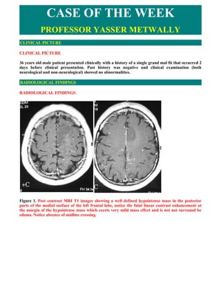
  • 1. CASE OF THE WEEK PROFESSOR YASSER METWALLY CLINICAL PICTURE CLINICAL PICTURE 36 years old male patient presented clinically with a history of a single grand mal fit that occurred 2 days before clinical presentation. Past history was negative and clinical examination (both neurological and non-neurological) showed no abnormalities. RADIOLOGICAL FINDINGS RADIOLOGICAL FINDINGS   Figure 1. Post contrast MRI T1 images showing a well defined hypointense mass in the posterior parts of the medial surface of the left frontal lobe, notice the faint linear contrast enhancement at the margin of the hypointense mass which exerts very mild mass effect and is not not surround be edema. Notice absence of midline crossing.
  • 2. Figure 2. The mass is diffusely hyperintense of the MRI T2 images. Figure 3. The mass is best delineated on FLAIR images Stereotactic biopsy showed a mixed grade II, grade III diffuse astrocytoma. Table 1 shows the clinical, radiological and pathological characteristic of grade II diffuse astrocytoma.
  • 3. Table 1. Diagnostic criteria of low grade (grade II) astrocytoma   Mild clinical disability (if any), with long history before clinical presentation  The lesions are well defined, oval or rounded with minimal mass effect, and not surrounded by edema  The lesions appear diffusely hypodense on CT scan, hypointense on precontrast T1 MRI images and hyperintense on T2 MRI images, with no postcontrast enhancement. The mere existence of contrast enhancement should shift the tumor grade to grade III astrocytoma (Mixed grade II and grade III astrocytoma). While the existence of central necrosis, perilesional edema or tumor hemorrhage should shift the tumor grade to grade IV astrocytoma (glioblastoma multiforme).  The lesions are better delineated by FLAIR imaging  The lesions are frequently misinterpreted as old infarctions, however they can easily be differentiated from infarctions by the following criteria  The existence of definite, though subtle positive mass effect  The lesions are not in the distribution of a known blood vessel  The clinical picture of the patients is not consistent with cerebrovascular disorders  The lesions are oval or rounded in shape and purely subcortical while embolic infarctions are wedge shaped cortical and subcortical In fact The existence of a such a lesion ( hypodense of CT scan, hypointense on MRI T1 images and hyperintense on on MRI T2 images with minimal mass effect and no postcontrast enhancement) in a patient presented clinically with fits ( in any age and especially in adult age) should always warrant biopsy and the clinician should not jump to the diagnosis of old infarctions, encephalomalacia or similar useless terminologies. Diagnosis of low grade astrocytomas at a younger age is very important because with the passage of time diffuse low grade astrocytomas (grade II) have a peculiar tendency to change its grade into a higher grade (grade III,IV or anaplastic astrocytomas and glioblastomas). Diffuse astrocytoma is a pathological spectrum that starts at a younger age as grade II and with time it changes its grade to grade III and IV astrocytoma. Chance for survival is undoubtedly greater when the neoplasm is diagnosed when at grade II. [53] Genetically primary glioblastomas (those that start as glioblastomas from the very beginning) are different from secondary glioblastomas (those that start as astrocytomas grade II at a younger age and change to glioblastomas at an older age). Genetic lesions associated with the development and malignant transformation of diffuse astrocytomas have been well described in the cytogenetic literature. To date, three distinct clinical, histologic, and genetic patterns of glioblastoma multiforme have been characterized. In younger patients, most diffuse astrocytomas are believed to begin as low-grade astrocytoma, with progression to glioblastoma multiforme through a stepwise acquisition of genetic lesions. These secondary glioblastoma multiforme often contain areas of well-differentiated residual tumor. The most frequent chromosomal abnormality identified in diffuse astrocytomas is the abnormal gain of chromosome 7 with an associated loss of one of the sex chromosomes. Additionally, allelic loss or
  • 4. mutation of 17p, resulting in critical alterations of the TP53 gene, has been targeted as an essential step in the early development of glioma. [53] Mutant TP53, identified in at least one third of all astrocytomas, may contribute to the formation of these tumors by inhibiting programmed cell death. glioblastoma multiforme in older patients are usually primary-that is, they occur as glioblastoma multiforme from their inception, without progression from a lower- grade tumor. In this group, the development of glioblastoma multiforme involves a parallel sequence of genetic alterations, including amplifications and deletions, that up-regulate growth factor receptors and drive cell proliferation. [53] Table 2. Diagnostic criteria of glioblastoma multiforme [53] In general three zones are identified in glioblastomas (1) A Radiologically glioblastomas are different from low grade central zone (hypointense on the T1 images, hyperintense on astrocytomas in the following points the T2 images and hypodense on CT scan) (2) A peripheral enhanced rim with multiple enhanced mural nodules and (3) 1-Short history, marked clinical disability An ill-defined diffuse large zone surrounding the first two zones. (hypointense on the T1 images, hyperintense on the T2 images and hypodense on CT scan). The first zone corresponds 2-Significant peritumoral edema with marked mass effect to the necrotic tumour tissues, the second zone corresponds to the viable tumour tissues, while the third zone corresponds to 3-The existence of enhancement (patchy, irregular or ring edema, malignant glial cell infiltrations and reactive gliosis. enhancement) The mere presence of a necrotic center in any glioma shifts the pathological grade from one with low grade malignancy to the 4-The existence of central necrosis highly malignant glioblastoma. FLAIR MRI imaging was done to the patient and proved very useful in delineating the tumour. A brief overview on FLAIR imaging is present in table 3 Table 3. FLAIR MRI imaging [53]  Fast fluid-attenuated inversion-recovery is a sequence that produces heavily T2-weighted images with cerebrospinal fluid nulling by using an inversion pulse placed at the cerebrospinal fluid nullpoint followed by a long echo time readout.  FLAIR provides superior tumor delineation by more clearly demonstrating infiltrating tumor and edema because of prominent T2 weighting and reduced contrast between gray and white matter from mild TI weighting. That mild Tl weighting is also responsible for reducing the signal of tumor on FLAIR relative to proton density- and T2-weighted sequences enabling better delineation between tumor, which is often isointense or even hypointense, and surrounding edema, which is markedly hyperintense.  Fast fluid-attenuated inversion-recovery is useful in primary tumor characterization. It can delineate the infiltrating portion of tumor better than conventional T2-weighted images. Common pathological characteristics of diffuse astrocytomas  Diffuse astrocytomas are tumors predominantly composed of astrocytes. Unless otherwise indicated, the term usually applies to diffusely infiltrating neoplasms (WHO grades II through IV).  Diffuse astrocytoma is unusual in the first decade of life and most commonly presents in older children or young adults up to the age of 40 to 45.  All diffuse astrocytomas, particularly the diffusely infiltrating variety, have a tendency toward progression to more malignant forms. Diffuse astrocytomas have a peculiar tendency to change its
  • 5. grade over time into the next higher grade of malignancy and the condition is age dependant. A change in the grade of diffuse astrocytoma is more likely to occur in the older age group.  Diffuse astrocytomas commonly start as grade II at a younger age group then gradually change its grade over time into the next higher grade until they ultimately dedifferentiate into glioblastomas (secondary glioblastoma multiforme), on the other hand, glioblastoma multiforme in older patients are usually primary-that is, they occur as glioblastoma multiforme from their inception, without progression from a lower- grade tumor.  Diffuse astrocytomas appear to form a continuum of both biological and histological aggression. They vary from lesions with almost normal cytology (grade I and grade II astrocytomas) through intermediate stages (grade III, anaplastic astrocytomas) and up to the most aggressive of all human brain tumours (grade IV astrocytomas or glioblastoma multiforme). [53]  Diffuse astrocytoma often spreads widely through the brain but without destruction and also without interruption of normal function. Microscopically, tumor cells infiltrate between myelinated fibers in a nondestructive manner (perineuronal satellitosis). The local spread of diffuse astrocytomas (forming gliomatosis cerebri and butterfly gliomas) does not mean that the tumour grade is grade IV (glioblastoma multiforme), local spread can occur in grade II and grade III and in the author experience gliomatosis cerebri and butterfly gliomas are much more commonly seen in grade II astrocytomas and has not been encountered in grade III (anaplastic astrocytomas) and grade IV (glioblastoma multiforme). It takes a long time for a diffuse astrocytoma to cross the corpus callosum to the opposite hemisphere to form a butterfly glioma. Patients harbouring glioblastomas have a much shorter life span for their tumours to form butterfly gliomas, however cases were reported for glioblastomas forming butterfly tumours. [53]  These glioma cells migrate through the normal parenchyma, collect just below the pial margin (subpial spread), surround neurons and vessels (perineuronal and perivascular satellitosis), and migrate through the white matter tracks (intrafacicular spread). This invasive behavior of the individual cells may correspond to the neoplastic cell's reacquisition of primitive migratory behavior during central nervous system development. The ultimate result of this behavior is the spread of individual tumor cells diffusely over long distances and into regions of brain essential for survival of the patient. The extreme example of this behavior is a condition referred to as gliomatosis cerebri, in which the entire brain is diffusely infiltrated by neoplastic cells with minimal or no central focal area of tumor per se. Furthermore, 25% of patients with GBM have multiple or multicentric GBMs at autopsy. Although GBMs can be visualized on MRI scans as mass lesions that enhance with contrast, the neoplastic cells extend far beyond the area of enhancement. Fig. 2 illustrates a typical result of quot;gross total resectionquot; of a temporal lobe GBM followed 6 months later by recurrence at the surgical margin and elsewhere. Even with repeat surgeries for tumor recurrences, the patients die from tumor spread into vital regions of the brain. [53]  In practice considerable histological heterogeneity in astrocytic tumours is found ( i.e., low grade areas with Rosenthal fibers and calcification can be intermixed with with frankly malignant ones).  The differences in histologic features, potential for invasiveness, and extent of progression likely reflect genetic differences acquired during astrocytoma growth.  Grade IV astrocytomas (glioblastoma multiforme) differ from diffuse astrocytoma grade II and grade III (anaplastic astrocytomas) in the presence of gross necrosis, and microscopically in the presence of vascular endothelial hyperplasia and tumour hemorrhage. [53] DIAGNOSIS: DIAGNOSIS: DIFFUSE LOW GRADE MIXED ASTROCYTOMA (GRADE II-III)
  • 6. DISCUSSION DISCUSSION Following an initial peak in the first years of life, the incidence of epilepsy remains relatively stable for decades before again increasing after the age of 60 years. Fist seizure in adult age is an uncommon occurrence and is commonly secondary to a structural brain pathology as seen in the current case, diffuse low grade astrocytoma is the prototype of that pathology. From the histopathological point of view diffuse low grade astrocytomas consist of relatively normal-appearing astrocytes, but there are just too many of them. In the past, this appearance created the impression of some type of developmental or hamartomatous change. They are not characterized by any significant vascular changes, and those that occur are limited to the capillaries. The tumors derive their nutrition from the preexisting normal vessels. Because the vessels are normal, the blood- brain barrier is intact. Mitosis, hemorrhage, vascular proliferation, endothelial changes, anaplasia and necrosis are notably absent in these neoplasms. The lesion infiltrates through the brain, usually by following the path of white matter tracts and the infiltrated brain region is diffusely expanded and hypercellular. These tumors often may spread through the brain without causing destruction or functionally significant damage, so that symptoms are not an early feature. These tumors may attain a a large size without causing any significant clinical disability. It is interesting to note that the tumor, in the current patient, was not causing any signs or symptoms, apart from a single file, despite being large enough. In low grade diffuse astrocytomas, the brain can be extensively infiltrated before any symptoms occur. These tumours widely spread without causing neural destruction or interruption of normal function. The very slow rate of growth of these tumours allows the brain to move functionality from one region to anther and this process of brain remapping, which has been shown to occur at all ages, is partially responsible for the relatively late appearance of symptoms in low grade diffuse astrocytomas. [53] Radiologically low grade diffuse astrocytomas are usually identified by diffuse enlargement of the involved brain region, abnormal signal intensity on MR or abnormal attenuation on CT. The lesions typically have precontrast CT attenuation and MRI signal changes suggesting increased water content and lower than normal specific gravity (lower CT scan densities with MRI T1 hypointensities and diffuse MRI T2 hyperintensities). It is tempting to consider that these changes represent edema. The question then arises: Is this vasogenic edema or cytotoxic edema? Because the blood-brain barrier is intact in these tumors, vasogenic edema is unlikely. The cells are not dead or dying, so that cytotoxic edema is also unlikely. Perhaps the edema results from the increased number of astrocytic cells that spread apart the normal myelinated axons of the white matter. The presence of significant amount of normal appearing astrocytes results in total increase in the water content of the affected brain area. These cells may merely have different physical and chemical properties than the normal tightly packed bundles of axons that traverse through the involved brain area. [53] As the blood brain barrier is intact in low grade diffuse astrocytomas (grade II astrocytomas according to the WHO), no significant enhancement occurs, either on MRI or CT scan. Enhancement is characteristic of the more aggressive anaplastic astrocytomas (grade III) or glioblastoma multiforme. [53] The accurate diagnosis and management of patients who have infiltrating low-grade gliomas (LGGs) is increasing in importance. Recent advances in molecular characterization, imaging, and treatment of these tumors underscore this current focus of investigations. LGGs are primary brain tumors arising within the brain parenchyma and rarely disseminate outside the central nervous system. Pathologically, they include astrocytomas, oligodendrogliomas, and mixed oligoastrocytomas, all classified as grade 2 by the World Health Organization (WHO) grading system [1]. Although considered low grade with a low proliferate rate and homogeneous cellular morphology by histologic evaluation, these tumors often undergo malignant transformation to a
  • 7. higher-grade glial tumor; therefore, they no longer are considered benign neoplasms [2,3]. Typically, LGGs are well demonstrated on routine MRI as a nonenhancing mass that is hypointense on T1 sequences and hyperintense on T2 or fluid-attenuated inversion-recovery (FLAIR) imaging (as shown in Fig. 3) [4]. These lesions are not as well defined on CT of the brain, likely accounting for the increase in early diagnosis of these lesions over the past 2 decades commensurate with the improved availability of MRI. The new era of earlier diagnosis has led some physicians to advocate early intervention with surgical resection, radiation, or chemotherapy. The hope of this early approach is that with limited disease, cure may be possible. Conversely, others advocate observation, given the slow growth of the tumor and the potential morbidities associated with radical surgical resection and radiotherapy. Early diagnosis has not been shown clearly to alter prognosis but does complicate interpretation of treatment results because of lead-time bias; those patients diagnosed early may seem to live longer even if the natural history of the tumor is not altered by the therapeutic interventions. Furthermore, there are recent data suggesting that a subset of patients may benefit from chemotherapy, a treatment previously considered ineffective. Although controversy exists regarding the optimal management of patients who have LGGs, recent randomized trials and large cooperative group efforts are beginning to provide some guidelines. Additionally, recent advances in molecular profiling of these tumors are helping to define prognosis and likelihood of response to therapy better. These important discoveries, paralleling the findings in WHO grade 3 gliomas, are changing individual patient treatment and having an impact on clinical trial designs by changing tumor classification and stratification [5,6]. This article reviews the recent results and discusses some of the controversies in management.  Histologic and molecular classification Primary brain tumors rarely spread outside the central nervous system. Therefore, use of the conventional T (tumor size), N (nodal involvement), M (metastases) system commonly used for solid tumors is of little benefit for staging primary brain tumors, such as glial malignancies. Instead, grading systems traditionally have used histologic features to stratify patients for prognostic and treatment purposes. Although several systems exist, the one developed by the WHO is used most commonly [1]. Glial tumors are graded from I to IV with grade I restricted to noninfiltrative tumors, such as pilocytic astrocytoma, often curable by surgical resection. Grade II tumors, in which LGGs are classified, are significantly different from grade I in that they are infiltrative, rarely curable by surgical resection, and have the potential to undergo malignant transformation to a higher-grade neoplasm. Within the grade II group, the most common tumors are glial in origin. This includes astrocytomas, oligodendrogliomas, and the mixed oligoastrocytomas. This subclassification is based on the morphologic appearance of the tumor cells and has prognostic and therapeutic importance. Most series demonstrate a median survival of 6 to 8 years for astrocytomas and 9 to 12 years for oligodendrogliomas [7]. Patients who have mixed oligoastrocytomas have a reported outcome that falls between the astrocytomas and pure oligodendrogliomas. In all three histologic subtypes, malignant transformation to a higher-grade tumor occurs in the majority of cases and accounts for the subsequent short survival [2,3]. Recently, molecular changes have been uncovered in LGGs that have allowed more accurate subclassification. The most prominent has been the association between the loss of heterozygosity of the 1p and 19q chromosomes (1p19q LOH) and prognosis in oligodendrogliomas. A recent study demonstrated that the 1p19q LOH is mediated by a translocation t(1;19)(q10;p10) [8] Patients harboring tumors with this codeletion have a far better prognosis than those who have oligodendrogliomas without these changes (reviewed by Aldape and colleagues [9]). 1p 19q LOH has been associated with a marked increase in treatment response in anaplastic (grade III) oligodendrogliomas; however, this has not been studied extensively for grade II tumors. Mutation of the p53 tumor suppressor gene is seen in more than 60% of astrocytomas and is related inversely to
  • 8. the presence of 1p19q LOH [10]. Loss of heterozygosity at 6q, 13q and 22q also is reported.  Clinical prognostic factors Several studies have attempted to determine prognostic factors, recognizing that grade II gliomas have a wide prognostic range with survival as short as 1 year to greater than 10 to 12 years. Pignatti and colleagues determined five factors, including age, histology, presence of neurologic deficits before surgery, tumor diameter, and tumor crossing the midline as significant determinants of outcome. There was a direct correlation of the number of poor factors with survival [11]. As shown in Table 4, patients who had no poor prognostic factors had a median survival of 9 years, whereas patients who had all 5 poor prognostic factors had a median survival of less than 1 year. The study by Bauman and colleagues [12] performed a recursive partitioning analysis of potential prognostic factors on data from 401 patients who had LGG. Four groups were identified with distinct survival differences (shown in Table 5). Table 4.Clinical prognostic factors for low-grade glioma Negative prognostic factors  Age >40 years  Largest diameter of tumor >6 cm  Tumor crosses midline  Histology is astrocytoma  Neurologic deficits are present Median survival by score (sum of negative prognostic factors) Median survival in years 0 9.2 1 8.8 2 5.8 3 3.5 4 1.9 5 0.7 Data from Pignatti F, van den Bent M, Curran D, et al. Prognostic factors for survival in adult patients with cerebral low-grade glioma. J Clin Oncol 2002;20:2076,-84. Table 5. Results of a recursive partitioning analysis demonstrating the impact of clinical factors on survivall Group KPS Patient age Tumor enhancement Median survival 1 <70 >40 years NS 12 months 2 >/=70 >40 years Yes 46 months 3 <70 18,-40 years NS 87 months >/=70 >40 years No 4 >/=70 18,-40 years NS 128 months Abbreviations: KPS, Karnofsky Performance Status score; NS, presence or absence of enhancement was not a significant factor. Data from Bauman G, Lote K, Larson D, et al. Pretreatment factors predict overall survival for patients with low-grade glioma: a recursive partitioning analysis. Int J Radiat Oncol Biol Phys 1999;45:923-9.
  • 9. As described previously, molecular factors also have a great impact on prognosis, as illustrated by 1p19q LOH. Other analyses have found a correlation between measures of tumor proliferation, such as Ki-67 and proliferative cell nuclear antigen levels and survival [13]. Those tumors with a higher proliferative rate were associated with a worse prognosis.  Signs and symptoms Patients who have brain tumors usually present with generalized or focal signs or symptoms, although occasionally the diagnosis is made coincidentally. Generalized symptoms, such as headaches, nausea, vomiting, and altered level of consciousness, arise as a consequence of increased intracranial pressure or seizures. Focal signs often are the consequence of tumor infiltration or dysfunction arising from displacement of brain parenchyma by the tumor mass. Although 30% of patients who have primary brain tumors (all types) present with seizures, 50% to 90% of patients who have LGGs present with seizures. This high rate of seizures may be the result of the frequent involvement of the cerebral cortex by these low-grade tumors. Headaches or focal neurologic deficits are present in approximately one third of patients. Cognitive or behavioral changes and papilledema are reported at presentation in approximately 10% of patients. The differential diagnosis of a nonenhancing abnormality noted on MRI is extensive. These include multiple sclerosis, stroke, infectious diseases (including viral encephalitis, toxoplasmosis, and cysticercosis), and other idiopathic conditions, such as sarcoidosis and Behçet's disease. As recently reviewed by Omuro and coworkers [14], imaging, including advanced technology (such as magnetic resonance spectroscopy, diffusion-weighted imaging, apparent diffusion coefficient maps, and positron emission tomography) often cannot distinguish reliably many of these diseases from gliomas. Investigations looking for systemic processes or other manifestations of the disease may be critical in excluding these illnesses. Additionally, since the advent of MRI, there is a cohort of patients found to have incidental nonenhancing abnormalities that frequently are unrelated to the primary reason for the imaging study. For example, some patients who have head trauma or a new-onset headache syndrome undergo a brain MRI that reveals a small, nonenhancing lesion that is unlikely related to the trauma or causing symptoms, such as headache. In these patients, the decision regarding the timing of proceeding with an extensive diagnostic evaluation, including a surgical procedure, is difficult. Often, serial imaging evaluations are performed and the decision to proceed with tissue sampling procedure, biopsy, or tumor resection is based on growth of the lesion or a change in the imaging appearance, such as the development of contrast enhancement.  Patient management Patients who have clinical or imaging-based evidence of increased intracranial pressure often benefit from the administration of corticosteroids. Dexamethasone is used most commonly because of the low level of mineralicorticoid effects combined with potent glucocorticoid effects. Patients presenting with seizures require treatment with anticonvulsants. The choice of anticonvulsant may have an important impact on the treatment of the tumor. Several anticonvulsants used commonly, such as phenytoin, carbamazepine, and phenobarbital, are well known to increase activity within the hepatic cytochrome P450 system (CYP-450) [15]. This augmentation of the CYP-450 system markedly may alter the pharmacokinetics of certain cancer chemotherapy agents, such as paclitaxel, irinotecan, and the newer tyrosine kinase signal transduction modulators, such as imatinib, gefitinib, and tipifarnib. Valproic acid has been found to inhibit CYP-450 enzymes, although the decrease in chemotherapy clearance often is not clinically significant. Anticonvulsants may have an additional impact on cancer treatment. Carbamazepine and valproic acid are associated with myelotoxicity manifest primarily as leukopenia with carbamazepine and thrombocytopenia with valproic acid. The newer generation of anticonvulsants, such as leviteractam, lamotigen, and topiramate, do not alter CYP-450 activity and rarely are associated with myelotoxicity. Recent studies are demonstrating efficacy of these newer agents in the treatment of tumor-associated
  • 10. seizures [16]. Prophylactic use of anticonvulsants in patients who have brain tumors remains controversial. A consensus statement from the American Academy of Neurology was based on a metaanalysis of four randomized studies comparing prophylactic use of anticonvulsants with no treatment [17]. Prophylactic administration of anticonvulsants did not reduce the incidence of seizures. The conclusions of this meta-analysis may not be directly applicable to patients who have LGGs, however, because (1) the studies used for the analysis included patients who had brain metastases and who had a much lower likelihood of developing seizures; (2) early generation anticonvulsants were used without documentation of adequate blood concentrations; and (3) each study included a relatively small number of patients. The impact of the newer generation of anticonvulsants on seizure prevention has not been studied adequately in patients who have brain tumor, particularly those who have LGGs where the incidence of seizures is high.  Treatment  Surgery Surgery is critical to the management of LGGS, as it is necessary for establishing a diagnosis. There remains debate, however, about the extent of surgery on the outcome of patients who have LGGs. The choice of aggressively removing diffuse LGGs to achieve a gross total or subtotal resection, rather than performing a stereotactic biopsy, has been one of the most controversial debates among neurosurgical oncologists. The decision to proceed with resection versus a biopsy often is dependent on location and growth characteristics of the lesion. Biopsies often are undertaken for deep-seated lesions (eg, tumors within the brainstem or thalamus), those confined to eloquent cortex (eg, a lesion located directly in the motor cortex), or widespread infiltrative lesions, particularly if they extend across the midline. Even for patients who have tumors in surgically accessible locations, the debate of biopsy versus tumor resection continues, however. This debate is heated, particularly for lesions in regions of the brain where surgical resection is feasible but may be associated with significant morbidity, such as the insula cortex [18]. No level I evidence exists that supports either approach. The invasive nature of diffuse LGGs and the frequent juxtaposition to or within critically “eloquent” brain regions often precludes complete tumor removal and certainly prevents resection of a margin. Because even aggressive resections may leave tumor cells behind, many neurosurgeons opt against any intervention more than a biopsy; however, other neurosurgeons advocate radical removal, or at least subtotal removal, to minimize tumor burden and reduce symptoms, in particular seizures. Over the past decade, there has been an increasing trend to more extensive tumor resection. This may be related partly to advances in imaging technologies, including functional imaging that provide preoperative localization of motor, sensory, visual, and language function. Additionally, advances in intraoperative navigational systems improve the ability to define tumor margins and the advent of intraoperative imaging, such as MRI, may result in more extensive tumor resection by identifying residual tumor that is not apparent on visual inspection. Recent studies suggest that these technologic advances have permitted increased tumor resection while reducing surgical morbidity and mortality [19]. Although improved survival has not been proved by more extensive tumor resection, there is evidence that radical resection improves the accuracy of diagnosis. A study of 81 patients who underwent a biopsy for diagnosis and then underwent an extensive resection revealed that in 38% of cases the grade of tumor was changed after a larger tissue sample was obtained at surgery [20]. The new diagnosis changed prognosis in all 31 patients and altered the treatment regimen in 21 of the patients studied. In situations where extensive resection is not safe or feasible and where biopsy is indicated, use of multivoxel spectroscopy may increase the likelihood of sampling the most malignant region of the tumor by targeting the voxel with the highest choline to creatine ratio [21].
  • 11. There also is evidence that extensive tumor resection may have a positive impact on patients' quality of life. Although only a few studies have addressed this area, there are data supporting a decrease in seizure frequency [22]. Additionally, anecdotal experience suggests that patients who have a complete or near complete resection have better palliation of symptoms and less postoperative edema than patients who undergo biopsy or only a partial resection. The impact of surgical removal of LGGs on patient survival remains unclear. For high-grade gliomas, two recent phase III studies have indicated that radical resection improves patient survival compared with biopsy or subtotal resection [23,24]. Whether or not these results translate to low- grade tumors is not known and there is no randomized trial in LGGs that addresses this question specifically. There are, however, many reports that used retrospective analyses to examine this issue [3,25,26,27,28,29,30]. These studies have been summarized in several reviews, the most comprehensive of which was by Keles and colleagues [31]. They identified 30 English-language articles on LGGs published between 1970 and 2000 that incorporated statistical analyses and addressed the issue of extent of resection. The investigators found that there was a general trend for more of the recently published articles to support extensive resections, whereas many of the older articles showed no difference between gross total resection and biopsy. To reduce known biases, the investigators then eliminated studies that included pediatric patients, that contained WHO grade I astrocytomas, or that evaluated small numbers of patients (<75). They were left with only five articles they deemed valid studies, all of which demonstrated that extent of resection was a statistically significant variable in univariate analyses, and in four of the five studies it was a significant variable in multivariate analyses. Even in these studies, however, the extent of tumor resection often was not determined precisely. Extent of resection is measured best by comparing preoperative and postoperative MRI, ideally using volumetric computational analyses. Few studies have used postoperative images to document the extent of resection [3,26,28,29,30]. In one of the better studies in the literature, Berger and colleagues [28], used a computerized image analysis technique to determine the preoperative and postoperative radiographic tumor volumes in a series of 53 patients who had diffuse LGGs. The investigators found that the extent of resection influenced significantly the incidence of recurrence and the time to tumor progression. Specifically, none of the patients who had undergone a total resection (N = 13) recurred during a mean follow-up of 54 months. In contrast, 14.8% of patients who had residual tumor whose volume was less than 10 cm3 (N = 27) recurred and 46% of patients who had residual tumor whose volume was greater than 10 cm3 recurred. Current data suggest that biopsy and radical resection are appropriate options for treating patients who have diffuse LGGs. Because of the potential advantages of maximal removal in terms of diagnosis, symptom control, and cytoreduction, however, there is an increasing trend to recommend maximal tumor removal, especially in patients who have large tumors that cause neurologic deficits, as long as neurologic function is preserved. Effective surgery requires maximal use of surgical adjuncts for defining the spatial limits of the tumor (ie, computer-assisted surgery or intraoperative ultrasound) and for assessing functional brain (ie, cortical and subcortical mapping techniques usually with awake craniotomy).  Radiation therapy Radiation therapy has been the standard treatment of LGGs for several decades. Despite widespread acknowledgment that this modality may result in tumor stabilization or reduction, however, controversy exists about optimal dosing and timing of treatment. The recent publications of results from three large international cooperative group trials and the recent completion of a fourth large randomized trial provide guidance for developing treatment algorithms.  Radiotherapy dosing The European Organization for Research and Treatment of Cancer (EORTC) in their trial, EORTC 22,844, compared low-dose radiation with high-dose radiation [32]. This study accrued 379
  • 12. patients who were randomized to receive 45 Gy over 5 weeks or 59.4 Gy over 6.6 weeks of conventional, limited-field, external beam radiotherapy. The two arms were well matched by age, gender, and percent of patients who had oligodendrogliomas or mixed oligoastrocytomas. No difference in overall survival or 5-year progression-free survival (PFS) was detected between the two treatment arms. Similarly, an intergroup study with involvement of the North Central Cancer Treatment Group, the Radiation Therapy Oncology Group (RTOG), and the Eastern Cooperative Oncology Group (ECOG) randomized 211 patients to 50.5 Gy in 28 fractions or 64.8 Gy in 36 fractions using conventional, limited-field, external beam radiotherapy [33]. The two treatment arms were well matched for histology, age, gender, and performance status. Similar to the EORTC trial, no differences in survival or PFS were noted. More radiation-induced neurotoxicity was noted with the higher-dose regimen. As a consequence of these studies, patients who have LGG typically are treated with a radiotherapy regimen that has a total dose of 50 to 54 Gy.  Timing of radiotherapy The typical slow growth of LGGs has led to a controversy regarding the timing of radiotherapy. Expectation of progressive tumor enlargement, albeit potentially over a long period of time, supports the view that early treatment should be undertaken. Conversely, given the variable growth rate of these tumors, others advocate serial observation, reserving initiation of treatment until there is clear evidence of tumor growth. A randomized clinical trial, EORTC 22,845, a collaboration between the EORTC and the Brain Tumor Working Group of the Medical Research Council of the United Kingdom, was launched in 1986 to assess the impact of early versus late initiation of radiotherapy [34,35]. The study accrued 311 patients who were randomized to undergo serial observation, reserving treatment for clear evidence of tumor progression, or receive immediate (within 8 weeks of surgery) treatment with radiation using a dosing schedule of 54 Gy over 6 weeks. The groups were well matched by extent of tumor resection. The results demonstrated a statistically significant difference in PFS with a 5-year PFS of 55% in the treated group versus 34.6% in the observation arm. There was no difference in overall survival, however, between the two groups. Unfortunately, this study did not include formal measures of quality of life using established quality of life instruments, measures of neurocognitive function, or assessment of symptom burden, including the impact of these symptoms on daily activities (interference items) [36,37,38]. An improvement in seizure control was noted for the group treated with radiotherapy and although no differences in performance status outcome were noted, concern remains that the patients treated early with radiotherapy are more likely to develop neurocognitive decline as a consequence of radiation toxicity and the development of leukoencephalopathy. More recently, an intergroup study led by the RTOG (RTOG 98-02) specifically identified patients believed to have a good prognosis to undergo only serial observation [39]. This group only included patients who were 40 years of age or younger who had undergone complete tumor resection, as estimated by a treating neurosurgeon. The initial analysis of this study arm (accrual = 111 patients) recently has been presented and submitted for publication. The overall survival rate at 5 years is 93% with 5-year PFS rate of 48%. Three factors were identified, however, that predicted outcome. Preoperative tumor diameter greater than 4 cm, astrocytic histology (versus mixed or oligodendroglial), and postoperative residual (on central review) greater than or equal to 1 cm was associated with a significantly higher risk for early progression. The group with all three poor prognostic factors had a 5-year PFS rate of 13% compared with the group with all favorable factors demonstrating a 5-year PFS rate of 70%. These results support an observation strategy for selected patients who have LGG but contemplation of additional clinical and tumor factors needs to be considered in making the decision.  Chemotherapy The role of chemotherapy in the management of LGGs is evolving. Historically, this group of tumors was considered poorly responsive to traditional cytotoxic chemotherapy. This lack of response is believed a consequence of a low rate of cell proliferation, similar to the low-grade, indolent
  • 13. lymphomas. Few studies have been performed to evaluate chemotherapy for newly diagnosed LGGs systematically. The Southwest Oncology Group performed the only published randomized trial [40]. Sixty-six patients who had LGGs and measurable tumor were randomized to external beam radiation or radiation plus lomustine (CCNU). There was no difference in survival between the two arms, and the radiation-only arm had a higher objective response rate (79% versus 54%). This study did not complete accrual, however, and there was a trend toward improved survival in the combined radiation and chemotherapy group. More recently, a series of phase II studies have been published demonstrating objective responses in patients who had recurrent LGGs. A summary of published studies is shown in Table 6 [41,42,43,44,45,46]. Objective response rates as high as 80% have been reported with the use of the combination of procarbazine, CCNU, and vincristine (PCV); complete responses, however, are rare. Similar results, presented in Table 6, also have been reported with the use of temozolomide, an oral alkylating agent that has a favorable toxicity profile. Careful review reveals that many of the tumors treated in these published studies were found to have contrast enhancement, an atypical feature for a LGG, raising the concern that the presumed recurrence was the consequence of malignant transformation or radiation-induced necrosis. Several of these studies, however, demonstrate clearly that oligodendroglial tumors and those with mixed oligoastrocytic histology are far more likely to respond to chemotherapy. Furthermore, similar to grade III oligodendrogliomas, tumors demonstrating allelic loss of the 1p and 19q arms are more likely to respond to a chemotherapy regimen [47]. Table 7. Trials of chemotherapy for low-grade glioma Prior radiation 1 No. of Enhancement Chemotherapy Response Study Pathology therapy or year patients (%) Regimen rate (%) chemotherapy PFS Buckner et 28 O, OA 46 No PCV 52 91 al [41] Soffieti et 26 O, OA 73 Yes PCV 62 80 al [42] Quinn et al 46 A, O, AA 70 Yes TMZ 61 76 [43] Pace et al 43 A, O, AA 60 Yes TMZ 47 39 [44] Brada et al 30 A, O, AA 0 No TMZ 10 >90 [48] Hoang- 60 O, OA 11 No TMZ 31 73 Xuan et al [47] Van den 32 O, OA 100 Yesa TMZ 22 11 Bent et al [45] Abbreviations: A, astrocytoma; O, oligodendrogloma; OA, oligoastrocytoma; TMZ, temozolomide. Data from Lang FF, Gilbert MR. Diffusely infiltrative low-grade gliomas in adults. J Clin Oncol 2006;24 (8):1236,-45. a All patients received prior PCV. Temozolomide has been used as the primary therapy in patients who have newly diagnosed LGG [48]. Twenty-nine patients who had incompletely resected LGG were enrolled on the study. Nineteen patients had tumors with an astrocytic or mixed histology; the remaining 10 patients had an oligodendroglioma. The overall objective response rate was 58%, combining partial and minor responses. Although progression during treatment was rare, the 3-year PFS rate was only 66%,
  • 14. suggesting that chemotherapy alone was not a sufficient treatment for patients who had residual LGG after partial resection. No data regarding the 1p 19q chromosomal status were provided for these patients. There are ongoing efforts to define further the subgroup of patients who have LGG who are likely to benefit from chemotherapy. Methylguanine-methyltransferase (MGMT) is a DNA repair enzyme, and high expression of this enzyme has been correlated with resistance to chemotherapy, including temozolomide. A recent study demonstrated a significant correlation of hypermethylation of the promoter region of the MGMT gene with improved outcome in patients who had glioblastoma who were treated with a combination of radiation therapy and temozolomide [49]. Methylation of the promoter region prevents gene transcription and tumor cells with methylated promoter have low levels of MGMT activity. A recent study correlated response to temozolomide in patients who had newly diagnosed LGG with the methylation status of the promoter region of the MGMT gene [50]. A total of 68 patients were enrolled with 62% having an oligodendroglioma and the remainder an astrocytoma (12%) or a mixed histology tumor (26%). There was combined allelic loss of 1p and 19q in 36% and intact chromosome in the remaining 64%. MGMT was hypermethylated in 63 of the 68 patients (92%). Despite the large numeric imbalance, there was a statistically significant difference in progression-free survival between the methylated and unmethylated MGMT groups (29.5 months versus 6 months, respectively; P<.0001). Allelic status of 1p and 19q also demonstrated a statistical difference in PFS. Those with allelic loss had a PFS of 35 months compared with 23 months for those without loss (P = .04). There was no statistical difference in PFS when comparing outcome based on histology. Several prospective randomized clinical trials currently are underway to address the role of chemotherapy for patients who have LGG. RTOG 98-02 is a three-arm trial that completed accrual in 2002. In addition to the observation arm for good risk patients (described previously), patients who have residual tumor or those over 40 years of age were randomized to receive radiation therapy (54 Gy) plus 6 cycles of adjuvant PCV or radiation alone. More than 250 patients were accrued to the randomized component and the results of the trial are maturing. Tumor tissue was collected on the majority of patients and it is anticipated that 1p 19q LOH status and MGMT promoter methylation data will be determined and used in the analysis of outcome. The RTOG is completing a phase II study of concurrent radiation plus daily temozolomide followed by 6 months of adjuvant temozolomide for patients who have poor-risk LGG. Using the prognostic factors defined by Pignatti and colleagues [11] (described previously), patients who have three or more poor prognostic factors are eligible. The EORTC recently opened an important study, protocol 22,041, a randomized trial comparing upfront radiation therapy (50.4 Gy) with upfront chemotherapy using temozolomide. Patients will be stratified on the basis of 1p allelic loss with the primary outcome measure of PFS with treatment-related changes in quality of life measures and neurocognitive function as the secondary objectives. The role of chemotherapy in the management of patients who have LGG remains to be defined. Although some clinical trials demonstrate a high response rate, this has not yet been shown to have an impact on patient outcome as symptomatic improvement or prolongation of survival. Much of the impetus to develop an effective chemotherapy regimen results from the concern regarding the late toxicities associated with radiotherapy to the brain. Furthermore, caution should be exercised in proceeding with more intensive treatments, such as the concurrent chemotherapy and radiotherapy regimen. Although this treatment has proved superior to radiation alone for patients who have glioblastoma, early reports suggest that the incidence of treatment-related brain injury may be higher than with radiation therapy alone [51,52]. This increased risk for late toxicity may be an important consideration, particularly in patients who have favorable prognosis LGG. The additional studies (described previously) and continued improvements in molecular classification should assist in optimizing treatment of patients who have LGG.
  • 15. SUMMARY The demonstrated case highlighted the clinical dilemma of first seizure in adult age and diffuse low grade astrocytoma. First seizure in adult age is not frequently seen and is commonly symptomatic to a serious diagnosis and structural brain lesion is commonly demonstrated by MRI examination in these patients. MRI examination must be done to every patient presented with the first seizure in adult age (between 25 to 65 years of age). Different modalities of treatment of diffuse low grade astrocytoma are discussed. In general the prognosis of diffuse low grade astrocytoma is not very good despite great advances in the management of these tumors. The most common causes of first seizure in adult age is demonstrated in table 8. Table 8. Causes of epilepsy in the adult age group Disease process Description Seizures are more common in benign intracranial tumours (meningiomas) and tumors of low grade malignancy (low grade diffuse astrocytoma (grade II,III), oligodendrogliomas, gangliogliomas), these tumours are more common in the adult age Brain tumours, brain abscess group (between 30-50 years). About 40 % of brain tumours present initially with seizures in the adult age group. More malignant glioblastomas and brain metastasis are more common in the elderly age group, seizures are not common in malignant brain tumours patients due to short life expectancy of these patients Arteriovenous malformation, cavernomas, subarachnoid Cerebrovascular disorders haemorrhage Meningocerebritis, other Tuberculous, herpetic encephalitis, cysticercosis brain infection Head trauma Seizures could be the first manifestation of systemic lupus Autoimmune erythematosus Uremia, hyponatremia, hypoglycemia, hepatic failure, Metabolic disorders hypocalcemia, hypoparathyroidism Alcohol withdrawal, drug intoxication  Addendum  A new version of this PDF file (with a new case) is uploaded in my web site every week (every Saturday and remains available till Friday.)  To download the current version follow the link quot;http://pdf.yassermetwally.com/case.pdfquot;.  You can also download the current version from my web site at quot;http://yassermetwally.comquot;.  To download the software version of the publication (crow.exe) follow the link:
  • 16. http://neurology.yassermetwally.com/crow.zip  The case is also presented as a short case in PDF format, to download the short case follow the link: http://pdf.yassermetwally.com/short.pdf  At the end of each year, all the publications are compiled on a single CD-ROM, please contact the author to know more details.  Screen resolution is better set at 1024*768 pixel screen area for optimum display.  For an archive of the previously reported cases go to www.yassermetwally.net, then under pages in the right panel, scroll down and click on the text entry quot;downloadable case records in PDF formatquot; REFERENCES References [1] [1]In: Kleihues P, Cavenee WK editor. Pathology and genetics of tumours of the nervous system. Lyon (France): IARC Press; 2000;. [2] [2]Vertosick FT, Selker RG, Arena VC. Survival of patients with well-differentiated astrocytomas diagnosed in the era of computed tomography. Neurosurgery. 1991;28(4):496–501. [3] [3]McCormack BM, Miller DC, Budzilovich GN, et al.. Treatment and survival of low-grade astrocytoma in adults–1977–1988. Neurosurgery. 1992;31(4):636–642[discussion: 642]. [4] [4]Henson JW, Gonzalez RG. Neuroimaging in glioma therapy. Expert Rev Neurother. 2004;4 (4):665–671. [5] [5]Cairncross G, Berkey B, Shaw E, et al.. A phase III trial of chemotherapy plus radiotherapy (RT) versus RT alone for pure and mixed anaplastic oligodendroglioma (RTOG 9402): an intergroup trial by the RTOG, NCCTG, SWOG, NCIC CTG and ECOG. J Clin Oncol. 2006;80:27– 35. [6] [6]van den Bent MJ, Carpentier AF, Brandes AA, et al.. Adjuvant PCV improves progression free survival but not overall survival in newly diagnosed anaplastic oligodendrogliomas and oligoastrocytomas: a randomized EORTC phase III trial. J Clin Oncol. 2006;. [7] [7]Ohgaki H, Kleihues P. Population-based studies on incidence, survival rates, and genetic alterations in astrocytic and oligodendroglial gliomas. J Neuropathol Exp Neurol. 2005;64(6):479– 489. [8] [8]Jenkins RB, Blair H, Ballman KV, et al.. A t(1;19)(q10;p10) mediates the combined deletions of 1p and 19q and predicts a better prognosis of patients with oligodendroglioma. Cancer Res. 2006;66(20):9852–9861. [9] [9]Aldape K, Burger PC, Perry A. Clinicopathologic aspects of 1p/19q loss and the diagnosis of oligodendroglioma. Arch Pathol Lab Med. 2007;131(2):242–251. [10] [10]Okamoto Y, Di Patre PL, Burkhard C, et al.. Population-based study on incidence, survival rates, and genetic alterations of low-grade diffuse astrocytomas and oligodendrogliomas. Acta Neuropathol (Berl). 2004;108(1):49–56. [11] [11]Pignatti F, van den Bent M, Curran D, et al.. Prognostic factors for survival in adult patients with cerebral low-grade glioma. J Clin Oncol. 2002;20(8):2076–2084.
  • 17. [12] [12]Bauman G, Lote K, Larson D, et al.. Pretreatment factors predict overall survival for patients with low-grade glioma: a recursive partitioning analysis. Int J Radiat Oncol Biol Phys. 1999;45(4):923–929. [13] [13]Maiuri F, Del Basso De Caro ML, Iaconetta G, et al.. Prognostic and survival-related factors in patients with well-differentiated oligodendrogliomas. Zentralbl Neurochir. 2006;67 (4):204–209. [14] [14]Omuro AM, Leite CC, Mokhtari K, et al.. Pitfalls in the diagnosis of brain tumours. Lancet Neurol. 2006;5(11):937–948. [15] [15]Gilbert MR, Supko JG, Batchelor T, et al.. Phase I clinical and pharmacokinetic study of irinotecan in adults with recurrent malignant glioma. Clin Cancer Res. 2003;9(8):2940–2949. [16] [16]Newton HB, Goldlust SA, Pearl D. Retrospective analysis of the efficacy and tolerability of levetiracetam in brain tumor patients. J Neurooncol. 2006;78(1):99–102. [17] [17]Glantz MJ, Cole BF, Forsyth PA, et al.. Practice parameter: anticonvulsant prophylaxis in patients with newly diagnosed brain tumors. Report of the Quality Standards Subcommittee of the American Academy of Neurology. Neurology. 2000;54(10):1886–1893. [18] [18]Lang FF, Olansen NE, DeMonte F, et al.. Surgical resection of intrinsic insular tumors: complication avoidance. J Neurosurg. 2001;95(4):638–650. [19] [19]Sawaya R, Hammoud M, Schoppa D, et al.. Neurosurgical outcomes in a modern series of 400 craniotomies for treatment of parenchymal tumors. Neurosurgery. 1998;42(5):1044–1055 [discussion: 1055–6]. [20] [20]Jackson RJ, Fuller GN, Abi-Said D, et al.. Limitations of stereotactic biopsy in the initial management of gliomas. Neuro oncol. 2001;3(3):193–200. [21] [21]Hall W, Truwit C. 1.5 T Spectroscopy-supported brain biopsy. Neurosurg Clin N Am. 2005;16:165–172. [22] [22]Ammirati M, Vick N, Liao YL, et al.. Effect of the extent of surgical resection on survival and quality of life in patients with supratentorial glioblastomas and anaplastic astrocytomas. Neurosurgery. 1987;21(2):201–206. [23] [23]Stummer W, Novotny A, Stepp H, et al.. Fluorescence-guided resection of glioblastoma multiforme by using 5-aminolevulinic acid-induced porphyrins: a prospective study in 52 consecutive patients. J Neurosurg. 2000;93(6):1003–1013. [24] [24]Vuorinen V, Hinkka S, Farkkila M, et al.. Debulking or biopsy of malignant glioma in elderly people–a randomised study. Acta Neurochir (Wien). 2003;145(1):5–10. [25] [25]Janny P, Cure H, Mohr M, et al.. Low grade supratentorial astrocytomas. Management and prognostic factors. Cancer. 1994;73(7):1937–1945. [26] [26]Leighton C, Fisher B, Bauman G, et al.. Supratentorial low-grade glioma in adults: an analysis of prognostic factors and timing of radiation. J Clin Oncol. 1997;15(4):1294–1301. [27] [27]Shibamoto Y, Kitakabu Y, Takahashi M, et al.. Supratentorial low-grade astrocytoma. Correlation of computed tomography findings with effect of radiation therapy and prognostic variables. Cancer. 1993;72(1):190–195.
  • 18. [28] [28]Berger MS, Deliganis AV, Dobbins J, et al.. The effect of extent of resection on recurrence in patients with low grade cerebral hemisphere gliomas. Cancer. 1994;74(6):1784–1791. [29] [29]Claus EB, Horlacher A, Hsu L, et al.. Survival rates in patients with low-grade glioma after intraoperative magnetic resonance image guidance. Cancer. 2005;103(6):1227–1233. [30] [30]Yeh SA, Ho JT, Lui CC, et al.. Treatment outcomes and prognostic factors in patients with supratentorial low-grade gliomas. Br J Radiol. 2005;78(927):230–235. [31] [31]Keles GE, Lamborn KR, Berger MS. Low-grade hemispheric gliomas in adults: a critical review of extent of resection as a factor influencing outcome. J Neurosurg. 2001;95(5):735–745. [32] [32]Karim AB, Maat B, Hatlevoll R, et al.. A randomized trial on dose-response in radiation therapy of low-grade cerebral glioma: European Organization for Research and Treatment of Cancer (EORTC) Study 22844. Int J Radiat Oncol Biol Phys. 1996;36(3):549–556. [33] [33]Shaw E, Arusell R, Scheithauer B, et al.. Prospective randomized trial of low- versus high- dose radiation therapy in adults with supratentorial low-grade glioma: initial report of a North Central Cancer Treatment Group/Radiation Therapy Oncology Group/Eastern Cooperative Oncology Group study. J Clin Oncol. 2002;20(9):2267–2276. [34] [34]Karim AB, Afra D, Cornu P, et al.. Randomized trial on the efficacy of radiotherapy for cerebral low-grade glioma in the adult: European Organization for Research and Treatment of Cancer Study 22845 with the Medical Research Council study BRO4: an interim analysis. Int J Radiat Oncol Biol Phys. 2002;52(2):316–324. [35] [35]van den Bent MJ, Afra D, de Witte O, et al.. Long-term efficacy of early versus delayed radiotherapy for low-grade astrocytoma and oligodendroglioma in adults: the EORTC 22845 randomised trial. Lancet. 2005;366(9490):985–990. [36] [36]Heimans JJ, Taphoorn MJ. Impact of brain tumour treatment on quality of life. J Neurol. 2002;249(8):955–960. [37] [37]Meyers CA, Brown PD. Role and relevance of neurocognitive assessment in clinical trials of patients with CNS tumors. J Clin Oncol. 2006;24(8):1305–1309. [38] [38]Armstrong TS, Mendoza TR, Gring I, et al.. Validation of the M.D., Anderson Symptom Inventory Brain Tumor Module (MDASI-BT). J Neurooncol. 2006;in press. [39] [39]Shaw EG, Berkey B, Coons SW, et al. Recurrence following neurosurgeon-defined gross total resection of adult supratentorial low-grade glioma. Submitted for publication 2007. [40] [40]Eyre HJ, Crowley JJ, Townsend JJ, et al.. A randomized trial of radiotherapy versus radiotherapy plus CCNU for incompletely resected low-grade gliomas: a Southwest Oncology Group study. J Neurosurg. 1993;78(6):909–914. [41] [41]Buckner JC, Gesme D, O'Fallon JR, et al.. Phase II trial of procarbazine, lomustine, and vincristine as initial therapy for patients with low-grade oligodendroglioma or oligoastrocytoma: efficacy and associations with chromosomal abnormalities. J Clin Oncol. 2003;21(2):251–255. [42] [42]Soffietti R, Ruda R, Bradac GB, et al.. PCV chemotherapy for recurrent oligodendrogliomas and oligoastrocytomas. Neurosurgery. 1998;43(5):1066–1073. [43] [43]Quinn JA, Reardon DA, Friedman AH, et al.. Phase II trial of temozolomide in patients
  • 19. with progressive low-grade glioma. J Clin Oncol. 2003;21(4):646–651. [44] [44]Pace A, Vidiri A, Galie E, et al.. Temozolomide chemotherapy for progressive low-grade glioma: clinical benefits and radiological response. Ann Oncol. 2003;14(12):1722–1726. [45] [45]van den Bent MJ, Chinot O, Boogerd W, et al.. Second-line chemotherapy with temozolomide in recurrent oligodendroglioma after PCV (procarbazine, lomustine and vincristine) chemotherapy: EORTC Brain Tumor Group phase II study 26972. Ann Oncol. 2003;14(4):599–602. [46] [46]Lang FF, Gilbert MR. Diffusely infiltrative low-grade gliomas in adults. J Clin Oncol. 2006;24(8):1236–1245. [47] [47]Hoang-Xuan K, Capelle L, Kujas M, et al.. Temozolomide as initial treatment for adults with low-grade oligodendrogliomas or oligoastrocytomas and correlation with chromosome 1p deletions. J Clin Oncol. 2004;22(15):3133–3138. [48] [48]Brada M, Viviers L, Abson C, et al.. Phase II study of primary temozolomide chemotherapy in patients with WHO grade II gliomas. Ann Oncol. 2003;14(12):1715–1721. [49] [49]Hegi ME, Diserens AC, Gorlia T, et al.. MGMT gene silencing and benefit from temozolomide in glioblastoma. N Engl J Med. 2005;352(10):997–1003. [50] [50]Everhard S, Kaloshi G, Criniere E, et al.. MGMT methylation: a marker of response to temozolomide in low-grade gliomas. Ann Neurol. 2006;60(6):740–743. [51] [51]Stupp R, Mason WP, van den Bent MJ, et al.. Radiotherapy plus concomitant and adjuvant temozolomide for glioblastoma. N Engl J Med. 2005;352(10):987–996. [52] [52]Chamberlain MC, Glantz MJ, Chalmers L, et al.. Early necrosis following concurrent Temodar and radiotherapy in patients with glioblastoma. J Neurooncol. 2007;82(1):81–83. [53]Metwally, MYM: Textbook of neurimaging, A CD-ROM publication, (Metwally, MYM editor) WEB-CD agency for electronic publishing, version 9.1a January 2008