
Recomendados
Mais conteúdo relacionado
Mais procurados
Mais procurados (19)
Semelhante a Management of bilateral_brachial_artery PRS
Semelhante a Management of bilateral_brachial_artery PRS (20)
Mais de W. Thomas McClellan, MD FACS
Mais de W. Thomas McClellan, MD FACS (20)
Último
Último (20)
Management of bilateral_brachial_artery PRS
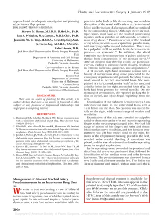
- 1. Plastic and Reconstructive Surgery • January 2012 approach and the adequate investigation and planning potential to be limb or life threatening, occurs when of perforator flap options. disruption of the vessel wall leads to extravasation of DOI: 10.1097/PRS.0b013e3182365e9c blood and formation of a hematoma that is contained Warren M. Rozen, M.B.B.S., B.Med.Sc., Ph.D. by the surrounding tissues.1 Although there are mul- tiple causes, most cases are the result of penetrating Iain S. Whitaker, M.A.Cantab., M.B.B.Chir., Ph.D. injuries, such as gunshot or stab wounds, or intrave- Jeannette W. C. Ting, M.B.B.S., Grad.Dip.Surg.Anat. nous drug abuse.1,3 Pseudoaneurysm of the brachial G. Gleda Ang, M.B.B.S., B.Med.Sc. artery often presents as an expanding, painful mass, with overlying erythema and induration. There may Rafael Acosta, M.D. be a palpable thrill or audible bruit, decreased tem- Jack Brockhoff Reconstructive Plastic Surgery Research perature, or cyanosis.1,3,4 In addition, the distal Unit extremity may demonstrate loss of pulses and pares- Department of Anatomy and Cell Biology thesias from compression of the median nerve.1–5 University of Melbourne Parkville, Victoria, Australia Arterial thrombi may develop within the pseudoan- eurysm, leading to embolic events distally, resulting Correspondence to Dr. Rozen in terminal ischemia, gangrene, and amputations.4 Jack Brockhoff Reconstructive Plastic Surgery Research A 42-year-old, right-hand-dominant woman with a Unit history of intravenous drug abuse presented to the Room E533 emergency department with pulsatile bleeding from a Department of Anatomy University of Melbourne small wound in her left antecubital fossa. She com- Grattan Street plained of a dark, raised, tender “scab” in this area, and Parkville 3050, Victoria, Australia a similar lesion in the right antecubital fossa (Fig. 1); warrenrozen@hotmail.com both had been present for several months. On the morning of presentation, she reported picking the le- DISCLOSURE sion on the left, and blood began “squirting” from the There was no source of funding for this article. The area. authors declare that there is no source of financial or other Examination of the right arm demonstrated a 3-cm support or any financial or professional relationships that subcutaneous mass in the antecubital fossa with a might pose a competing interest. 1-cm lesion on the skin. No cyanosis was noted dis- tally on the right, with normal range of motion and REFERENCES sensibility. 1. Hartrampf CR, Scheflan M, Black PW. Breast reconstruction Examination of the left arm revealed no palpable with a transverse abdominal island flap. Plast Reconstr Surg. radial or ulnar pulse at the wrist and cyanotic-appearing 1982;69:216–224. fingers to the metacarpophalangeal joint. She had full 2. Ribuffo D, Marcellino M, Barnett GR, Houseman ND, Scuderi range of motion of her fingers and wrist and dimin- N. Breast reconstruction with abdominal flaps after abdomi- ished median nerve sensibility, and her forearm com- noplasties. Plast Reconstr Surg. 2001;108:1604–1608. partment was soft but tender distal to the mass. Re- 3. Jandali S, Nelson JA, Wu LC, Serletti JM. Free transverse rectus moval of the left pressure dressing, placed in the field, abdominis myocutaneous flap for breast reconstruction in revealed active arterial bleeding. A tourniquet was patients with prior abdominal contouring procedures. J Re- constr Microsurg. 2010;26:607–614. placed, and she was taken immediately to the operating 4. Karanas YL, Santoro TD, Da Lio AL, Shaw WW. Free TRAM room for surgical exploration. flap breast reconstruction after abdominal liposuction. Plast In the operating room, control of the proximal and Reconstr Surg. 2003;112:1851–1854. distal brachial artery was performed first, followed by 5. Rozen WM, Garcia-Tutor E, Alonso-Burgos A, Corlett RJ, Tay- identification of the median nerve and volar forearm lor GI, Ashton MW. The effect of anterior abdominal wall scars fasciotomy. The pseudoaneurysm was dissected from a on the vascular anatomy of the abdominal wall: A cadaveric very friable and adherent vascular bed. The lesion was and clinical study with clinical implications. Clin Anat. 2009; 5 cm in diameter and eroded, with an associated thick- 22:815–822. Management of Bilateral Brachial Artery Supplemental digital content is available for Pseudoaneurysms in an Intravenous Drug User this article. Direct URL citations appear in the Sir: printed text; simply type the URL address into W e write to you concerning a case of bilateral brachial artery pseudoaneurysms secondary to intravenous drug abuse, one of which required emer- any Web browser to access this content. Click- able links to the material are provided in the HTML text of this article on the Journal’s Web gent repair for uncontained rupture. Arterial pseu- site (www.PRSJournal.com). doaneurysm, a rare but serious condition with the 200e
- 2. Volume 129, Number 1 • Viewpoints Fig. 1. Preoperative photograph depicting the mass in the right ante- cubital fossa. ened and stiff vascular wall extending 1 cm on either side. The lesion stopped 1 cm proximal to the bifur- cation of the radial and ulnar arteries. The diseased artery was resected, the bed was debrided, and an 8-cm ´ saphenous vein graft was used for bypass. After 90 minutes of tourniquet time, she had return of distal pulses and resolution of cyanosis. (See Video, Supplemental Digital Content 1, which shows a ruptured pseudoaneurysm of the left brachial artery, http://links.lww.com/PRS/A443. The video demonstrates preoperative physical examina- tion findings, operative repair of a left brachial artery pseudoaneurysm, and a postoperative arteriogram.) An arteriogram obtained 4 weeks postoperatively demonstrated a patent left bypass graft and a tortuous pseudoaneurysm of the right brachial artery (Fig. 2), which was repaired electively in a similar fashion several Video 1. Supplemental Digital Content 1 shows a ruptured pseudo- weeks later. (See Video, Supplemental Digital Content aneurysm of the left brachial artery, http://links.lww.com/PRS/A443. 2, which shows a pseudoaneurysm of the right brachial The video demonstrates preoperative physical examination findings, artery, http://links.lww.com/PRS/A444 . The video dem- operative repair of a left brachial artery pseudoaneurysm, and a post- onstrates preoperative physical examination findings, a operative arteriogram. preoperative arteriogram, and operative repair of the right brachial artery pseudoaneurysm.) DOI: 10.1097/PRS.0b013e3182365e84 Fig. 2. Preoperative arteriogram demonstrating the right brachial artery pseudoaneurysm. 201e
- 3. Plastic and Reconstructive Surgery • January 2012 and tumors. If a direct tensionless coaptation cannot be achieved, other techniques can be used to obtain the best possible functional outcome. Traditionally, autog- enous nerve grafts have been the criterion standard for bridging such defects.1 However, it has been demon- strated that, when the gap is less than 3 cm, vein conduit grafts yield excellent results without the comorbidities associated with harvesting a donor nerve.2 The theory behind the success of the vein graft is the creation of a patent conduit that allows a neurotrophic matrix to collect, allows axons to migrate, and prevents scar tissue ingrowth. Meticulous alignment of the nerve and tubulization of the vein graft underpin this theory and are the aims of repair.3,4 Video 2. Supplemental Digital Content 2 shows a pseudoaneu- However, accurate tubulization and alignment re- rysm of the right brachial artery, http://links.lww.com/PRS/A444. quire the surgeon to be equipped with advanced mi- The video demonstrates preoperative physical examination find- crosurgical skills and, ideally, to be operating with an assistant. Here, we outline the transluminal stay stitch, ings, a preoperative arteriogram, and operative repair of the right a technique that can provide excellent support in sit- brachial artery pseudoaneurysm. uations where there is no assistant and also help those trainees with limited experience in microsurgery. Ryan M. Wilson, M.D. The stitch works by achieving temporary alignment and stability between the venous conduit and the nerve. W. Thomas McClellan, M.D. This stability then allows anastomosing sutures to be Department of Surgery precisely performed. West Virginia University School of Medicine Morgantown, W.Va. Step 1: Align the two ends of the nerve correctly. Using Correspondence to Dr. McClellan an 8-0 nylon suture, take an epineural bite of the Morgantown Plastic Surgery Associates proximal nerve ending. 1085 Van Voorhis Road, Suite 350 Step 2: Pass the suture through the lumen of the vein Morgantown, W.Va. 26505 graft. It may be necessary to straighten the needle wtmcclellan@hotmail.com first and use the forceps to grasp the needle from inside the lumen. DISCLOSURE Step 3: Take a bite through the epineurium of the The authors have no financial interests in this research corresponding point on the distal portion of the project or in any of the techniques or equipment used in this study. nerve ending so that the correct alignment is achieved (Fig. 1). REFERENCES Step 4: Tie the two ends of this suture and apply 1. Gow KW, Mykytenko J, Patrick EL, Dodson TF. Brachial artery appropriate tension to stabilize the vein graft. pseudoaneurysm in a 6-week-old infant. Am Surg. 2004;70:518–521. Step 5: Place 9-0 nylon anastomosing sutures to 2. Yetkin U, Gurbuz A. Post-traumatic pseudoaneurysm of the achieve tubulization according to normal tech- brachial artery and its surgical treatment. Tex Heart Inst J. nique. 2003;30:293–297. Step 6: Cut the transluminal stay stitch and pull the 3. Siu WT, Yau KK, Cheung YS, et al. Management of brachial suture out (Fig. 2). artery pseudoaneurysms secondary to drug abuse. Ann Vasc Surg. 2005;19:657–661. 4. Tan KK, Chen K, Chia KH, Lee CW, Nalachandran S. Surgical management of infected pseudoaneurysms in intravenous drug abusers: Single institution experience and a proposed algorithm. World J Surg. 2009;33:1830–1835. 5. Wahlgren CM, Lohman R, Pearce BJ, Spiguel LR, Dorafshar A, Skelly CL. Metachronous giant brachial artery pseudoan- eurysms: A case report and review of the literature. Vasc En- dovasc Surg. 2007;41:467–472. Interposition of Autogenous Venous Nerve Conduit: The Transluminal Stay Stitch Sir: P eripheral nerve neurotmesis is a common finding for the plastic surgeon. It can occur following acute trauma injury or after surgical excision of neuromas Fig. 1. Step 3: take a bite through the epineurium of the corre- sponding point on the distal portion of the nerve ending, so that the correct alignment is achieved. 202e