

Recomendados
Mais conteúdo relacionado
Mais procurados
Mais procurados (20)
Destaque
Destaque (20)
Semelhante a Sc04 frank's small lungs
Semelhante a Sc04 frank's small lungs (20)
Sc04 frank's small lungs
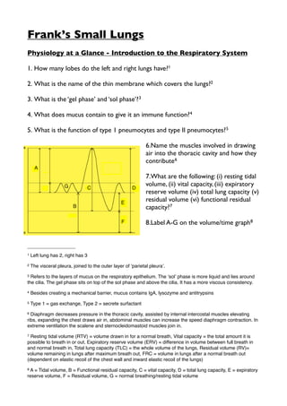
- 1. Frank’s Small Lungs Physiology at a Glance - Introduction to the Respiratory System 1. How many lobes do the left and right lungs have?1 2. What is the name of the thin membrane which covers the lungs?2 3. What is the ‘gel phase’ and ‘sol phase’?3 4. What does mucus contain to give it an immune function?4 5. What is the function of type 1 pneumocytes and type II pneumocytes? 5 6.Name the muscles involved in drawing air into the thoracic cavity and how they contribute 6 7.What are the following: (i) resting tidal volume, (ii) vital capacity, (iii) expiratory reserve volume (iv) total lung capacity (v) residual volume (vi) functional residual capacity?7 8.Label A-G on the volume/time graph8 1 Left lung has 2, right has 3 2 The visceral pleura, joined to the outer layer of ʻparietal pleuraʼ. 3 Refers to the layers of mucus on the respiratory epithelium. The ʻsolʼ phase is more liquid and lies around the cilia. The gel phase sits on top of the sol phase and above the cilia. It has a more viscous consistency. 4 Besides creating a mechanical barrier, mucus contains IgA, lysozyme and antitrypsins 5 Type 1 = gas exchange, Type 2 = secrete surfactant 6 Diaphragm decreases pressure in the thoracic cavity, assisted by internal intercostal muscles elevating ribs, expanding the chest draws air in, abdominal muscles can increase the speed diaphragm contraction. In extreme ventilation the scalene and sternocleidomastoid muscles join in. 7 Resting tidal volume (RTV) = volume drawn in for a normal breath, Vital capacity = the total amount it is possible to breath in or out, Expiratory reserve volume (ERV) = difference in volume between full breath in and normal breath in, Total lung capacity (TLC) = the whole volume of the lungs, Residual volume (RV)= volume remaining in lungs after maximum breath out, FRC = volume in lungs after a normal breath out (dependent on elastic recoil of the chest wall and inward elastic recoil of the lungs) 8A = Tidal volume, B = Functional residual capacity, C = vital capacity, D = total lung capacity, E = expiratory reserve volume, F = Residual volume, G = normal breathing/resting tidal volume
- 2. 9. Why is air sucked into the pleural space, causing the chest wall to expand during a pneumothorax?9 10.What kinds of diseases affect elastic recoil and how? 10 11.What is ‘dead space’?11 Physiology at a Glance - Lung Mechanics 1. What is the difference between static compliance and dynamic compliance? 12 2. What is hysteresis? 13 3. What is Laplace’s law? 14 4. What is Darcy’s law?15 5. What physiological factors might decrease the radius of the airway? 16 6. What do the terms ‘effort dependent’ and effort independent’ mean? 17 7. Which parts of a single full breath out are effort dependent and effort independent?18 9 Because intrapleural pressure (pressure within the pleural cavity) is negative (-0.2 to -0.5kPa) 10 Fibrosis increases elastic recoil whereas emphysema reduces it 11Anatomical dead space is the volume of airways that does not actively engage in gas exchange. Alveolar dead space is brought on by disease and is the volume of alveoli which can no longer engage in gas exchange because of damage. 12Static compliance is the change in volume per unit of of change in distending pressure. Dynamic compliance is the compliance of the lung at any given time during actual movement of air. 13The normal difference between a curve showing pressure/volume for breathing in compared to one showing breathing out. 14 Pressure is proportional to (surface tension/radius). The most important variable clinically here is radius. 15 Flow = (P1-P2)/R (P1 = alveolar pressure, P2 = mouth pressure, R = airway resistance to flow 16Parasympathetic stimulation (sympathetic stimulation of beta-2 increases radius). Inflammatory mediators, histamine, prostaglandins, leukotrienes. 17Effort dependent = increased force of breaths increases flow, Effort independent = increased force does not increase flow 18 The breath out goes from being totally effort dependent at the start to being totally effort independent at the end, i.e. when lungs are down to residual capacity no amount of force will empty them further. They switch over half way through the breath, when intra-pleural pressure becomes positive, effort makes no difference to rate of flow out.
- 3. 8. Why does effort independent breathing not happen during normal breathing?19 9. What exactly does a spirometer measure? 20 10.What is peak expiratory flow rate (PEFR) and what would happen to it in the case of obstructive lung disease?21 11.How does restrictive lung disease affect FEV1 and FVC? 22 12.How does asthma affect FEV1 and FVC?23 Physiology at a Glance - Transport and The Gas Laws 1. What are Boyles Law and Charles’ law?24 2. How much volume does 1 mol of an ideal gas occupy at 1 atm?25 3. What is Henry’s law?26 4. Regarding gasses dissolving in fluid, what is the relationship between temperature and solubility?27 5. How does solubility of CO2 in water compare to that of oxygen?28 19 During normal breathing intra-pleural pressure is negative throughout 20 Lung volume, measures the total amount of air breathed out of a full exhalation 21The maximum speed of exhalation, measured using a peak flow metre. Peak flow decreases if airway resistance increases, such as in obstructive lung disease. 22In restrictive lung disease, FEV and FVC are low but FEV1 is normal or possibly a little high because the lung fibrosis increases elastic recoil of the lungs. 23Asthma blocks the airways rather than the alveoli, so FEV1 tends to be lower. In emphysema (floppy lungs) and COPD airway resistance is also higher but the elastic recoil in lungs is lower so FEV1 is dramatically lower. 24Boyles law = volume is inversely proportional to the pressure. Charlesʼ law = volume is proportional to the absolute temperature 25 22.4 Litres 26 (for gasses dissolved in fluid) dissolved gas concentration = partial pressure of gas above fluid x solubility of that gas in that fluid. So solubility differs for different liquids. 27solubility of gas decreases with a rise in temperature, the maximum saturated water vapour pressure depends on the temperature and pressure 28 The solubility of Co2 in water is 20x that of O2.
- 4. 6. What does partial pressure mean?29 7. What two factors influence ‘diffusing capacity’/‘transfer factor’ (DL) of a gas in the lungs?30 8. What is the distinction between ‘diffusion limited’ and ‘perfusion limited’?31 Scenario Presentation 1.Wheezing is generated by turbulent flow, but why might you hear crackles when listening to a patient’s chest with a stethoscope32 2.Label features A-K on the normal chest x-ray33 Gaseous Diffusion and Transport Parts 1 & 2 1.What is Dalton’s Law?34 2.What is the average barometric (atmospheric) pressure at sea level? 35 3. Convert this into mmHg 36 29 Partial pressure is the pressure that would be required to dissolve that amount of gas in the fluid according to Henryʼs law. Movement of gasses between gas and fluid states is therefore related to partial pressures not necessarily concentration. 30Diffusing capacity of a gas is the permeability of the lungs to given gas x area. It is basically a measure of alveolar/capillary membrane function. 31Diffusion limited = limited only by the rate of diffusion across the alveolar-capillary membrane, because the gas binds so avidly to haemoglobin (e.g. CO) the rate of transfer at low concentrations is not dependent on blood flow. Perfusion limited = for a less soluble gas, transfer is limited by the amount of haemoglobin (blood flow), increased blood flow will increase transfer. 32Crackles are generated by airways that have collapsed during expiration snapping open as pressure equalizes in inspiration. The fineness or coarseness varies with the size of the airway. The timing on inspiration varies with the lung condition. 33A = liver, B = anterior rib, C = clavicle, D= scapula, E = bronchus, F = aortic knob, G = left bronchus, H = descending aorta, I = posterior rib, J = heart/right atrium , K = gastric air bubble 34The partial pressure of a gas in a mixture is the pressure it would exert if it occupied the volume alone. Expressed as fractional concentration (FO2) x barometric pressure (atm) 35 101kPa 36 1 kPa = 7.5 mmHg, so 101kPa = 760mmHg
- 5. 4. What will an increase in partial pressure of a gas mixed with liquid cause the gas to do?37 5. What happens if you put two different immiscible liquids together, one contains a greater amount of dissolved gas than the other but both have the same partial pressure? 38 6. What difference does a gas passing over moist surfaces for a long enough time have compared to passing over dry surfaces? 39 7. What are the typical PO2 and PCO2 values for alveolar air vs atmospheric air?40 8. What factors affect the rate of diffusion across the alveolar membrane? 41 9. What is the oxygen diffusing capacity?42 10.What substance would you use to measure the accurate oxygen diffusing capacity?43 11.How many and what types of protein chain does a normal haemoglobin have? 44 12.What form does iron need to be in order to bind with oxygen?45 13.What is the difference between oxygen saturation in the pulmonary vein and oxygen saturation in arterial blood?46 37More would dissolve in the liquid (bubbles in bottled coke/beer when opened). Partial pressure difference between liquid (read: blood) and air (read: alveolar air), defines the direction of movement. 38There is no movement of gas because the partial pressure is the same. It is partial pressure NOT concentration that determines movement. 39Passing over wet surfaces will eventually make the gas 100% saturated. This is why it rains more in the west of england. 40 Atmospheric air: PO2 (21kpa), PCO2 (0kpa), Alveolar gas: PO2 (13.5kpa), PCO2 (5.3kpa) 41 Fickʼs and Grahamʼs laws are based on thickness and area (large area with thinnest membrane = highest diffusion). Area/thickness 42 (a.k.a Transfer factor) oxygen uptake from the lungs/alveolar O2 pressure - capillary O2 pressure 43Carbon Monoxide (CO) because it is taken up 100% by the capillaries using the same mechanism as oxygen, whereas less than 100% of alveolar oxygen will be taken up. 44 Two alpha and two beta chains. 45 Ferrous (Fe2+) NOT Ferric (Fe3+) 46 100% in pulmonary vein, 100-97% in arteries as contaminated by some deoxygenated blood by this stage.
- 6. 14.What is the haemoglobin concentration in normal blood?47 15.What does ‘mixed venous blood’ mean?48 16.Why does the oxygen-haemoglobin dissociation curve get steeper above 25% oxygen saturation?49 17.Why does the curve slow down and start to plateau above 75% O2 saturation?50 18.What causes oxygen to leave the haemoglobin once it reaches the tissues?51 19.What is the oxygen saturation of mixed venous blood?52 20.Why does hyperventilation or breathing pure oxygen not send a lot more oxygen to your tissues? 53 21.What are the main differences between HbA and HbF?54 22.What is myoglobin?55 23.What is cyanosis?56 24.What oxygen saturations are signs of cyanosis?57 47 150g/l 48 Blood in the pulmonary artery, because venous blood in different veins varies hugely in how much oxygen it had left in it. Pulmonary artery blood can be said to be the best ʻaverageʼ. 49Because when the first O2 molecule binds to haemoglobin, its changes the haemoglobinʼs configuration to give it a higher affinity for further oxygen binding. 50 Because the haemoglobin binding sites are starting to get taken up, there are fewer of them. 51The PO2 is lower, temperature is higher, CO2 and acid levels are higher (acid levels may also increase with lactate) both these things decrease the affinity of haemoglobin for oxygen. 52 75% at rest 53 PO2 is raised but oxygen delivery to tissues is not increased because oxygen content of the blood is not increased, haemoglobin is already fully saturated so the only difference that can be made is a small increase in oxygen dissolved in the plasma. If the PO2 is lower, such as at high altitude or in somebody who is hypoxic, raising PO2 will help (because you start on the steep bit of the curve). 54HbA is normal adult haemoglobin and has 2 alpha and 2 beta chains, HbF is fetal haemoglobin and has 2 alpha and 2 gamma protein chains, giving it a higher affinity for oxygen and allowing it to pick up oxygen across the placenta. 55 Single binding site, only releases single oxygen bound to it when pO2 is very low, found in muscles. 56Blue-grey colour of skin, associated with a high (absolute) concentration of deoxygenates haemoglobin. Peripheral cyanosis (only seen on skin), central cyanosis (seen by blue tongue, lips and buccal mucosa) 57 85-90%
- 7. 25.How does this change in the case of anaemia?58 26.How does the solubility of CO2 in the plasma compare to O2?59 27.What happens to the HCO3- produced from buffering CO2 in haemoglobin?60 28.Some of the Co2 in haemoglobin is converted into carb-amino compounds, give 2 examples of these?61 29.What is the Haldane effect? 62 30.What proportion of CO2 breathed out came from (i) dissolved CO2, (ii) carb- amino compounds, (iii) HCO3- from buffering63 31.Why is it meaningless to talk about % CO2 saturation? 64 32.How can you work out CO2 production?65 33.How do you define hyperventilation? 66 34.What is hypercapnia?67 58It will appear less easily in an anaemic person, because they have low haemoglobin so blue deoxygenated haemoglobin is less visible. Person will have to be extremely hypoxic before cyanosis visible. 59 CO2 is 20 times more soluble in the blood plasma than oxygen 60HCO3- moves out the cell down its concentration gradient, it is exchanged with Cl-. Haemoglobin buffers hydrogen ions. 61 Lysine and arginine 62At any given PCo2 the quantity of CO2 carried is greater in deoxygenated blood than in oxygenated blood. Hb forms carbamino compounds more readily when deoxygenated and Hb is a better buffer when deoxygenated (as there is a greater concentration of CO2) 63 dissolved (10%), carb-amino (30%), HCO3- (60%) 64Because although there are a limited number of binding sites for carb-amino compounds, a basically unlimited amount of CO2 can be buffered into HCO3- 65 CO2 production = alveolar CO2 fraction x alveolar ventilation (ml/min) 66 Alveolar ventilation which is higher than the CO2 being produced. 67 High PaCO2 due to hypoventilation