Cardiovascular System | Cardiology
•
91 gostaram•6,811 visualizações

Cardiovascular Medical Terminology

Recomendados
Mais conteúdo relacionado
Mais procurados
Mais procurados (20)
Destaque
Destaque (20)
Semelhante a Cardiovascular System | Cardiology
Semelhante a Cardiovascular System | Cardiology (20)
Mais de whitchur
Último
Último (20)
Cardiovascular System | Cardiology
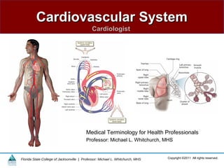
- 1. Cardiovascular SystemCardiovascular System Cardiologist Copyright ©2011 All rights reserved.Florida State College of Jacksonville | Professor: Michael L. Whitchurch, MHS Medical Terminology for Health Professionals Professor: Michael L. Whitchurch, MHS
- 2. Copyright ©2011 All rights reserved.Florida State College of Jacksonville | Professor: Michael L. Whitchurch, MHS Learning Objectives 1. Identify the structures of the cardiovascular system. 2. Describe the process of circulation. 3. Describe common cardiovascular diseases and conditions, laboratory and diagnostic procedures, medical and surgical procedures, and drug categories. 4. Give the medical meaning of word parts related to the cardiovascular system. 5. Build cardiovascular words from word parts and divide and define cardiovascular words. 6. Spell and pronounce cardiovascular words. 7. Analyze the medical content and meaning of a cardiology report.
- 3. Copyright ©2011 All rights reserved.Florida State College of Jacksonville | Professor: Michael L. Whitchurch, MHS Cardiology • The medical specialty that studies the anatomy and physiology of the cardiovascular system and uses diagnostic tests, medical and surgical procedures, and drugs to treat cardiovascular diseases.
- 4. Copyright ©2011 All rights reserved.Florida State College of Jacksonville | Professor: Michael L. Whitchurch, MHS Cardiovascular system Pulmonary system
- 5. Copyright ©2011 All rights reserved.Florida State College of Jacksonville | Professor: Michael L. Whitchurch, MHS The Cardiovascular System • A continuous, circular body system that includes the heart and the vascular structures (blood vessels such as arteries, capillaries, and veins) • Moves blood throughout the body and transports oxygen, carbon dioxide, nutrients, and wastes in the blood
- 6. Copyright ©2011 All rights reserved.Florida State College of Jacksonville | Professor: Michael L. Whitchurch, MHS Anatomy of the Cardiovascular System • Heart – A muscular organ that contracts at least once every second to pump blood through the body – Has an extensive electrical system that initiates and coordinates its contractions
- 7. Copyright ©2011 All rights reserved.Florida State College of Jacksonville | Professor: Michael L. Whitchurch, MHS Figure 5-2 Surface of the heart
- 8. Copyright ©2011 All rights reserved.Florida State College of Jacksonville | Professor: Michael L. Whitchurch, MHS Anatomy of the Cardiovascular System • Heart Chambers – The heart contains four chambers, two on the top and two on the bottom. – Each small upper chamber is an atrium. – Each large lower chamber is a ventricle. – The septum, a center wall, divides the heart into right and left halves. – The inferior tip of the heart is the apex.
- 9. Copyright ©2011 All rights reserved.Florida State College of Jacksonville | Professor: Michael L. Whitchurch, MHS Figure 5-3 Chambers and valves of the heart
- 10. Copyright ©2011 All rights reserved.Florida State College of Jacksonville | Professor: Michael L. Whitchurch, MHS Figure 5-4 Aortic valve
- 11. Copyright ©2011 All rights reserved.Florida State College of Jacksonville | Professor: Michael L. Whitchurch, MHS Anatomy of the Cardiovascular System • Heart Muscle – Mycardium — composed of cardiac muscle – Responds to electrical impulses generated by a node within the heart itself – Contracts in a coordinated way to pump blood – Thickest on the left side of the heart
- 12. Copyright ©2011 All rights reserved.Florida State College of Jacksonville | Professor: Michael L. Whitchurch, MHS Table 5-1 Layers and Membranes of the Heart
- 13. Copyright ©2011 All rights reserved.Florida State College of Jacksonville | Professor: Michael L. Whitchurch, MHS Figure 5-5 Layers and membranes of the heart
- 14. Copyright ©2011 All rights reserved.Florida State College of Jacksonville | Professor: Michael L. Whitchurch, MHS Anatomy of the Cardiovascular System • Thoracic Cavity – Contains the lungs and the mediastinum, an irregularly shaped central area between the lungs
- 15. Copyright ©2011 All rights reserved.Florida State College of Jacksonville | Professor: Michael L. Whitchurch, MHS Anatomy of the Cardiovascular System • Mediastinum – Contains the heart and parts of the great vessels (aorta, superior vena cava, inferior vena cava, pulmonary artery, and veins), as well as the thymus, trachea, and the esophagus
- 16. Copyright ©2011 All rights reserved.Florida State College of Jacksonville | Professor: Michael L. Whitchurch, MHS Figure 5-6 Mediastinum
- 17. Copyright ©2011 All rights reserved.Florida State College of Jacksonville | Professor: Michael L. Whitchurch, MHS Anatomy of the Cardiovascular System • Blood Vessels – Vascular channels through which blood circulates in the body – Have a central opening or lumen through which the blood flows – Lined with endothelium, a smooth inner layer that promotes the flow of blood
- 18. Copyright ©2011 All rights reserved.Florida State College of Jacksonville | Professor: Michael L. Whitchurch, MHS Anatomy of the Cardiovascular System • There are three kinds of blood vessels in the body, each performing a different function: – Arteries – Capillaries – Veins
- 19. Copyright ©2011 All rights reserved.Florida State College of Jacksonville | Professor: Michael L. Whitchurch, MHS Anatomy of the Cardiovascular System • Arteries – Large blood vessels – Smaller branches of an artery are arterioles
- 20. Copyright ©2011 All rights reserved.Florida State College of Jacksonville | Professor: Michael L. Whitchurch, MHS Anatomy of the Cardiovascular System • All arteries share some important characteristics and functions: – They carry blood away from the heart to the body. Exception: pulmonary arteries – They carry bright red blood that has a high level of oxygen. – Most arteries lie deep beneath the skin. – All arteries have smooth muscle in their walls.
- 21. Copyright ©2011 All rights reserved.Florida State College of Jacksonville | Professor: Michael L. Whitchurch, MHS Figure 5-7 Vasoconstriction and vasodilation
- 22. Copyright ©2011 All rights reserved.Florida State College of Jacksonville | Professor: Michael L. Whitchurch, MHS Anatomy of the Cardiovascular System • Capillaries – Smallest blood vessels in the body – The lumen of a capillary is so small that blood cells must pass through in single file.
- 23. Copyright ©2011 All rights reserved.Florida State College of Jacksonville | Professor: Michael L. Whitchurch, MHS Anatomy of the Cardiovascular System • Veins – Capillaries combine to form small veins known as venules, which then combine to form the largest veins.
- 24. Copyright ©2011 All rights reserved.Florida State College of Jacksonville | Professor: Michael L. Whitchurch, MHS Anatomy of the Cardiovascular System • All veins share some important characteristics and functions: – They carry blood from the body back to the heart. – They carry dark red-purple blood with a low level of oxygen.
- 25. Copyright ©2011 All rights reserved.Florida State College of Jacksonville | Professor: Michael L. Whitchurch, MHS Anatomy of the Cardiovascular System • All veins share some important characteristics and functions: – The largest veins have valves that keep the blood flowing in one direction―toward the heart. – Many veins are near the surface of the body and can be seen just under the skin as bluish, sometimes bulging lines.
- 26. Copyright ©2011 All rights reserved.Florida State College of Jacksonville | Professor: Michael L. Whitchurch, MHS Anatomy of the Cardiovascular System • Aorta – Largest artery in the body – Receives blood from the left ventricle of the heart
- 27. Copyright ©2011 All rights reserved.Florida State College of Jacksonville | Professor: Michael L. Whitchurch, MHS Figure 5-9 Arteries and veins around the heart
- 28. Copyright ©2011 All rights reserved.Florida State College of Jacksonville | Professor: Michael L. Whitchurch, MHS Figure 5-10 Arteries in the body
- 29. Copyright ©2011 All rights reserved.Florida State College of Jacksonville | Professor: Michael L. Whitchurch, MHS Anatomy of the Cardiovascular System • Arteries – Coronary artery – Carotid artery – Subclavian artery – Axillary artery (armpit) – Brachial artery (upper arm) – Radial artery (thumb side of the lower arm) – Ulnar artery (little finger side of the lower arm)
- 30. Copyright ©2011 All rights reserved.Florida State College of Jacksonville | Professor: Michael L. Whitchurch, MHS Anatomy of the Cardiovascular System • Thoracic Aorta and Arterial Branches – The thoracic aorta travels inferiorly through the thoracic cavity. – It branches into arteries that bring blood to the esophagus, muscles between the ribs, diaphragm, upper spinal cord, and the back.
- 31. Copyright ©2011 All rights reserved.Florida State College of Jacksonville | Professor: Michael L. Whitchurch, MHS Anatomy of the Cardiovascular System • Abdominal Aorta and Arterial Branches – Brings oxygenated blood to the stomach, liver, pancreas, spleen, gallbladder, small and large intestines, adrenal glands, kidneys, ovaries, testes, and the lower spinal cord – Abdominal aorta ends where right and left iliac arteries begin
- 32. Copyright ©2011 All rights reserved.Florida State College of Jacksonville | Professor: Michael L. Whitchurch, MHS Anatomy of the Cardiovascular System • Abdominal Aorta and Arterial Branches – Iliac artery – Femoral artery (upper leg) – Popliteal artery (near the knee joint) – Tibial artery (front and back of the lower leg) – Peroneal artery (little toe side of the lower leg)
- 33. Copyright ©2011 All rights reserved.Florida State College of Jacksonville | Professor: Michael L. Whitchurch, MHS Anatomy of the Cardiovascular System • Pulmonary Arteries – Originate from the pulmonary trunk, which comes from the right ventricle of the heart, not from the aorta
- 34. Copyright ©2011 All rights reserved.Florida State College of Jacksonville | Professor: Michael L. Whitchurch, MHS Anatomy of the Cardiovascular System • Two major veins of the body – Superior vena cava – Inferior vena cava • Other major veins – Jugular vein – Portal vein – Saphenous and femoral veins
- 35. Copyright ©2011 All rights reserved.Florida State College of Jacksonville | Professor: Michael L. Whitchurch, MHS Anatomy of the Cardiovascular System • Circulation – The cardiovascular system or circulatory system circulates blood through the blood vessels. – Systemic circulation includes the arteries, capillaries, and veins everywhere in the body, except in the lungs. – Pulmonary circulation includes the arteries, capillaries, and veins going to, within, and coming from the lungs.
- 36. Copyright ©2011 All rights reserved.Florida State College of Jacksonville | Professor: Michael L. Whitchurch, MHS Figure 5-11 Circulation of the blood
- 37. Copyright ©2011 All rights reserved.Florida State College of Jacksonville | Professor: Michael L. Whitchurch, MHS Physiology of a Heartbeat • Heart contracts and relaxes in a regular rhythm coordinated by the conduction system – Sinoatrial node (SA node), or pacemaker of the heart, initiates the electrical impulse that begins each heartbeat
- 38. Copyright ©2011 All rights reserved.Florida State College of Jacksonville | Professor: Michael L. Whitchurch, MHS Physiology of a Heartbeat • Heart contracts and relaxes in a regular rhythm coordinated by the conduction system – Atrioventricular node (AV node) receives the impulse to contract from the SA node – Purkinje fibers, a network of nerves, cause both ventricles to contract simultaneously
- 39. Copyright ©2011 All rights reserved.Florida State College of Jacksonville | Professor: Michael L. Whitchurch, MHS Figure 5-12 Conduction system of the heart
- 40. Copyright ©2011 All rights reserved.Florida State College of Jacksonville | Professor: Michael L. Whitchurch, MHS Physiology of a Heartbeat • Two Heartbeat Phases – Systole (contraction) – Diastole (resting period)
- 41. Copyright ©2011 All rights reserved.Florida State College of Jacksonville | Professor: Michael L. Whitchurch, MHS Diseases and Conditions • Myocardium – Acute coronary syndrome – Angina pectoris – Cardiomegaly – Cardiomyopathy – Congestive heart failure (CHF) – Myocardial infarction (MI)
- 42. Copyright ©2011 All rights reserved.Florida State College of Jacksonville | Professor: Michael L. Whitchurch, MHS Figure 5-14 Peripheral edema Antonia Reeve/Photo Researchers, Inc.l
- 43. Copyright ©2011 All rights reserved.Florida State College of Jacksonville | Professor: Michael L. Whitchurch, MHS Diseases and Conditions • Heart Valves and Layers of the Heart – Endocarditis – Mitral valve prolapse (MVP) – Murmur – Pericarditis – Rheumatic heart disease Vegetation on the mitral valve Abrahas/Custom Medical Stock Photo, Inc
- 44. Copyright ©2011 All rights reserved.Florida State College of Jacksonville | Professor: Michael L. Whitchurch, MHS Figure 5-15 Vegetation on the mitral valve Abrahas/Custom Medical Stock Photo, Inc.
- 45. Copyright ©2011 All rights reserved.Florida State College of Jacksonville | Professor: Michael L. Whitchurch, MHS Diseases and Conditions • Conduction System – Arrhythmia Bradycardia Fibrillation Flutter Heart block (Arrhythmias on an EKG tracing)
- 46. Copyright ©2011 All rights reserved.Florida State College of Jacksonville | Professor: Michael L. Whitchurch, MHS Figure 5-16 Arrhythmias on an EKG tracing
- 47. Copyright ©2011 All rights reserved.Florida State College of Jacksonville | Professor: Michael L. Whitchurch, MHS Diseases and Conditions • Conduction System – Arrhythmia Premature contraction Sick sinus syndrome Tachycardia – Asystole – Palpitation
- 48. Copyright ©2011 All rights reserved.Florida State College of Jacksonville | Professor: Michael L. Whitchurch, MHS Diseases and Conditions • Blood Vessels – Aneurysm – Arteriosclerosis – Bruit – Coronary artery disease (CAD) – Hyperlipidemia – Hypertension (HTN) – Hypotension – Peripheral artery disease (PAD)
- 49. Copyright ©2011 All rights reserved.Florida State College of Jacksonville | Professor: Michael L. Whitchurch, MHS Figure 5-27 An aneurysm (b) Michael English, M.D./Custom Medical Stock Photo, Inc.
- 50. Copyright ©2011 All rights reserved.Florida State College of Jacksonville | Professor: Michael L. Whitchurch, MHS Aneurysm Video Click on the screenshot to view a video on the topic of aneurysms. Back to Directory
- 51. Copyright ©2011 All rights reserved.Florida State College of Jacksonville | Professor: Michael L. Whitchurch, MHS Figure 5-18 Mild atheromatous plaque SIU BioMed/Custom Medical Stock Photo, Inc.
- 52. Copyright ©2011 All rights reserved.Florida State College of Jacksonville | Professor: Michael L. Whitchurch, MHS Figure 5-19 Severe atherosclerotic plaque in an artery C. Abrahams, M.D./Custom Medical Stock Photo, Inc.
- 53. Copyright ©2011 All rights reserved.Florida State College of Jacksonville | Professor: Michael L. Whitchurch, MHS Diseases and Conditions • Blood Vessels (con’t) – Peripheral vascular disease (PVD) – Phlebitis – Raynaud’s disease – Varicose veins
- 54. Copyright ©2011 All rights reserved.Florida State College of Jacksonville | Professor: Michael L. Whitchurch, MHS Laboratory and Diagnostic Procedures • Blood Tests – Cardiac enzymes – C-reactive protein (CRP) – Homocysteine – Lipid profile – Troponin
- 55. Copyright ©2011 All rights reserved.Florida State College of Jacksonville | Professor: Michael L. Whitchurch, MHS Laboratory and Diagnostic Procedures • Diagnostic Heart Procedures – Cardiac catheterization – Cardiac exercise stress test – Electrocardiography (ECG, EKG) – Electrophysiologic study (EPS)
- 56. Copyright ©2011 All rights reserved.Florida State College of Jacksonville | Professor: Michael L. Whitchurch, MHS Figure 5-21 Treadmill exercise stress test Fotopic/Miles Simons/Phototake NYC
- 57. Copyright ©2011 All rights reserved.Florida State College of Jacksonville | Professor: Michael L. Whitchurch, MHS Figure 5-22 Electrocardiography Jupiter Images – PictureArts Corporation/Brand X Pictures-Royalty Free
- 58. Copyright ©2011 All rights reserved.Florida State College of Jacksonville | Professor: Michael L. Whitchurch, MHS Figure 5-23 An EKG tracing
- 59. Copyright ©2011 All rights reserved.Florida State College of Jacksonville | Professor: Michael L. Whitchurch, MHS Laboratory and Diagnostic Procedures • Diagnostic Heart Procedures – Holter monitor – Pharmacologic stress test – Telemetry
- 60. Copyright ©2011 All rights reserved.Florida State College of Jacksonville | Professor: Michael L. Whitchurch, MHS Laboratory and Diagnostic Procedures • Radiology and Nuclear Medicine Procedures – Angiography – Echocardiography Echocardiogram Custom Medical Stock Photo, Inc
- 61. Copyright ©2011 All rights reserved.Florida State College of Jacksonville | Professor: Michael L. Whitchurch, MHS Figure 5-25 Doppler ultrasonography Matt Meadows/Science Photo Library/Photo Researchers, Inc.
- 62. Copyright ©2011 All rights reserved.Florida State College of Jacksonville | Professor: Michael L. Whitchurch, MHS Laboratory and Diagnostic Procedures • Radiology and Nuclear Medicine Procedures Multiple- gated acquisition (MUGA) scan – Myocardial perfusion scan – Single-photon emission computed tomography (SPECT) scan Three consecutive multi-detector row CT images of an acute transmural infarct in short-axis orientation
- 63. Copyright ©2011 All rights reserved.Florida State College of Jacksonville | Professor: Michael L. Whitchurch, MHS Medical and Surgical Procedures • Medical Procedures – Auscultation – Cardioversion – Sclerotherapy – Vital signs Defibrillation Pearson Education/PH College
- 64. Copyright ©2011 All rights reserved.Florida State College of Jacksonville | Professor: Michael L. Whitchurch, MHS Figure 5-27 Pulse points
- 65. Copyright ©2011 All rights reserved.Florida State College of Jacksonville | Professor: Michael L. Whitchurch, MHS Figure 5-28 Carotid pulse Michal Heron/Pearson Education/PH College
- 66. Copyright ©2011 All rights reserved.Florida State College of Jacksonville | Professor: Michael L. Whitchurch, MHS Figure 5-29 Measuring the blood pressure
- 67. Copyright ©2011 All rights reserved.Florida State College of Jacksonville | Professor: Michael L. Whitchurch, MHS Medical and Surgical Procedures • Surgical Procedures – Aneurysmectomy – Cardiopulmonary bypass – Carotid endarterectomy F. Schussler/PhotoDisc/Getty Images
- 68. Copyright ©2011 All rights reserved.Florida State College of Jacksonville | Professor: Michael L. Whitchurch, MHS Medical and Surgical Procedures • Surgical Procedures – Coronary artery bypass graft (CABG) – Heart transplantation – Pacemaker insertion Pacemaker (left) English/Custom Medical Stock Photo, Inc. (right) Alvis Upitis/Jupiter Images-PictureArts Corporation/Brand X Pictures-Royalty Free
- 69. Copyright ©2011 All rights reserved.Florida State College of Jacksonville | Professor: Michael L. Whitchurch, MHS Medical and Surgical Procedures • Surgical Procedures – Percutaneous transluminal coronary angioplasty (PTCA) – Pericardiocentesis – Radiofrequency catheter ablation – Valve replacement – Valvoplasty
- 70. Copyright ©2011 All rights reserved.Florida State College of Jacksonville | Professor: Michael L. Whitchurch, MHS Figure 5-32 Balloon angioplasty
- 71. Copyright ©2011 All rights reserved.Florida State College of Jacksonville | Professor: Michael L. Whitchurch, MHS Figure 5-33 Stent
- 72. Copyright ©2011 All rights reserved.Florida State College of Jacksonville | Professor: Michael L. Whitchurch, MHS Figure 5-34 Valve replacement surgery Custom Medical Stock Photo, Inc.
- 73. Copyright ©2011 All rights reserved.Florida State College of Jacksonville | Professor: Michael L. Whitchurch, MHS Drug Categories • These categories of drugs are used to treat cardiovascular diseases and conditions: – Angiotensin converting enzyme inhibitor drugs – Antiarrhythmic drugs – Anticoagulant drugs – Antihypertensive drugs – Aspirin
- 74. Copyright ©2011 All rights reserved.Florida State College of Jacksonville | Professor: Michael L. Whitchurch, MHS Drug Categories • These categories of drugs are used to treat cardiovascular diseases and conditions: – Beta-blocker drugs – Calcium channel blocker drugs – Digitalis drugs – Diuretic drugs
- 75. Copyright ©2011 All rights reserved.Florida State College of Jacksonville | Professor: Michael L. Whitchurch, MHS Drug Categories • These categories of drugs are used to treat cardiovascular diseases and conditions: – Drugs for cardiac arrest – Drugs for hyperlipidemia – Nitrate drugs – Thrombolytic drugs
- 76. Copyright ©2011 All rights reserved.Florida State College of Jacksonville | Professor: Michael L. Whitchurch, MHS Abbreviations
- 77. Copyright ©2011 All rights reserved.Florida State College of Jacksonville | Professor: Michael L. Whitchurch, MHS Abbreviations
Notas do Editor
- The cardiovascular system, also called the circular system, is one of the busiest systems in the body and works constantly to transport nutrients, oxygen, and waste in and out of the body. Your heart never takes a break. To understand the complexities of the circulatory system, you need to understand the structures that compose the heart, other structures that are part of the system, and how they are all related. You must become familiar with the types of tests and procedures that are available to diagnose and treat cardiovascular problems, which type of cardiovascular specialists can treat the variety of problems that occur within this body system, and the specialized terms and abbreviations associated with the cardiovascular system. You'll see the combining form cardio-throughout this lesson. Several other commonly used prefixes are found in the lesson, such as myo-, endo-, peri-,and hyper-. Many vessels have names that are the same as bones, so you will already know some of these terms.
- The heart is a complicated organ. Despite being roughly the size of a clenched fist, your heart averages 70 beats per minute and more than 30 million beats per year to push blood throughout your body. This blood carries nutrients to every cell and then carries the waste products away for elimination. Without nutrients, cells cannot survive. Learning the anatomy of the cardiovascular system will help with your pursuit of a career in health or medicine.
- The heart is like a duplex apartment-divided into two parts with a definite wall in between the parts, with each part or apartment containing two rooms. Instead of apartment A and B, we have the right and left heart. Remember when looking at the heart, that you are looking at the patient, not at yourself. So the right side of the patient is on the left side of the illustration.In the heart, the rooms are called chambers, which is actually an old word for room. Two chambers are on the top of the heart, and two are on the bottom. Each top chamber is an atrium (atria is the plural). The two lower chambers are each a ventricle. The wall dividing the heart into the two apartments is the septum. You had this word in the Pulmonology chapter. Do you remember that it divided the nasal cavity into two parts?And the inferior point of the heart is the apex. Just as apartment rooms are separated by walls with doorways and doors, the chambers of the heart have openings and valves to make sure the blood flows only in one direction. If blood flowed in both directions, it would be like people trying to get on and off a bus through the same door at the same time. Instead, it's better if people get on using the front door, and get off through the back door. It's faster and more people can move where they need to go.The heart has a doorway or valve between the two chambers and at the exit of the lower chambers, like a front door. In the upper chambers, the "back doors" are always open. There are no valves there where blood enters the heart.
- Let's start on the right side of the heart. The valve between the right atrium and right ventricle is the tricuspid valve. It has three small cusps or leaflets of tissue that can open and close. The cusps allow the blood to flow from the atrium into the ventricle, but not in reverse.After blood has delivered its oxygen and nutrients to the body tissues, it picks up waste and carbon dioxide and brings it to the heart. This blood enters the right atrium, goes through the tricuspid valve into the right ventricle, and then leaves the ventricle to go to the lungs. However, the blood must pass through that "front door" the pulmonary valve. The name of the valve tells you where the blood is going-to the lungs to get rid of carbon dioxide and pick up fresh oxygen.
- After blood goes to the lungs, it returns to the heart with a fresh load of oxygen, and much less carbon dioxide. This fresh blood reenters the heart on the left side now.Blood enters the open "back door" of the left atrium, and goes through the mitral valve (or bicuspid valve) into the left ventricle. As you can tell from the word biscupid, this valve only has two cusps. Do you remember how many lobes there were in the right and left lungs? It's the same number as the number of cusps in the valves between the atria and ventricles. On the right side, there are three lobes and three cusps; on the left side, there are two lobes and two cusps in the mitral valve.Blood leaving the left ventricle must go through that front door," the aortic valve. Again, the name of this valve tells you where the blood is going-into the aorta.
- Heart Layers and MediastinumThe heart is a pump and it takes muscle tissue to make this pump work. This specialized tissue is called the myocardium which is made of cardiac muscle cells.With so much blood continuously flowing through the heart, it is important that the lining of the heart is very smooth and the endocardium is this smooth layer. Around the outside of the heart is the pericardium, which is composed of two layers, like the pleura of the lungs. And also like the pleura, the two layers of the pericardium create pericardial fluid between them, which prevents friction as the heart beats.The heart and pericardial sac lie between the lungs in the mediastinum. Also in this space are the large blood vessels that enter and leave the heart, the trachea, esophagus and thymus gland.
- Table is on page 6 of Ch. 5.
- Arteries All blood vessels that leave the heart and carry blood from the heart are arteries. Smaller arteries are arterioles. (Remember the ending –ioles?) Arteries carry bright red blood, rich in oxygen, away from the heart to most of the body, while the pulmonary artery carries darker blood with a higher percentage of carbon dioxide from the right ventricle to the lungs. Arteries have a fairly thick layer of muscle in the artery wall that allows the artery to constrict or relax. These movements are vasoconstriction and vasodilation. When arteries are constricted, blood pressure is increased, and blood pressure lowers when the arteries are dilated, opening the vessel lumen.The heartbeat can be felt in arteries that are close to the surface, such as the pulse in the wrist.
- CapillariesCapillaries in some ways are the most important blood vessels, and they are the smallest. They are so small that the walls are only one cell thick, and blood cells must march through in single file.Yet capillaries provide the place where transfer between blood and cells can take place, bringing food and oxygen to cells, and removing carbon dioxide waste.Capillaries are located between the arterioles and venules, completing a loop of circulation between the heart and the tissues of the body.
- VeinsVeins are blood vessels that return blood to the heart, and small veins are venules. Most of the "old" blood is returning from the tissues with more carbon dioxide, and it is dark red. Think about the color of the blood when you have a routine blood test done and blood is collected from a vein in your arm.But blood returning to the heart from the lungs, after the blood picks up a fresh load of oxygen, also uses veins. These are the pulmonary veins, and the "fresh" blood is bright red.Veins have only a small amount of muscle in their walls. Also, it is important that blood in your feet return to the heart efficiently, or you will have swollen ankles. In order to do this, veins have valves which prevent blood from flowing backwards toward your feet. It takes muscle action in your legs to move the blood upward.
- Textbook page 9 goes into greater detail about the ascending aorta, aortic arch, and arterial branches. I just summarized in slide 37.
- CirculationOkay, now let's trace blood in its journey through the body. Let's start at the vena cavae. From the heart to the body and back to the heart is called the systemic circulation. From the heart to the lungs and back to the heart is the pulmonary circulation.
- . The Heartbeat: ConductionThe heart is a muscle, and for muscle to contract-or the heart to pump-an electrical stimulus or nerve message must be sent to the muscle tissue. The heart has a special system of nervous tissue, called the conduction system, to make sure the heartbeats are constant.In the right atrium is a small area of nervous tissue that is the sinoatrial node (SA node), or the natural pacemaker. This node is responsible for starting every single heartbeat by itself. When the SA node fires, the stimulus causes the atria to contract, pushing blood through the one-way valves into the ventricles. But the stimulus isn't finished. It continues to the AV node (atrioventricular node). As you can tell by the name, it is located at the junction of the right atrium and ventricle.The electrical stimulus goes down the septum through the bundle of His and then separates to the right and left side, where a nerve ends at a Purkinje fiber. These fibers cause the ventricles to contract, sending blood to the lungs and throughout the body.
- Regulation of the HeartbeatWhen the heart is contracting, that period is called systole. Between each heartbeat is a short rest period called diastole, when the atria just passively fills with blood from the vena cavae and pulmonary veins.When not exercising or being anxious, your heart beats about 60–80 beats a minute (bpm). But obviously, if you are moving a lot, your body needs more oxygen, so the nervous system sends a message to increase the rate.Likewise, when you are resting, reading, or sleeping, the heart rate slows.
- The heart pumps blood throughout the body like a machine that never stops. As with any machine that is constantly active, many things can go wrong with your heart. It can experience muscle weakness or dysfunction, and circulation might be compromised because of problems with veins, arteries, valves, electrical impulses, or even diseases, such as infections. Congestive Heart Failure Hypertension or coronary heasrt disease can cause the heart to hypertrophy. This enlarged heart, known as cardiomegaly, cannot keep enlarging forever. The muscle will weaken and the heart cannot pump enough blood with each heartbeat. The result is congestive heart failure (CHF), a serious problem that can be life threatening.Peripheral edema is one symptom of CHF if the weakness is on the right side of the heart. Left-sided failure causes pulmonary edema. Myocardial InfarctionOne of the most severe myocardial disorders is myocardial infarction, which results in actual death of some of the muscle tissue. If enough of the tissue is affected, a person can die.Atherosclerosis or a blood clot can completely obstruct a coronary artery, which is known as an infarct. No blood gets through to the muscle tissue at all. Thus, the tissue first reacts with severe chest pain, and then the tissue dies. This is necrosis.While some TV ads say that the person should be quickly given an aspirin to prevent further clotting, first make sure the person is not allergic to aspirin!
- Infections of the Heart Layers The lining of the heart and blood vessels is the endocardium. This lining, especially on defective valves, can trap bacteria and become infected, resulting in acute or subacute bacterial endocarditis.Infection or inflammation of the pericardial sac, pericarditis, results in an increased amount of fluid in the sac. This fluid squeezes the heart, making it more difficult to pump efficiently. This problem is cardiac tamponade.Valves that do not close correctly cause an abnormal heart sound called a murmur, kind of like a squeaky door. Rheumatic Heart Disease While many people ignore getting treatment for strep throat, it is a common cause for rheumatic heart disease. The strep bacteria like to cause damage to the mitral and aortic valves, causing growth of vegetations and stenosis. This can be prevented with initial antibiotic therapy for the strep infection.Infection of the mitral valve or a congenital deformity is the cause of mitral valve prolapse. Regurgitation of blood makes an abnormal heart sound as the blood flows backward through the valve.
- Conduction Disorders Have you ever had a bad starter on your car? If so, you know that until it's fixed, you probably aren't going anywhere. Your battery can be okay, but if the starter isn't working, it seems like the car is "dead." The heart can have a similar problem. The starter in the heart is the SA node, and if it is not working well, the heart can have a number of problems.Like a "dead" car, a heart that receives no starting stimulus from the SA node for each heartbeat has asystole, also known as cardiac arrest. Perhaps, like a car starter that works intermittently (it always seems to work at the repair shop), the heart's pacemaker works erratically, resulting in bradycardia or tachycardia. Or even more interestingly, the heart can have sick sinus syndrome, in which these two disorders alternate, sometimes with bradycardia and sometimes with tachycardia.An electrical rhythm disorder, such as just described, is known as an arrhythmia or the alternate term, dysrhythmia. Do you know why the first term has two "r"s and the second term only has one? I'll tell you on the next page.
- Coronary Artery Disorders Hypertension or coronary heasrt disease can cause the heart to hypertrophy. This enlarged heart, known as cardiomegaly, cannot keep enlarging forever. The muscle will weaken and the heart cannot pump enough blood with each heartbeat. The result is congestive heart failure (CHF), a serious problem that can be life threatening.Peripheral edema is one symptom of CHF if the weakness is on the right side of the heart. Left-sided failure causes pulmonary edema. The coronary arteries are small arteries on the surface of the heart that are responsible for getting food and oxygen to the myocardium. These arteries can become clogged with plaque (atherosclerosis) or can spasm. Either way, less blood reaches the heart muscle and ischemia results.Angina pectoris causes severe, crushing pain in the chest that can spread into the neck or down the left arm. The primary treatment is nitroglycerin, an old ingredient in gunpowder, or another nitrate drug. This drug relaxes and opens the coronary arteries.A general term for any disease of the heart muscle is cardiomyopathy. Can you see the meaning in the word parts?
- Renumber as Fig. 5-21.
- Blood Tests Blood tests are crucial when diagnosing chest pain. Cardiac enzymes rise when myocardial tissue cells are damaged or die.A lipid profile test determines the levels of HDL and LDL in the blood. HDL is the "happy" or good cholesterol level that should be high, while the LDL is the "lousy" or harmful cholesterol level that should be low to be healthy.A C-reactive protein test reveals the presence of inflammation in the body. Inflammation of blood vessels can result from inflammation anywhere else in the body. Inflamed coronary blood vessel walls become rough, which is a good place for a blood clot to form, possibly leading to a myocardial infarction.
- Conduction Monitoring The most common procedure, which you might even have had at some time, is electrocardiography, abbreviated either as ECG or EKG. The "K" in EKG comes from the Greek word for heart, kardia. The printed record of electrical heart activity is an electrocardiogram. The printout is on long pieces of narrow paper, and sometimes a printout is called a rhythm strip.A Holter monitor is used to record a 24-hour electrocardiogram. This allows the physician to note differences in heart rate and rhythm during daily activities. The patient keeps a record of these activities as well as a diary of meals and symptoms.Telemetry is another method of recording heart rate and rhythm in a continuous format on a monitor. Telemetry is usually used in a hospital setting, such as Intensive Care, Coronary Care Unit, or even the Emergency Department.
- Diagnostic Heart Procedures The cardiac exercise stress test does indeed place stress on the patient's heart while walking on a treadmill. The heart and blood pressure are monitored during the test. The treadmill angle increases throughout the test, causing the heart to work harder.A more complex procedure is cardiac catheterization. A small plastic tubing, or catheter, is inserted into a vein and threaded through the vein into the heart. Pressure readings are taken and a dye is injected so that the heart chambers can be seen.If the catheter is inserted on the left side, the dye outlines the coronary arteries and their condition can be noted. Sometimes follow-up procedures are necessary if those arteries have any type of blockage.
- Medical Procedures The simplest medical procedure is one you have probably experienced-auscultation. The physician can learn a lot about the heart's rhythm, rate, and state of the heart valves just by listening with a stethoscope.Another procedure done for all patients who visit the doctor's office is the taking of vital signs, which includes TPR (temperature, pulse, and respirations) and blood pressure (BP or B/P). Typically, the pulse part of TPR is taken at the radial artery in the wrist, although the pulse can be taken at other pulse points, such as the carotid pulse used at times by paramedics. The apical pulse requires a stethoscope.The systolic (higher number when the heart is pumping) and diastolic (lower number when the heart is resting) blood pressures are measured with a sphygmomanometer.Cardioversion is a much more serious procedure used to restore a normal rhythm for a person with an arrhythmia. Defibrillation is a procedure using a defibrillator to stop the rapid and ineffective pulse of ventricular fibrillation.
- Renumber as Fig. 5-30.
- .
- Arterial Surgical Procedures The term for removing a dangerous aneurysm is aneurysmectomy. Sometimes an artificial artery is used to replace the defective segment.While plaque in the coronary arteries is dangerous, because it could lead to a heart attack, plaque in the carotid arteries restricts blood flow to the brain. Carotid endarterectomy is an effective surgical procedure for enlarging the lumen of these important arteries.Other methods for opening the lumens of plaque-filled arteries are balloon angioplasty and inserting a stent.
- Surgical Procedures Do you remember the danger involved with pericarditis-that of cardiac tamponade? The surgical procedure, pericardiocentesis, removes excess fluid from the pericardial sac using a needle. This gives the heart more room to fill with blood and to pump more effectively.Valve replacement is a good solution when a diseased valve does not open sufficiently or close completely, which interferes with proper blood flow. Usually, an artificial valve is placed in the heart.Sometimes, a valve just needs a bit of reconstructive surgery, called valvoplasty.