SlideShare uma empresa Scribd logo
1 de 33
Baixar para ler offline
Unexplained Cattle Deaths and the
Emergence of a Transmissible Spongiform Encephalopathy (TSE)
                  Epidemic in North America

                         National Institute for Discovery Science
                                4975 South Polaris Ave.
                                  Las Vegas, NV 89118
                                       nids@anv.net

Abbreviations
TSE — transmissible spongiform encephalopathy, a general term for infection in humans
    and all animals
CWD — chronic wasting disease, refers to specific TSE in deer and elk.
BSE — bovine spongiform encephalopathy, refers to specific TSE in cattle, also known as
    mad cow disease
Scrapie — specific TSE in sheep
Kuru — specific TSE in humans (overlaps with CJD=Creutzfeldt-Jakob Disease, also a
    human TSE).

Abstract
    We present evidence that a correlation exists between reports of animal mutilation and
the emergence of a Transmissible Spongiform Encephalopathy (TSE) epidemic in North
America.
   •   We show that sharp instruments are used in animal mutilations. Our data contradict
       the conclusions of the 1980 Rommel Report that claimed predators and scavengers
       could explain reports of cattle mutilations.
   •   Using data obtained from a NIDS nationwide survey of bovine veterinarian
       practitioners, we show that certain organs are preferentially removed during animal
       mutilations.
   •   We focus attention on the temporal and geographical overlaps between the animal
       mutilation and TSE epidemics in NE Colorado. The most highly publicized TSE
       epidemic in North America, chronic wasting disease (CWD), emerged in NE
       Colorado in the late 1960s.
   •   We show evidence that patterns of animal mutilations conform to covert but
       classical wild life sampling methodologies for infectious diseases.
   •   We show evidence in support of an epidemic of prion disease that is both sub-
       clinical in cattle and clinical in deer/elk in North America.
   •   We describe evidence from two laboratories that a number of prion diseases in
       humans are misdiagnosed as Alzheimer’s disease and therefore currently escape
       detection.
National Institute for Discovery Science


    •   The historical record shows that high levels of infectious TSEs were imported from
        New Guinea into research facilities at Fort Detrick and Bethesda, Maryland after
        1958 and were used for intensive cross-species infectivity experiments.
    •   We hypothesize that animal mutilations represent both a TSE-disease sampling
        operation on domestic animals AND a graphic warning that the beef and venison
        food chain is compromised.

    Overall, the evidence suggests that animal mutilations are a long-term, covert, prion
disease sampling operation by unknown perpetrators who are aware of a substantial
contamination of the beef and venison food supply. Although this paper presents evidence
in favor of a motive for animal mutilations, there is still insufficient evidence to identify
the perpetrators.
        The hypotheses described in this paper yield a number of testable predictions.
Examining these predictions in the coming months and years is increasingly urgent
because they have considerable public health implications. Secondly the recent (May 2003)
announcement of a case of mad-cow disease in Alberta, Canada has brought the issue of
the contamination of the human food chain into sharper focus.

Introduction
        Cases of unexplained cattle deaths, also known as animal mutilations, are
characterized by the deliberate removal of organs from domestic and wild animals by
unknown perpetrators. Testimony from veterinary pathologists, law enforcement officials
and cattle inspectors clearly distinguish animal mutilations from death of domestic and
wild animals by infectious disease, predation, and other natural causes (1). The
phenomenon has been unsuccessfully investigated by law enforcement and by a variety of
researchers since the early 1970s. Animal mutilations emerged into the glare of media
attention beginning in the late 1960s, intensified in the 1970s and since then have waxed
and waned in intensity. Although animal mutilation research has been immersed in a
miasma of wild speculation, false claims and unscientific methodology, there is
considerable evidence that the phenomenon is real.
        The two central and unanswered questions that have dogged research into this
phenomenon are (a) Who and (b) Why? The purpose of the present paper is to focus on the
second question and to review evidence suggesting a link between the intense animal
mutilation waves of the 1970s/1980s and the emergence of an epidemic of infectious
disease in North America during and after this period. The issue of the contamination of
the North American food supply by an infectious prion agent has come into sharper focus
since the announcement in May 2003 by Canadian authorities of a confirmed case of mad
cow disease in Alberta, Canada. This paper hypothesizes that patterns of animal
mutilations are consistent with a covert infectious disease monitoring operation in the


2
Unexplained Cattle Deaths & TSE


United States and elsewhere. It is not the purpose of this paper to ask the question: “who is
killing and mutilating the animals”?

Results-Discussion

Lines of Evidence Suggesting the Animal Mutilation-TSE Linkage

1. NE Colorado was a Major Animal Mutilation Epicenter 1975-1977
        In a two-year period, (1975-77) in two Colorado counties alone, there were nearly
two hundred reports of mutilated cattle (1). Governor Richard D. Lamm flew to Pueblo
Colorado on Sept 4, 1975 to confer with the executive board of the Cattlemen’s
Association about the mutilations, which he called “one of the greatest outrages in the
history of the western cattle industry.” The Governor added, “it is no longer possible to
blame predators for the mutilations” (2). In the 1970s, in addition to the scores of cases in
NE Colorado, hundreds, perhaps thousands, of animal mutilation reports were investigated
by local law enforcement with cases occurring in 15 states, from South Dakota and
Montana to New Mexico and Texas.

2. Since 1981, NE Colorado has been the epicenter of a CWD epidemic
        Chronic wasting disease (CWD) is a prion associated neurodegenerative disease
that is a part of a larger family of fatal conditions that include Bovine Spongiform
Encephalopathy (BSE) in cattle, scrapie in sheep and sporadic and variant Creutzfeldt-
Jakob Disease (sCJD or vCJD)/kuru in humans (3). Prion diseases are described in more
detail below.
        CWD afflicts mule deer, white-tailed deer and American elk (waipiti) in several
states and in Canada. CWD was first seen in 1967 in captive deer at a Colorado State
University research station in Fort Collins, Colorado. Shortly after the animal mutilation
epidemic in NE Colorado died down, beginning in 1981, cases of CWD were found in
free-ranging deer and elk, initially only in areas in northeastern Colorado and southeastern
Wyoming. Recently, however, surveys in Colorado, Wyoming and Nebraska have
documented an astoundingly high CWD prevalence in some wild deer herds (3). By
February 2003, additional CWD cases have been documented in Kansas, Minnesota, Utah,
Montana, New Mexico, Oklahoma, South Dakota, Wisconsin, and Saskatchewan and
Alberta, Canada. In spite of strenuous culling efforts by state wildlife agencies, CWD
appears to be spreading rapidly.

3. Predators, Scavengers or Sharp Instruments?
       An oft-cited study throughout the history of animal mutilation investigations has
been the famous Rommel Report. Written in 1980 by retired FBI officer Ken Rommel, the
report purports to show that simple predator and scavenger activity was mistaken by
ranchers and law enforcement officials for “mutilations” (19). There have been numerous


                                                                                           3
National Institute for Discovery Science


criticisms of this report, not least of which was a complete lack of forensic or veterinary
pathology expertise on the part of the senior investigator. In fact, before being appointed to
head Operation Animal Mutilation, Rommel’s prior career expertise had focused on the
investigation of bank robberies.
        Rommel purportedly investigated about 20 mutilations in the state of New Mexico
during a six month period in 1979. Given that NIDS has (so far) spent seven years
continuously investigating cattle mutilations, arguably with far greater resources than those
given to the Rommel investigation, the tiny scope and extremely short time frame of the
Rommel study demolished it’s credibility as a serious scientific study. Nevertheless, the
NIDS investigations of animal mutilations in northern New Mexico have confirmed at
least one aspect of the results published in the Rommel Report. Using veterinary pathology
and bacteriology analysis, NIDS found a majority of reported animal mutilations in
northern New Mexico 1996-2002 were false positives. Animals had died of Clostridial
infection (blackleg), under-nourishment, as a result of the inappropriate application of the
organophosphate insecticide Warbex (64) or from other natural causes.
        The NIDS investigations found that scavengers had subsequently attacked and
devoured parts of these carcasses. Where the Rommel Report and the NIDS analysis part
ways is that NIDS has also conducted field investigations, including necropsies and tissue
sampling, of animal mutilations in other states, including Utah, Montana, Nebraska,
California, Oregon, Washington etc. Where the Rommel investigation used a few isolated
cases of false positives in New Mexico that were gathered over an extremely truncated
time frame to generalize that all animal mutilations in the United States were simply the
result of predator or scavenger activity, it could be argued that the NIDS approach has been
more scientific. It is also noteworthy that the Rommel Report has been widely cited by
some law enforcement groups (although, as time goes on, increasingly fewer), university
laboratories, and veterinary groups as justification for not expending resources on
investigations into animal mutilations.
        NIDS has investigated several mutilation cases where evidence for the use of sharp
instruments was documented using veterinary pathology techniques (20 and references
therein). In this report, we will cite just two examples; others can be found in (20).

Case #1: Circumstances and Preliminary Investigation
March 10, 1997 — 10:00 AM:   Two ranchers on a remote pasture in NE Utah began the daily
tagging of calves born the night before. The weather was bright and sunny, temperatures in
the 50s. The ranchers estimated they tagged and weighed the 87-pound animal about 100
yards from the fence line. There was a ring of snow surrounding the pasture where they
tagged the animal.
       After tagging the animal, they walked about 300 yards west to another newborn
animal and went through the process of weighing and tagging that animal. The two were


4
Unexplained Cattle Deaths & TSE


accompanied by their blue heeler dog. About 10:45 AM, the heeler began to growl and act
strangely with a focus on the area they had just left.
March 10 — 10:45 AM:    The blue heeler began snarling in earnest and arching his back.
Without warning, the animal ran west across the fields, away from the direction he had
been looking. The heeler was never seen again.
March 10 — 10:50 AM:    The rancher and his wife, looking back, then noticed a grown cow
running frantically back and forth towards the fence line while dragging her leg. Both then
walked back to investigate. The rancher reported seeing the recently tagged newborn calf
lying eviscerated in the field (see photos), close to where it had been tagged about 45
minutes previously. In a 45-minute period in daylight, 100 yards from any cover, with the
rancher about 200-300 yards away, the calf had most of its body weight removed,
including entrails, and appeared to have been placed carefully on the ground with no blood
present on or near the animal.
March 10 — 4:00 PM:    In one of the most rapid turn-around times in NIDS’ investigative
history, two NIDS scientific investigators and a veterinarian were standing over the dead
calf only a few hours after receiving the call from the rancher. The photo below (Fig 1) is
an accurate representation of how the animal was found:


Figure 1. The animal was found spread-eagled on the grass with no blood on or underneath.


                                                                                         5
National Institute for Discovery Science


        The investigators confirmed the eviscerated calf as reported by the rancher. The
veterinarian began the necropsy. Investigators videotaped the necropsy and photographed
the procedure. As the veterinarian performed the necropsy, he said a sharp instrument,
possibly a knife, had been used to remove the ear. He also reported there may have been
evidence of chewing on the animal. The initial observation made by the veterinarian
regarding the use of a sharp instrument on the animal’s ear (see photograph below) was
later confirmed by an independent veterinary pathology lab.


Figure 2: The animal’s left ear had been cleanly cut with a sharp instrument.


6
Unexplained Cattle Deaths & TSE


Figure 3. Close-up of ear.

         A close-up of the ear (Figure 3) showed that the cartilage, hide and all connective
tissue had been cleanly sliced to remove the ear. A detached femur bone from the animal
was sent to one of the top forensic pathologists in the country who confirmed that two
separate sharp instruments had been used on the bone: a heavy machete-like instrument
and a smaller scissors-like instrument.
         Within 24 hours, an experienced tracker who makes a living tracking game animals
arrived and quartered an area nearly a mile radius from the dead calf. No tracks were
found.
         No blood was found on or near the animal. The veterinarian who conducted the
necropsy opined the animal had been exsanguinated very effectively. In order to test the
hypothesis that blood may have seeped from the animal into the soil, NIDS obtained about
3 liters of fresh blood (the approximate blood volume of the exsanguinated animal, using
the standard assumption that blood is approx 7% of body weight) from the local
slaughterhouse. The blood was poured on the ground where the calf was found. Videotapes
and photographs were recorded of the blood on the ground at regular intervals for 48 hours
following the initiation of the experiment. Even 48 hours after the blood was poured, the
bright red stain of hemoglobin was very obvious on the grass.


                                                                                          7
National Institute for Discovery Science


Case #2
         At 1600 hours on the afternoon of October 16, 1998, the owner called NIDS to
report that his best cow was dead on his property, possibly mutilated. The owner had seen
the animal, an expensive registered polled Hereford, in perfect health the previous day.
The animal was lying in a waterlogged area of his pasture about 20 feet from a paved road
that is used by many local residents (see photograph, Figure 4).


Figure 4. Shows the position the animal was lying in when found by the owner.

        To preserve anonymity of the owner, we can say that the property is located in the
Uinta Basin, Utah. (The neighbors were subsequently interviewed and it was determined
that there was nothing unusual noticed by them in the previous or subsequent days that the
dead animal was discovered.) Immediately, two NIDS investigators, both experienced
ranchers and animal mutilation investigators, were dispatched to the scene. They arrived as
it was beginning to get dark, less than two hours after the initial call. According to the
investigators, the animal was lying on its front (sternal recumbency) with front legs tucked
in under and rear legs splayed behind. Within feet of the head and sides of the animal the
ground was waterlogged. There were no signs of struggle and no visible tracks. Using a
compass, the investigator found that the animal was lying in a north-south axis with its
head pointing north. The animal’s left ear had been cut off and its left eye was missing
together with a half-inch diameter piece of tissue around the top of the eye. The cut around
the eye from visual inspection of photographs appeared to have been made with a sharp
instrument (see Figure 5).


8
Unexplained Cattle Deaths & TSE


Figure 5. Sharp looking cut around the eye.

         Both investigators and the owner, also noticed an unusual bluish colored gel
substance around the eye of the animal (Figure 5) as well as on its anus/vagina and a small
amount on its ear. The investigator sampled some of the bluish gel from the anus area into
a test tube and within an hour placed the tube in the freezer (-10 °C). He also took a sample
of the bluish gel from the eye together with a tissue sample. Finally, he removed a part of
the ear that contained the cuts for subsequent histological analysis. A local veterinarian
was immediately contacted to conduct a full necropsy. NIDS was informed that because of
the lateness of the hour the necropsy would be conducted the next day.
         The following morning, October 17, the veterinarian arrived under contract to
NIDS to conduct the necropsy. He found:
• The animal appeared to have died instantly on the spot, since there were no signs of
    struggle.
• Cardiac tissue that was almost unrecognizable. The pericardium was intact.
    Investigator described the heart as “shredded”. There was no blood in the pericardial
    sac.
• Enlarged uterus which on palpation yielded no fetus.
• Hemorrhaging around the neck area.
• CUT AROUND THE EYE: There was a half-inch diameter cut from around the upper
    eye. Photographic evidence, showing hair that was obviously cut, suggested strongly
    that the cut was made with a sharp instrument.


                                                                                           9
National Institute for Discovery Science


Figure 6. Microscopic image of the hair around the eye (x 6.5).

         This was confirmed by a veterinary pathologist from Purdue State University and
by the NIDS staff veterinarian using a Wesco dissecting microscope equipped with an
Olympus digital camera. The photograph (Fig 6) indicate that under low microscopic
power, the hair around the eye appeared to have been cut, rather than torn by a scavenger’s
teeth. It was further established histologically that there was no high heat or cautery used
in making the cuts according to veterinary pathologists from Purdue State University and
Colorado State University. These two opinions were confirmed by the NIDS staff
veterinarian. In summary it was established by three independent experts that the cuts were
made with a sharp instrument and not by a predator/scavenger and that no high heat was
used to make the cuts.
         Therefore, from these two cases studies, involving analysis by multiple veterinary
pathologists and forensic specialists, it can be established that sharp instruments were used.
Thus, the conclusions of the Rommel Report, while perhaps justified for a few cases in
northern New Mexico in 1979, cannot be generalized. Sharp instruments, and by definition
unknown perpetrators, are involved in at least a subset of cattle mutilations.

4. Animal Mutilation Patterns Resemble Wildlife Sampling Procedures
    Wildlife experts have frequently pointed out the uncanny resemblance between animal
mutilation patterns and standard wildlife sampling techniques for monitoring the spread of
infectious agents in the wild (see for example, 30 and references therein). The following is


10
Unexplained Cattle Deaths & TSE


a summary of the parallels between eyewitness reports of animal mutilations and wildlife
sampling:
   (a) The use of helicopters: Dozens, if not hundreds of anecdotal reports claim the
       presence of black or unmarked helicopters in the area of animal mutilations. A
       NIDS study of a particularly well-documented animal mutilation wave in the
       vicinity of Malmstrom AFB that was investigated by the Cascade County sheriff’s
       department showed a statistically significant link between the time and locations of
       animal mutilations and the presence of helicopters/unidentified aircraft (31). In
       wildlife research, some of the most successful stalking of free-ranging animals is
       done from helicopters either during the day or at night with vehicles equipped with
       spotlights (32). In 1993, for example, anthrax caused significant mortality in
       wildlife west of the Great Slave Lake in Canada’s Northwest Territories.
       Researchers located 55% of the carcasses visually using remote infrared sensing
       cameras mounted externally on a helicopter (30, 33). On occasion, field biologists
       have used the downdraft from helicopter rotors to flush animals from brushy hiding
       places and helicopter maneuverability is exploited effectively to get gunners within
       dart range of selected animals (30, 32).
   (b) Formaldehyde: Anecdotal evidence indicates that predators and scavengers
       sometimes avoid mutilated carcasses. Their avoidance may be the result of
       unnatural chemical odors lingering on the body. For example, in the Canadian
       anthrax study, formaldehyde was applied to infected carcasses to prevent
       scavenging which would further transmit the disease (30, 33). As a result of
       chemical analysis of an unusual blue gel found on a mutilated cow in NE Utah in
       1998, NIDS found low levels of formaldehyde in the gel (34). It should be noted
       that formaldehyde is extremely labile in air and that initial formaldehyde
       concentrations in the gel were probably considerably higher than those reported.
   (c) Tranquilizers and sedatives are routinely used to immobilize and euthanize wildlife
       prior to tissue sampling. Although not definitive, NIDS investigations of a small
       number of mutilations using gas chromatography mass spectrometry (GCMS) of
       the animal’s eye-fluid (35), have found evidence for much higher levels of oxindole
       in the eye-fluid from the mutilated animal than in the eye-fluid from a control
       (sham mutilated) animal (35). Oxindole at low levels is a metabolic byproduct of
       tryptophan, but at high levels has been shown to be an extremely effective sedative
       in rats, humans and dogs, causing decrease in blood pressure, loss of muscle tone
       and loss of consciousness (36,37). Investigations of other mutilation cases using
       high-resolution GCMS techniques, have uncovered a range of phenolic compounds,
       many of which could be metabolic breakdown products of sedatives (38). Further,
       preliminary evidence from veterinary toxicology analysis of some mutilated
       animals in Argentina indicate the presence of sedative compounds (39). The


                                                                                        11
National Institute for Discovery Science


        Argentina wave of mutilations in summer 2002 and their link to infectious disease
        monitoring will be discussed below. Finally, a large-gage needle was found under
        the mutilated carcass of an animal near Great Falls Montana and veterinarians
        reported puncture marks in the jugular of another mutilated animal in Montana
        (40). The presence of medical hardware at the mutilation scene appears supportive
        of a sampling operation, rather than cult activity.

5. Specific Organs are Preferentially Removed During Animal Mutilation.

        A vast body of anecdotal literature exists in the local newspaper archives from
1975-1980 across the nation that documents hundreds, perhaps thousands of animal
mutilation reports. Few methodical studies have been conducted, but anecdotal evidence
suggests that the eye, ear, tongue, lips, reproductive organs and anus are most commonly
reported missing in these animals (1). In order to obtain data on animal mutilations, NIDS
conducted a survey among 3849 veterinarian bovine practitioners in the United States (27).
Out of the 189 returned questionnaires (see Figure 7), 92 reported mutilations and
practitioners who replied to the survey reported 39 cases in which the tongue had been
removed, 54 cases documented the removal of an eye, and 70 each for the removal of
reproductive organs or rectum.


Figure 7. Bovine Practitioner Survey conducted by NIDS indicating the frequency of organs
found missing during animal mutilations.


12
Unexplained Cattle Deaths & TSE


       Hence, the NIDS survey suggested the four most common organs removed were
rectum, reproductive organs, tongue and eye, thus confirming the anecdotal evidence.

6. Kuru, CJD, BSE and Scrapie -the Prion Diseases

        Transmissible spongiform encephalopathy (TSE) describes a broad class of
neurological diseases that until recently, had been rare and even obscure. D. Carleton
Gajdusek’s brilliant investigation of kuru, a mysterious and fatal neurological disease that
afflicted over 2000 members of the Fore tribe in New Guinea, together with his discovery
of characteristic striking similarities in brain histopathology between kuru and an obscure
human disease known as Creutzfeldt-Jakob disease (CJD), earned him the Nobel prize in
1976 (22). Gajdusek and others also linked kuru and CJD with scrapie, an endemic sheep
neuropathological condition. Although scrapie had been endemic in British and European
sheep for over two centuries, it has only been sporadically documented in the United States
since the 1950s. Scrapie became endemic in multiple areas in the U.S. by the mid-1980s,
mostly because of the abandonment of a previous USDA policy of slaughtering the entire
infected sheep flock. BSE emerged in the United Kingdom in 1985/1986 and as of June 30,
2002, 179,300 cases had been confirmed in British cattle, but the estimated number
infected was around 1 million (41). According to USDA officials, BSE has never been
detected in the United States, although there is evidence to contradict this assertion (see
below).
        Although Gajdusek maintained kuru was caused by the passage of a “slow virus” as
a result of the Fore’s cannibalistic practice of eating their relatives who had succumbed to
kuru, biochemical work by Prusiner in the 1980s suggested that TSEs were caused by the
aberrant folding of a protein, known as a prion. Prusiner received a Nobel prize in 1997 for
his work, although there is still considerable controversy regarding the causative agent of
TSEs (see below). In the 1980s, an outbreak of BSE, known as “mad cow disease”, in
Great Britain was linked to the practice of feeding rendered cow-parts and other rendered
animals back to cows, a process that was later dubbed “high-tech cannibalism” by the
media. The common practice of feeding rendered ruminants to other ruminants made
herbivore cattle and deer into cannibals and drew parallels with the cannabalism of the
Fore tribe.
        Rhodes describes Gajdusek’s first encounter with kuru in two Fore women in
March 1957: “When he first saw the Fore women they could no longer walk. They shivered
uncontrollably, not from cold but from brain damage. Their arms and legs pulsed as well
with slow, continuous, involuntary tremors-“athetoid movements”, Gajdusek called them.
Their speech was blurred. Silly smiles, with grimacing, were prominent. Gadjusek coined
the phrase “pathological laughter” for the exaggerated emotionalism of kuru” (22).
Gajdusek described the course of kuru: “one month of unsteady gait followed by tremors
and athetosis and blurred speech in the second month and in the third month almost
complete incapacitation” (22).


                                                                                         13
National Institute for Discovery Science


         Prior to arriving in New Guinea, Gajdusek had traveled extensively in South
America, the Middle East, and Central and S.E. Asia, under army funding obtained by Dr.
Joe Smadel, Director/Chair of the U.S. Armed Forces Commission on Viral and Rickettsial
Diseases at United States Army Walter Reed Hospital. Gajdusek’s purpose was to
document epidemic bacterial and viral infectious diseases, with an emphasis on
hemorrhagic fever viruses, that were “of military importance” (61). The full title of
contract DA-49-007-MD-77 that funded Gajdusek’s travels was: “Field Studies on the
Control of Infectious Disease of Military Importance.” Beginning in 1958, Dr Joe Smadel
had moved from the United States Army Walter Reed Hospital to the nearby National
Institutes of Health (NIH) campus, had set up a sophisticated lab with extensive
neuropathology facilities and staff and had begun to receive a steady stream of autopsied
kuru brains from the wilds of New Guinea, all sent by Carleton Gajdusek (22). Although
the origin of kuru is presently unknown, Gajdusek’s frequent written correspondence with
Smadel (61) mentions the acceleration in kuru infection that apparently occurred in the
1940s, although there is evidence from interview testimony with elderly Fore tribes people
(62) that the first kuru cases were remembered in the 1920s.
         The Fore were notoriously vague about time (see for example 63), so attempts to
specifically delineate the time when kuru emerged as a full blown epidemic in New Guinea
are fraught with error, although Gajdusek appears to estimate the largest leap in the
epidemic numbers occurring sometime in the 1940s (61). Nevertheless, it can be
unequivocally stated that the importation of dozens of kuru brains and their storage at Fort
Detrick, Maryland (63) together with subsequent infectious testing in multiple species (44)
at NIH during the 1960s, represented a huge introduction and spread of infectious TSE’s
into the United States. Primates (Chimps, Gibbons, Old World monkeys, New World
monkeys), guinea pigs, mink, goats, sheep, mice etc were injected with human kuru brain
extracts. It is intriguing that the first documented case of CWD appeared in 1967 at a
Colorado State University wildlife research facility in Fort Collins, Colorado, within a few
years of this intensive cross species kuru testing that originated from experimental
facilities at Fort Detrick and Bethesda.

7. Is the Human Food-Chain in United States already compromised?

        Variant CJD (vCJD) is the only certain example of transmission of animal prion
disease to humans. Since 1994, more than 130 people have been diagnosed with vCJD in
the UK (4). Recent evidence in transgenic mice (5) suggests that the more common
sporadic CJD may also be linked to BSE, in which case animal to human transmission is
easier than previously assumed. It has been conclusively shown in Britain and Europe, that
BSE emerged in cattle because of the practice of feeding cattle rendered carcasses that
included rendered road kill and rendered sheep infected with scrapie. Additional evidence
from a team led by Adriano Aguzzi of the University Hospital Switzerland reported a
sudden twofold increase in sporadic CJD figures in 2001 and suggested BSE might be to


14
Unexplained Cattle Deaths & TSE


blame (6). More worryingly, the demonstration of asymptomatic subclinical prion diseases
in several mammalian species (7 and references therein) indicates that animals can harbor
high levels of prion associated infectious agent without displaying detectable sickness. In
this regard, senior author Collinge (7) goes on to say: “The demonstration of animals
which can harbor high levels of prion infectivity and detectable levels of PrPSc (infectious
prion) without exhibiting any clinical signs of prion disease challenges our understanding
of the pathogenic mechanisms involved in these diseases.”
        BSE according to USDA officials does not and has not existed in the United States.
But the recent paradigm-altering laboratory data described by Collinge and his group (7),
raise two questions: (a) Is there evidence of transmission to humans of CWD in the United
States? And (b) Is there evidence of sub-clinical BSE infection in the United States? In
answer to the first question, CJD predominantly occurs in elderly patients. Recent reports
describe several cases of CJD in young people in the United States, some of whom are
known to have consumed venison (8,9). Perhaps the most definitive answer to the question
regarding sub-clinical infection in the United States arose from an unexplained outbreak of
a TSE in mink in Michigan in 1985. The mink disease became known as transmissible
mink encephalopathy (TME). There had been another outbreak of TME in 1947 in
Michigan, killing all animals on a farm and killing 125 mink on a ranch in Minnesota.
During the latter epidemic, over the course of a five-month period in 1985, over 4000 mink
died of an unexplained TSE. Investigation revealed that the mink had all been fed the
infectious agent between June 1 and July 17 1984, since the rancher kept excellent written
records. The rancher had fed the mink rendered downer cows with about five percent
horsemeat according to an investigation conducted by Dr Richard Marsh, a veterinarian at
the University of Wisconsin (11).
        This outbreak of TME was never explained but the most obvious explanation was a
sub-clinical BSE infection in downer cows. Indeed veterinarian Marsh insisted for years
that a sub-clinical BSE infection existed in United States cattle. Marsh’s finding was by
and large ignored. Nevertheless, his data strongly implied that in the 1980s, while the BSE
epidemic was beginning in Britain, an unidentified sub-clinical BSE infection existed in
cattle in the United States. Dr Marsh went one step further. He took brains from infected
mink and injected them into two calves and 19 months later the calves showed evidence of
a form of TSE that was different from the classical symptoms of what we all know as “mad
cow” disease or BSE. Marsh’s two calves became lethargic and then fell over. They
exhibited symptoms of “downer cow” syndrome. Finally, when Marsh took the brains
from the downer calves and injected them back into mink, all of the mink came down with
TME, thus confirming that the infectious agent was TME (11). “The real surprise of this
experiment is that the clinical signs were quite different from what we’ve seen in Great
Britain” Marsh said. “This is what’s changed our perspective on a surveillance of BSE in
the United States. We thought BSE in the U.S. would look like BSE in Great Britain-a mad


                                                                                         15
National Institute for Discovery Science


cow type of disease where the animal would have behavioral changes, become aggressive
and look very much like a rabies infection does in cows” (52).
        In another telling experiment, in 1979 U.S. Department of Agriculture researchers
in Mission, Texas inoculated 10 cows with sheep scrapie (50). Three of the 10 animals
developed neurological symptoms that were more like “downer cow” symptoms than the
classic British “mad cow” BSE. Dr Clarence Gibbs, Acting Chief of the Laboratory of
Central Nervous Diseases described the symptoms in the three animals: “progressive
difficulty in rising, a stiff-legged gait, incoordination, abnormal tail position, disorientation
and terminal recumbency” (50). This experiment refuted the long held assumptions that
scrapie was not transmissible to either cattle or humans. Dr. Gibbs bluntly declared
“Susceptibility of cattle to scrapie further suggests the possibility that sporadic cases of
BSE (mad cow disease) may have occurred in the United States under the clinical picture
of the downer cow syndrome….” (50).
        In yet another confirmatory series of experiments (51), Dr Randall Cutlip from
USDA Ames Iowa, repeated the Gibbs experiment, obtained the same results with cows
injected with scrapie and described the results as follows: “All calves kept longer than one
year became severely lethargic and demonstrated clinical signs of motor neuron
dysfunction that was manifest as progressive stiffness, posterior paresis, general weakness
and permanent recumbency” (51). This constituted an almost identical litany of symptoms
to the earlier Gibbs experiment (50). Why are the similarities in symptoms between the
Marsh, Gibbs and Cutlip experiments and the symptoms exhibited by downer cows
important? There are two reasons: (a) Every year some 100,000 cows in the United States
die of “downer cow” syndrome, a catch-all description that observes the animals died of an
undiagnosed ailment, and before 1997 these downer cows were routinely “recycled” via
the rendering industry into ruminant feed and (b) Dr. Marsh’s strongly held belief in the
possibility that downer cow syndrome has, and is, masking an undetected BSE outbreak in
United States cattle. Therefore, even if a small fraction of the annual crop of 100,000
downer cows had BSE, their recycling could have accelerated the spread of the TSE
infectious agent through the United States cattle (and captive deer) populations. However,
given all this bad news, why has the incidence of CJD (human TSE) apparently remained
the same in the United States for the past several decades?

CJD is misdiagnosed as Alzheimer’s Disease
       The official CDC figures suggest that the incidence of CJD in the United States
population is extremely low, citing 4751 CJD deaths between 1979-1998 (12). Therefore,
according to USDA, the presence of BSE or infectious TSE is not a cause for worry. Is
there evidence in humans that prion diseases might be more widespread than previously
thought? The answer comes from an intriguing study by one of the top neurodegenerative
disease scientists in the world. At Yale University, Dr. Laura Manuelidis examined the
post mortem brains of 46 Alzheimer disease patients and found six cases (13%) where the


16
Unexplained Cattle Deaths & TSE


patients had died of Creutzfeld-Jakob disease (10). The six patients had been misdiagnosed
as Alzheimers patients. A second study examined the brains of 54 presumed Alzheimer
patients and found 3 cases of CJD (5.5%). The most recent figures for Alzheimer’s in the
United States indicate there are 4 million people in the United States who have the disease.
These two studies on misdiagnosed CJD imply a range of 200,000-400,000 CJD cases in
the United States-an alarming epidemic. Finally, there is no mandatory reporting of CJD
cases in most of the states in the U.S., so many CJD cases undoubtedly slip through the
cracks. Added to this is the difficulty in correctly diagnosing CJD, as well as the profound
reluctance on the part of medical professionals to conduct autopsies on suspected cases of
CJD since iatrogenic transfer of CJD from surgical instruments to other people has been
documented several times (42). Recently reported research (February 2003) from Italy,
showing the detection of significant levels of pathologic prion protein in the olfactory
epithelium in sporadic CJD have raised new concerns and questions about the potential
infectiousness of CJD (53). In their New England Journal of Medicine paper, the authors
warn: “…our data have important implications regarding both the in vivo diagnosis of the
condition (CJD) and the risk of infection from living patients” (53).
         According to Carleton Gajdusek, long considered the father of TSE’s, the
infectiousness of Alzheimer’s has not been entirely excluded. In his Nobel prize
acceptance speech in 1976 (44), Gajdusek described his experiments involving the
injection of brain matter from Alzheimer’s patients into primates. Two primates came
down with neurodegenerative plaques, both had been inoculated with brain extracts from
patients with familial Alzheimers. These data have never been fully explained. According
to Gajdusek: “The diseases transmitted (from Alzheimer’s patients) to primates were
clinically and pathologically typical subacute spongiform virus encephalopathies, and did
not have pathological features of Alzheimer’s disease in man”. The majority of primates
did not come down with disease. These often cited experiments, however, have been
criticized (54) since 50% of the “untransmitted” cases used only a single primate for
inoculation. More definitive experiments with more primates per innoculum are needed to
fully address whether Alzheimer’s is transmissible or not. Other studies have noted an
induction of αβ plaques in primates injected with brain homogenates from a patient with
Alzheimer’s disease (55). Thus, there is some evidence that Alzheimer’s disease, like CJD,
is transmissible.
         Finally, Han and coworkers at MIT noticed “a striking sequence similarity between
NAC, the carboxyl terminus of the beta-amyloid protein, and a region of the scrapie prion
protein (PrP) which has been implicated in amyloid formation” (45). Therefore, there are
intriguing similarities between the plaques found in the brains of sheep dead from scrapie
and the amyloid plaques found in the brains of Alzheimer’s patients.
The Remarkable Increase in Alzheimer’s Disease Deaths In United States 1979-2003.
        Table I, taken from official CDC statistics for Alzheimers Disease (48) illustrates
the catastrophic rise of Alzheimer’s Disease in the United States since 1979.


                                                                                              17
National Institute for Discovery Science


                                Senile and presenile
             Alzheimer's         organic psychotic
     Year      disease               conditions          Senility        Total
     1979           653                    1,435          1,229           3,317
     1980         1,037                    1,644          1,223           3,904
     1981         1,473                    2,022          1,062           4,557
     1982         2,261                    2,208          1,178           5,647
     1983         4,039                    2,624          1,266           7,929
     1984         5,846                    3,083          1,261          10,190
     1985         7,819                    3,737          1,343          12,899
     1986         8,984                    4,296          1,286          14,566
     1987        10,877                    5,471          1,191          17,539
     1988        11,842                    6,608          1,411          19,861
     1989        12,736                    7,967          1,271          21,974
     1990        13,391                    8,750          1,303          23,444
     1991        13,768                    9,957          1,412          25,137

Table 1. Alzheimer Annual Mortality Rates 1979-1991 taken from (48).


        In acknowledging a 20-fold increase in mortality from 1979-1991 from
Alzheimer’s Disease, the CDC explains the reasons: “The increasing trend may reflect
improvements in diagnosis, awareness of the condition within the medical community, and
other unidentified factors rather than substantial changes in the risk of dying from
Alzheimer’s Disease” (48). In conversations with several Alzheimer’s groups in the United
States, NIDS learned that since Alzheimer’s is still not a mandatory reportable disease,
there is a large uncertainty factor in determining the number of cases. There appears to be
widespread concurrence in the Alzheimer’s Disease volunteer community that annual
rates, as published by the CDC, are probably considerably under-reported. The consensus
appears to be that the true figures are unknown. Further, only projections, based on
national census data are available, for state-by-state Alzheimer’s figures (see for example
49). Few true epidemiological studies have been conducted to establish whether a
particular state, or region within a state, might harbor clusters of Alzheimer’s cases.
        Regardless of the degree of under-reporting, Table 1 shows the timing of the
dramatic rise in Alzheimer’s deaths after 1979 in the United States is coincident with (a)
the spread of CWD throughout the wildlife population, (b) the announcements from Dr.
Marsh regarding a subclinical infection of BSE in United States cattle and (c) the cessation
(around 1980/1981) of the nationwide wave of animal mutilations. Are these three waves
of apparently discordant datasets somehow connected? We will discuss the possible
connection at the end of the paper. Suffice it to say, it seems difficult to imagine that the
extraordinary rise of Alzheimer’s disease since 1979 can simply be explained away by
improvements in diagnosis of the disease and by “unidentified factors”. We suggest that
one of the unidentified factors in the Alzheimer’s equation may be an unidentified
infectious agent within the human food chain. Finally, there is considerable uncertainty


18
Unexplained Cattle Deaths & TSE


nationally regarding the precise numbers of “early onset” Alzheimer’s Disease in the
United States. According to NIDS’s conversations with Alzheimer’s organizations, the
conservative estimate for early onset Alzheimer’s varies between 5-10% of total
Alzheimer’s cases. The early onset subgroup is defined as those patients who succumbed
to symptoms before age 65. Therefore, if the figure of a total of 4 million Alzheimer’s
Disease patients is taken as accurate, between 200,000 and 400,000 of those patients
showed symptoms before age 65. There are insufficient epidemiological data to answer the
question whether the number of “early onset” patients is, or has been, on the rise.

8. A TSE Expert Sounds a Warning.

Speaking at the Days of Molecular Medicine conference in La Jolla in March 2002 (65),
European prion expert Adriano Aguzzi issued a strong warning against underestimating the
dangers of CWD in the United States:
“For more than a decade, the U.S. has by-and-large considered mad cows to be an
exquisitely European problem. The perceived need to protect U.S. citizens from this alien
threat has even prompted the deferral of blood donors from Europe,” he said. “Yet, the
threat-from-within posed by CWD needs careful consideration, since the evidence that
CWD is less dangerous to humans than BSE is less-than-complete”. Aguzzi went on to
point out that CWD is arguably the most mysterious of all prion diseases. “Its horizontal
spread among the wild population is exceedingly efficient, and appears to have reached a
prevalence unprecedented even by BSE in the UK at its peak. The pathogenesis of CWD,
therefore, deserves a vigorous research effort. Europeans also need to think about this
problem, and it would be timely and appropriate to increase CWD surveillance in Europe
too.”

9. Wisconsin’s CWD Epidemic

        In contrast to Dr Aguzzi’s sober assessment, on September 13, 2002, James
Kazmierczak, a Wisconsin Division of Public Health epidemiologist in an interview with
Milwaukee Magazine (21) said: “You could live on a diet of deer brains and never get
sick. There is either no, or very low, potential for infection in humans”. The remarkable
interview was conducted as a part of an investigation into the growing evidence of
widespread CWD infections in Wisconsin deer and in an atmosphere of increasing alarm
on the part of hunters. Going into the fall 2002 hunt, Wisconsin state officials estimated
there were 1.6 million wild deer, but the number that had tested positive for CWD, as of
May 2003 was 210.
        Wisconsin of course is well known as a national center for Transmissible Mink
Encephalopathy. Four out of five known TME outbreaks since 1947 have occurred in
Wisconsin. This, combined with the huge density of wild deer in the state prompted Allen
Boynton, wildlife biology manager for the Virginia Department of Game and Inland
Fisheries to state: “If Wisconsin doesn’t stop CWD, it will move all the way across the


                                                                                       19
National Institute for Discovery Science


country. This will become a national story” (21). The common practice of feeding
herbivore deer back to other deer, especially in the captive deer population is seen as a
major route for spreading CWD. In 1995 alone, Wisconsin Department of Natural
Resources (DNR) records show that 26,488 road-killed Wisconsin deer were rendered into
feed. This feed was used to feed other ruminants, notably cattle and other deer. Since there
was nothing remarkable about 1995, this figure can be taken as an annual road-kill average
in Wisconsin. If any of the 26,488 animals was infected with CWD, then CWD was passed
via the rendering process to both deer and cattle. And, lest there be doubt that CWD can
transmit to cattle, a recently published experimental study has unequivocally demonstrated
transmission of the CWD agent to cattle by intra-cerebral inoculation (26).
        NIDS interviewed a prominent and high profile epidemiologist with the Center of
Disease Control (CDC) who had investigated three deaths of young people who had
consumed venison (8). The epidemiologist informed NIDS that there simply was
insufficient evidence to draw a cause and effect relationship between CWD and the three
hunters who ate venison. He went on to recommend an in-depth epidemiological study of
the hunters in N.E. Colorado who had consumed venison from a known CWD endemic
area for many years, but the epidemiologist lamented the lack of political will in the
Colorado governor’s office to launch such a study, mainly because of “privacy concerns”.
Since sporadic CJD is still not a reportable disease in most states, and because nobody has
succeeded in firing up momentum in Colorado to conduct the definitive epidemiological
study, even in June 2003, there are no data to either support or negate a direct link between
CJD occurrence in humans and the eating of CWD-infected venison. However, it could be
simply stated that there is no link because nobody has looked.
        The situation in the United States with a possible subclinical BSE infection in cattle
and an overt CWD epidemic in deer and elk, is reminiscent of the decade-long refusal on
the part of the government of the United Kingdom to acknowledge that BSE was capable
of jumping the “species barrier” to humans. In January 1996, British Agriculture minister
John Gummer appeared on TV to feed his young daughter Cordelia a hamburger to
demonstrate that beef was safe. Cordelia refused the hamburger on live TV. The
subsequent deaths of over 120 young people from new variant CJD (nvCJD) since 1994 in
the UK have long since undermined the existence of the BSE-CJD “species barrier”. The
deaths served to brutally highlight the UK government’s stonewalling on BSE.

10. Prion or Virus?

        Although mostly irrelevant, with one important exception, to the arguments in the
present paper, it should be emphasized that there is substantial experimental evidence to
indicate that the causative agent of BSE/CJD may in fact contain nucleic acids (13) either
in addition to, or even separate from, prions. Many mainstream scientists remain extremely
doubtful about an infectious agent (the so-called prion) that can transmit itself without
nucleic acid (DNA/RNA). Indeed, during some of the initial observations about kuru, CJD


20
Unexplained Cattle Deaths & TSE


and BSE experiments failed to show an immune response during infection following
transmission. This failure appeared to mitigate against a viral etiology. But recently, these
early findings, which appear to demonstrate the “protein only” hypothesis for prion
infection, have been shown to be wrong (14). Further, it has been well documented in
multiple instances that transmission of the BSE/TSE agent can occur in the absence of
detectable prions (17) implying it may be premature to focus on detection of prions only
when looking for TSEs.
        It should also be noted that there was an unprecedented storm of outrage
immediately following the announcement that Stanley Prusiner had won the 1997 Nobel
Prize for his work on prions. Indeed, renowned TSE researcher Bruce Chesebro made the
following statement shortly after Prusiner was awarded his Nobel prize (18): “Although
the notion that “protein only” can account for the infectious agent has received
considerable publicity as a result of the Nobel prize award to S. Prusiner for the discovery
of prions, the fact remains that there are no definitive data on the nature of prions. Prions
continue to be vaguely defined, and for the most part this term is used as an operational
term for the transmissible agent, but without structural implications”. In the present paper,
we take Chesebro’s advice and we use the word prion only as an operational definition of
the infectious TSE agent. The putative viral or nucleic acid etiology of BSE and other
prion diseases may have important implications for the current widespread testing for
CWD in wildlife and for BSE in cattle in the United States. Confusion, controversy or
uncertainty regarding the infectious agent strongly increases the likelihood of false
negatives in these widespread testing programs. In other words, if the focus in testing
remains exclusively on prions, these data suggest that a putative other infectious agent may
go undetected. For example, the recent tendency towards live testing for CWD in Unites
States deer and elk by sampling tonsils from live animals using immunohistochemical
techniques may dramatically increase the likelihood of false negatives. Another example
may be the well-publicized failure (as of June 2003) on the part of investigators in Alberta
and Saskatchewan to find any other instances of BSE in the herds from which the first
confirmed case of mad cow disease appeared in May 2003.
        In summary, the historical record, especially the work of Dr Richard Marsh
suggests (a) there may already be a sub-clinical infection of BSE in the United States cattle
herds since the 1980s and (b) a spreading infection from BSE-infected meat and CWD-
infected venison may lie undetected in the rapidly increasing Alzheimer’s disease
population in the United States.

11. Organs Taken in Mutilations Potentially Contain High Levels of Prions

        As discussed, both anecdotal evidence and the NIDS survey of bovine practitioners
indicate that the most common organs harvested in mutilations are the reproductive organs,
anus/large intestine, eye and tongue. Recent experimental data suggests that accumulation
of aberrant prions in uterine-placental epithelial cells in the placentome of sheep (28), as


                                                                                          21
National Institute for Discovery Science


well as in the tongue in hamsters (23) and the eye in humans (24). Further, scrapie is
perhaps the most studied of the prion diseases since it has been around the longest. A
recent study of the pathogenesis of natural scrapie infection of sheep showed a rapid
accumulation of prions in Peyer's patches of the caudal jejunum and ileum, the so-called
gut-associated lymphoid tissues (GALT) during the early stages of scrapie infection (29).
Thus, recent studies have shown that all of the most commonly removed organs in animal
mutilations can harbor high levels of prions. Thus, if animal mutilations were a prion
sampling operation, these very organs would preferentially need to be removed for
laboratory analysis.

12. Other States Demonstrate a Correlation between Animal Mutilations and Subsequent
Outbreaks of CWD.

     •   Utah: NIDS investigation revealed over 20 animal mutilation reports that occurred
         in an area between Vernal and Roosevelt, Utah between 1985 and 2000. Many of
         them were investigated by the deputy sheriff of Uinta County (see map).


Figure 8. Locations of Animal Mutilations in NE Utah Investigated by NIDS and by NIDS
        Consultants 1985–1998.


22
Unexplained Cattle Deaths & TSE


    On February 19, 2003 the first case of CWD was reported in a sample from a mule
deer buck taken in fall 2002 on Diamond Mountain, just north of Vernal, Utah (47). It is
also of interest to note that several anecdotal reports received by NIDS indicate an intense
interest in animal mutilation investigations by Carl Whiteside, head of the Colorado
Bureau of Investigation in an area between Vernal, Utah and the Colorado border during
the late 1970s. Whiteside and his team used a helicopter to move quickly around the area
to take samples from mutilated animals in the field. A second case of CWD was reported
in May 2003 near Moab, Utah.

   •   Montana: As described, there was an intense wave of both helicopter/UFO
       sightings and animal mutilations that occurred within a 50-mile radius of Great
       Falls Montana from 1974-1977 (31). Two mutilations, however, were outliers and
       were reported a couple of hundred miles southwest of Great Falls. Within 50 miles
       of these mutilations, the only reported outbreak of CWD in Montana history (as of
       June 2003) occurred in captive elk in November 1999, in Phillipsburg Montana
       (58), a mere 50 miles away from the site of the two mutilations. Interestingly, the
       infected herd of deer were not incinerated but taken to a location north of Great
       Falls Montana and buried. In summer 2002, less than 100 miles North of this
       location NIDS received reports of the mutilation of more than 10 animals (35,60).
       It is also interesting to note that in a recent report (59) five bulls from the herd in
       Canada from which the BSE-infected animal originated were sent to a feedlot and a
       ranch Montana. Whether the intensive mutilation wave in the Dupuyer triangle in
       Montana of summer 2002 had anything to do with a covert search for evidence of
       BSE infection is not yet known, but the correlations are noteworthy.
   •   New Mexico: One of the highest profile early series of mutilations occurred near
       Soccorro, New Mexico. Socorro is located at the northern end of White Sands
       missile range. In late 2002/early 2003 out of 25 mule deer tested from White Sands,
       four tested positive. A further two tested positive in mid-February 2003, this time
       outside the White-Sands herd (46). The location at the southern end of the state of
       New Mexico is very far south from the known epicenter of the CWD epidemic,
       primarily located in NE Colorado, Nebraska and Wyoming. The finding of CWD
       near White Sands New Mexico raises questions about how long the CWD has been
       in the New Mexico wildlife. Since the main locus of animal mutilations in New
       Mexico occurred near Dulce, approximately three hundred miles north (lying
       between White Sands and NE Colorado), one has to ask the question whether any
       CWD has been identified in northern New Mexico? The official answer is no.
       Regardless, NIDS interviewed several CWD experts, as well as game and wildlife
       officials from the state of New Mexico, about the unexplained emergence of CWD
       near the White Sands missile range. All experts pronounced themselves mystified
       about how an outbreak could have occurred at such a great distance from the


                                                                                           23
National Institute for Discovery Science


         epidemic’s epicenter (NE Colorado). All of the usual possibilities (migration
         patterns, interstate transport of animals) had been examined and eliminated.
     •   Alberta-Saskatchewan Canada: Because of the recent (May 2003) announcement
         of a confirmed BSE case in a cow in Alberta, it is worth looking at the links
         between CWD, BSE and animal mutilations in some detail in Alberta, but
         especially in Saskatchewan, since the infected animal was imported from that
         province. Since 1999, about 40 separate outbreaks of CWD in farmed deer and elk
         have been documented primarily in a region West and NW of Saskatoon and
         centered around North Battleford, Saskatchewan. The ranch where the BSE
         positive cow spent considerable time before moving to Alberta was at Baldwinton,
         a town in NW Saskatchewan that lies in the heart of the CWD endemic area (see
         arrow on map below). In addition, a further nine cases of CWD in wild deer have
         been documented in 2002 and 2003 south of that area, primarily in Sask Landing
         Provincial Park, and the Sandhills area, although one case of CWD in a wild deer
         was reported in Paradise Hill, that lies in the heart of the CWD epidemic in farmed
         cervids. The region of NW Saskatchewan, the epicenter of CWD in farmed cervids
         in Saskatchewan is also the epicenter for about 18 separate cases of animal
         mutilations investigated since 1994 (see map, Figure 9). A fewer number of cattle
         mutilations have been reported in the southern area of Saskatchewan that
         corresponds to where the recent CWD cases have been found in wild deer.
         Therefore, there is considerable geographical overlap between the locations of the
         CWD epicenter in Saskatchewan and the locations where animal mutilations were
         reported since 1994. We hypothesize that the animal mutilations reported in NW
         Saskatchewan in the past several years may have been a covert prion sampling
         operation by perpetrators who knew that the TSE infectious agent was spreading
         from farmed elk/deer in Saskatchewan to wild deer (as has been documented) and
         thence to cattle. Many of the public pronouncements in the Canadian press in June
         2003 regarding the impossibility of the TSE agent jumping the species barrier from
         cervids to cattle in Saskatchewan, are eerily reminiscent of the same
         pronouncements made in the United Kingdom in 1995/1996, just prior to people
         dying from consuming tainted beef.


24
Unexplained Cattle Deaths & TSE


Figure 9. Geographical overlap between cattle mutilations reports (source: Fern Belzil) and
    the locations of known CWD outbreaks in Saskatchewan (Source: Canadian Food
    Inspection Agency map March 2003). The numbered circles are the locations of animal
    mutilation reports since 1995, stars represent the locations of CWD outbreaks in
    farmed cervids, and the arrowed spot represents Baldwinton, the location in
    Saskatchewan from where the BSE positive cow originated.


                                                                                        25
National Institute for Discovery Science


13. Monitoring an Occult Epidemic in Argentina

        On March 13, 2001, an outbreak of foot and mouth disease (FMD) Type A in
Argentina was reported to the OIE in the District of Rivadavia, Buenos Aires province.
Four additional outbreaks in neighboring provinces were reported in the days following. In
August of 2000, serologic evidence of FMD Type A was found in Argentina in areas
bordering Paraguay, Brazil, and Uruguay. These cases were linked to illegal imports of
cattle.
        Argentina’s fresh beef exports to the U.S. were suspended in August 2000 due to
the serological findings, and reopened in December 2000 with the requirement of
additional certification regarding their origin. In response to Argentina's March 13th OIE
report, the USDA issued a temporary ban on the importation of all live swine and
ruminants, and any fresh swine or ruminant meat (chilled or frozen) and other products of
swine and ruminants from Argentina. This ban affects any products processed on or after
February 19, 2001.
        As of March 31, 2001, Argentina had reported a total of 160 FMD outbreaks in 5
provinces (see map). The origin of infection was still under investigation.


Figure10. Darkest areas represent affected provinces/departments. Brazil and Uruguay both
    also reported FMD in the latter half of 2000; Brazil in August 2000 and Uruguay in
    October 2000. However, while the Argentina outbreaks were caused by type A virus,
    Brazil and Uruguay both reported outbreaks caused by type O virus.


26
Unexplained Cattle Deaths & TSE


     Unfortunately, convincing evidence emerged that Argentina had hidden the outbreak
of FMDV for several months before making the announcement. According to a March 17,
2001 Washington Post article: “Ranchers and beef industry officials said Argentine
authorities were aware of the outbreak by the end of last year and, largely in agreement
with the meat industry, hid the problem to avoid the panic sweeping Europe over "mad
cow” disease. By January, sources said, the government secretly began vaccinating
thousands of head of cattle in infected areas in at least three rural provinces” (15).
        It is noteworthy that (a) Argentina was deceptive in concealing an FMDV epidemic
in 2001and (b) a major animal mutilation wave began less than 12 months later in precisely
the same provinces that reported the FMDV epidemic (16). According to press reports, up
to 400 animals were reported mutilated in Argentina in summer 2002 (16). It is no secret
that in 2002, Argentina was desperately negotiating with the International Monetary Fund
(IMF) to secure an emergency bailout package for their economy, culminating in January
2003 with an agreement from the IMF to an $11.5 billion loan package (25). The very last
thing they needed was a second epidemic, perhaps this time of BSE or CWD. Argentina
has consistently and publicly affirmed that the country is “BSE free” and “CWD free”.
According to the numerous press reports emanating from Argentina in summer 2002, by
far the most common organs harvested from mutilated animals were tongues, eyes, ears,
lips and reproductive organs. Only recently, has it become obvious from laboratory
research that the tongue (23) and possibly the eye (24) are reservoirs of infectious prions. If
we hypothesize that the large Argentina animal mutilation epidemic of summer 2002 was
an emergency monitoring program to test for prion diseases in that nation’s cattle, then the
apparent overlap between the tissues taken (tongues and eyes) and the required scientific
knowledge to focus on these tissues, implies an extremely knowledgeable organization,
with access to unpublished data, who performed these field surgeries.
        One prediction for this hypothesis (see below) is that in the next few years,
confirmed cases of BSE/CWD will be detected and or reported in Argentina. Given
Argentina’s precarious economic position and their record of dissembling on the FMDV
epidemic, there may be a time lapse between detection and reporting.

Conclusion, Hypothesis and Predictions
        The primary conclusion of this paper is that the animal mutilation epidemic of
1970-2003 was and is a monitoring operation for an infectious agent that is spreading
through the human food chain (cattle, sheep and wild & farmed deer/elk). In North
America (Canada and United States) the infection comprises a full-blown CWD epidemic
in deer and elk and a sub-clinical BSE infection in cattle. The infectious agent, unlike all
known viruses and bacteria, is almost indestructible and the symptoms in people appear
very difficult to diagnose pre-mortem. In short this TSE agent is the perfect stealth killer.
        If the hypothesis is correct, animal mutilation operations are carried out by a
knowledgeable group that is cognizant of the biochemistry and infectious potential of prion


                                                                                            27
National Institute for Discovery Science


diseases and their fatal spread. How difficult is this knowledge to come by? Beginning in
1958, Dr. Carleton Gajdusek began mailing kuru brains from the wilds of New Guinea to
the central neuropathology facility at NIH in Bethesda and Fort Detrick. Hence, these fatal
neurodegenerative diseases have been known in the United States, but not highly
publicized, since the late 1950s or early 1960s. Only recently, due to the intensive prion
research carried in the past two decades has the extent of prion replication become obvious
in the eye, tongue, anus/large intestine and reproductive organs (see above) of animals. If
these specific tissues are indeed removed during animal mutilation for the purpose of prion
monitoring, this implies an intensive knowledge of prion physiology, biochemistry and
infectiousness, involving research results not published until relatively recently, on the part
of the perpetrators of animal mutilations.
        The identity of the mutilators remains unknown, but in addition to knowledge of
prion infectiousness research, a high level of technology, surgical skill and stealth has been
a feature of animal mutilations. It is a recurring theme in law enforcement circles that not a
single person has been caught or charged for the crime of animal mutilations in the thirty-
year history of the epidemic.

Why Leave the Body?

        This question has plagued investigators ever since the first well-publicized
investigations of mutilations began back in the early 1970s. As any reader familiar with the
animal mutilation topic will agree, a plethora of hypotheses have sprung up about the
perpetrators and their motives for animal mutilations. One of the most quoted hypotheses
involves a government operation to monitor radiation or biological warfare testing. But the
question “why leave the body?” has never been adequately answered by these hypotheses.
The government can just as easily test their own herds, the counter-argument goes, or
obtain carcasses from a slaughterhouse if they wish to covertly monitor radiation. Thus, for
this and many other reasons, the evidence points away from the government as perpetrators
of animal mutilations.
        Vallee (56) and Smith (57) have suggested intriguing hypotheses that leaving the
cow carcass on the ground constitutes a deliberate message. In common with both these
authors, we suggest that implicit in the deliberate lack of an attempt to conceal the carcass
on the part of the perpetrators of animal mutilation, is a brutal warning. We suggest that
attention is being deliberately focused on the mutilated animals. Further, we suggest the
warning is that the human food chain is compromised, probably with a prion-associated
infectious agent that still remains mostly undetected.


28
Unexplained Cattle Deaths & TSE


Figure 11. The semiotics of cattle mutilations. Was there a message intended? The eyeball
    (see foreground in photo) was excised from this animal in Northern California in
    November 2001 and carefully laid on the ground a few feet away. When found, all the
    eye-fluid in the excised eyeball was still intact.


Figure 12. The sharpness of the cut around the eye appears to convey a message that sharp
    instruments had been used. Even the hair had been cut (see section 3 of this report).
    The eye is also a known repository for infectious prions (24).


                                                                                        29
National Institute for Discovery Science


     As discussed above, some of the harrowing consequences of the spread of this TSE
infectious agent may lie in a subset of the epidemic of Alzheimer’s disease that is currently
ravaging the United States healthcare system. According to CDC estimates (43) there are
now 4 million Alzheimer’s patients in the United States, with annual health care costs
between $100-500 billion. With the aging population, this cost is projected to soon rise to
$1 trillion when 7-8 million have the disease (43). There is also the question of the
mysterious early onset Alzheimer’s, currently afflicting about 200,000 Americans. We
have presented the evidence that thousands of possible CJD cases may be misdiagnosed as
Alzheimer’s and therefore the extent of this CJD epidemic in the United States lies beneath
the public’s radar. We hypothesize that the animal mutilators know and have known of the
potential damage to humans of this infectious agent in the human food chain. Thus, we
hypothesize that animal mutilations serve as both a sampling operation AND a warning.
     A central implication of this paper is that animal mutilations serve two purposes: as
both a covert monitoring operation for the prion infectious agent and as a very graphic
public warning, a display that monitoring is being done. The body on the ground, with
glaring evidence of highly skilled surgery, serves as a calling card and a warning. From the
evidence presented in this paper, we believe the warning is: “A major human food source
(beef, elk, deer) is contaminated.”
    There are several predictions that arise out of the animal mutilation-prion monitoring
hypothesis, since the evidence seems to suggest that mutilations will be followed, years or
even decades later, by a TSE outbreak:
     (a) That a rather large outbreak of CWD/TSE will occur in the area around Great Falls
         Montana in the next several years.
     (b) Similar large outbreaks will occur in Argentina and in Northern New Mexico in the
         next few years (the first six cases of CWD were found on and near White Sands
         missile ground October 2002-February 2003).
     (c) In the next year or two, there will be an unambiguous link drawn between CWD
         and sporadic CJD in humans in the United States.
     (d) Even though the state of California has mandated a ban on importing elk and deer
         from other states, the highly intense animal mutilation (>30 animals mutilated in 5
         years) cluster on and near a ranch in N. California, predicts an outbreak of
         CWD/TSE in northern California in 5-10 years.
     (e) In the coming years, as new methods for distinguishing CJD from Alzheimer’s
         comes on line, there will be a dramatic increase in the incidence of “sporadic” CJD
         in the population of the United States.


30
Unexplained Cattle Deaths & TSE


Acknowledgements
     We thank members of the NIDS science advisory board for reviewing earlier versions
of this paper. Other people outside the board also contributed their expertise. They know
who they are. NIDS thanks Fern Belzil for providing unpublished data on animal
mutilations.

References
(1)      Onet G.E. (1997). Animal Mutilations: What we Know.
    http://www.nidsci.org/articles/animal1.html
(2)      The Meeker Herald, Meeker, Colorado, September 6, 1975.
(3)      Bosque P.J. (2002). Bovine Spongiform Encephalopathy, Chronic Wasting Disease,
    Scrapie and the Threat to Humans from Prion Disease Epizootics. Current Neurology and
    Neuroscience Reports. 2: 488-495.
(4)      http://www.cjd.ed.ac.uk/figures.htm
(5)      Asante, EA et al. (2002). BSE prions propagate as either variant CJD-like or sporadic CJD-
    like prion strains in transgenic mice expressing human prion protein EMBO J. 21: 6358-6366
(6)      Glatzel M et al (2002). Incidence of Creutzfeldt-Jakob disease in Switzerland. Lancet 360:
    139-141.
(7)      Hill AF & Collinge J (2002). Species-barrier-independent prion replication in apparently
    resistant species. APMIS. 110: 44-53.
(8)      Belay ED, et al.(2001). Creutzfeldt-Jakob disease in unusually young patients who
    consumed venison. Arch Neurol. 58(10): 1673-1678.
(9)      Peltier et al. (2002). Two cases of early onset sporadic Creutzfeld-Jakob disease in
    Michigan. Presented at American Academy of Neurology Annual Meeting, Denver Colorado.
(10)     Manuelidis E.E. and Manuelidis L. (1989), “Suggested links between different types of
    dementias: Creutzfeld-Jakob disease, Alzheimer disease, and retroviral CNS infections,”
    ALZHEIMER DISEASE AND ASSOCIATED DISORDERS Vol. 3, Nos. 1-2, pgs. 100-109.
(11)     Marsh RF et al. (1991). Epidemiological and experimental studies on a new incidence of
    transmissible mink encephalopathy. Journal Gen. Virol. 72, 589-594.
(12)     Gibbons RV et al. (2000). Creutzfeld-Jakob Disease in the United States: 1979-1998.
    JAMA 284: 2322-2323.
(13)     Manuelidis, L. (1997). Beneath the emperor’s clothes: The body of data in scrapie and
    CJD. Annales de L’Institut Pasteur. 8, 4, 311-326.
(14)     Baker CA & Manuelidis L. (2003). Unique inflammatory RNA profiles in microglia in
    Creutzfeldt-Jakob disease. Proc. Nat. Acad. Sci. 100: 675-679.
(15)     Faiola, A. (2001). Argentina’s Concealed Outbreak: Meat Exports Banned Months After
    Livestock Virus was Found. Washington Post. March 17. p A13.
(16)     Schuessler, J. (2002). Cattle Mutilations in Argentina-2002. Briefing Document. August
    2002.
(17)     Lasmezas C et al. (1997). Transmission of the BSE agent to mice in the absence of
    detectable prion protein. Science 275: 402-405.
(18)     Chesebro, B. (1998). BSE and Prions: Uncertainties About the Agent. Science 279: 42-43.
(19)     Rommel, KM. (1980). Operation Animal Mutilation: Report of the District Attorney First
    Judicial District State of New Mexico.


                                                                                                31
National Institute for Discovery Science


(20)    http://www.nidsci.org/articles/articles2.html
(21)    Van de Kamp Nohl, M. (2002). “The Killer Among Us: What state officials aren’t telling
   you about chronic wasting disease-the politics and blunders behind it’s spread and true
   dangers”. Milwaukee Magazine. December 2002.
(22)    Rhodes, R. (1997). Deadly Feasts: The “Prion” Controversy and the Public’s Health.
   Simon & Schuster. New York.
(23)    Bartz JC. et al. (2003). Rapid Prion Neuroinvasion following Tongue Infection. Journal
   Virology. 77(1):583-91
(24)    Head MW et al. (2003). Prion protein accumulation in eyes of patients with sporadic and
   variant Creutzfeldt-Jakob disease. Invest Ophthalmol Vis Sci. 44(1):342-6.
(25)    ABC News (2003). Massive IMF Loan to see Argentina through Poll.
   http://abc.net.au/news/newsitems/s769937.htm .January 25, 2003 newscast.
(26)    Hamir AN. et al (2001). Preliminary findings on the experimental transmission of chronic
   wasting disease agent of mule deer to cattle. J. Vet. Diagn. Invest. 13: 91-96.
(27)    NIDS Report. (1997). Results of a Survey Among Bovine Practitioners Concerning Animal
   Mutilation. http://www.nidsci.org/articles/bovinepractioners.html
(28)    Tuo, W. et al. (2002). Pregnancy status and fetal prion genetics determine PrPSc
   accumulation in placentomes of scrapie-infected sheep. Proc Natl Acad Sci U S A 99(9):6310-
   5.
(29)    Van Keulen LJ etal (2002). Early and late pathogenesis of natural scrapie infection in
   sheep. APMIS 110(1):23-32.
(30)    Brown LM (2001). Covert Microbiological Experimentation and Livestock Losses. Report
   submitted to NIDS.
(31)    NIDS Report (2002). Summary Report on a Wave of UFO/Helicopters and Animal
   Mutilations in Cascade County, Montana 1974-1977.
   http://198.63.56.18/pdf/wolverton_report.pdf February 2002.
(32)    Day G I. Et al. (1980). Capturing and Marking Wild Animals. P 61-88 in Wildlife
   management Techniques Manual. Sanford D. Shemnitz ed.
(33)    Gates, CC et al. (1995). Investigation, Control and Epizootiology of Anthrax in a
   Geographically Isolated, Free-Roaming Population in Northern Canada. Canadian Journal of
   Veterinary Research 59 (4) 256-264.
(34)    Kelleher CA et al. (1999). Final Report: Investigation of the Unexplained death of a cow in
   NE Utah, October 16, 1998. http://www.nidsci.org/articles/ucd_report1.html
(35)    NIDS Report. (2002). Investigation of a Report of Animal Mutilation in Dupuyer, Montana
   on June 27, 2001. http://198.63.56.18/pdf/montana_cattlemutilation.pdf
(36)    Mannaioni G. et al. (1998). Electrophysiological studies on Oxindole, a Neurodepressant
   Tryptophan Metabolite. Br. J. Pharmacol. 125: 1751-1760.
(37)    Orcutt JA et al. (1964). Some new compounds exhibiting selective CNS depressant
   activities. Part 1. Preliminary Observations. Arch. Int. Pharmacodyn. 152: 121-131.
(38)    NIDS Report (2002). Investigation of a Mutilation Report in Cache County, Utah.
   http://www.nidsci.org/pdf/cache_county_mutilationreport.pdf
(39)    Argentinian Veterinarian Pathologist. (2002). Pers. Comm.
(40)    Donovan R & Wolverton K (1976). Mystery Stalks the Prairie. Pub. THAR Institute,
   Raynesford Montana.
(41)    Anderson RM et al. (1996). Transmission dynamics and Epidemiology of BSE in British
   Cattle. Nature 382: 779-788.


32
Unexplained Cattle Deaths & TSE


(42)     Weber DJ & Rutala WA (2002). Managing the Risk of Nosocomial Transmission of Prion
   Diseases. Curr. Opin. Infect. Dis. 15: 421-425.
(43)     Costs of Alzheimer’s Disease. http://www.cdc.gov/diabetes/pubs/costs/figures.htm
(44)     Gajdusek DC (1976). Unconventional Viruses and the origin and Disappearance of Kuru.
   Nobel Lecture December 13, 1976. http://www.nobel.se/medicine/laureates/1976/gajdusek-
   lecture.pdf
(45)     Han H et al. (1995). The core Alzheimer's peptide NAC forms amyloid fibrils which seed
   and are seeded by beta-amyloid: is NAC a common trigger or target in neurodegenerative
   disease? Chem Biol 2(3):163-9
(46)     Reed, O (2003). Diseased Deer Outside Range Stuns Game Offical. Albuquerque Journal
   Feb 15, 2003. http://www.abqtrib.com/archives/news03/021503_news_deer.shtml
(47)     Knowles S. (2003). Wasting Disease hits Utah. Salt Lake Tribune. Feb 19, 2003.
(48)     Hoyert DL (1996). Mortality Trends for Alzheimer’s Disease 1979-1991.National Center
   for Health Statistics. Vital Health Stat 20(28).
(49)     Alzheimer’s Association (2002). 2000 Population and Alzheimer’s Disease Prevalence
   Projections. http://search.alz.org/Media/newsreleases/archived/currenttotalschart.htm
(50)     Gibbs, CJ et al. (1990). Experimental Transmission of Scrapie to Cattle. Lancet 335, 1275.
(51)     Cutlip RC et al. (1994). Intracerebral Transmission of Scrapie to Cattle. J. Infectious Dis.
   169: 814-820.
(52)     McNair J. (1990). Bypass Protein still Safe. Agri-View. Aug 9, 1990.
(53)     Zanusso, G. et al. (2003). Detection of Pathologic Prion Protein in the Olfactory
   Epithelium in Sporadic Creutzfeld-Jakob Disease. New Engl. J. Med. 348: 711-719.
(54)     Sigurdsson EM et al. (2002). Infectivity of Amyloid Diseases. Trends in Molecular
   Medicine. 8, 411-413.
(55)     Baker HF et al. (1994). Induction of β(A4)-amyloid in primates by injection of
   Alzheimer’s disease brain homogenate. Comparison with transmission of spongiform
   encephalopathy. Mol. Neurobiol. 8: 25-39.
(56)     Vallee, J. (1979). Messengers of Deception. And/Or Press Berkeley, California.
(57)     Smith, FW (1974). Cattle Mutilation: The Unthinkable Truth. Cedaredge, Freedland.
(58)     (1999) State considers emergency order to act on wasting disease. Billings Gazette 6
   November 99.
(59)     Gransbery, J. (2003). Bulls in BSE herd sold to Montana. Billings Gazette, June 5, 2003.
(60)     Oyan, K. (2002) Cattle mutilations back: Ranchers, lawmen baffled by crime wave. Great
   falls Tribune. January 3, 2002
(61)     Gajdusek, C (1955-1958). Correspondence on the Discovery and Original Investigations
   on Kuru Smadel-Gajdusek Correspondence, 1955-1958. DHEW Publication No. (NIH) 76-
   1168. Second Printing 1976.
(62)     Mead S. et al. (2003) Balancing selection at the prion protein gene consistent with
   prehistoric kurulike epidemics. Science. 25;300 (5619):640-3
(63)     Klitzman, R. (1998). The Trembling Mountain. A Personal Account of Kuru, Cannibals
   and Mad Cow Disease. Plenum. New York NY.
(64)     Onet, GE and Kelleher, C (1998). An Organophosphorus Insecticide Toxicosis in a Cow.
   Large Animal Practice, 19, 29-30.
(65)     Birmingham K. (2002). TSE Threat to USA Increases. Nat Med. 2002 May;8(5):431


                                                                                                   33

Mais conteúdo relacionado

Semelhante a NIDS - TSE and cattle mutilations full article

Emerging and Reemerging Viruses
 Emerging and Reemerging Viruses Emerging and Reemerging Viruses
Emerging and Reemerging VirusesBASITZESHAN
 
Mad Cow_Disease Research_JP
Mad Cow_Disease Research_JPMad Cow_Disease Research_JP
Mad Cow_Disease Research_JPJason Potter
 
Survival guide presentation online
Survival guide presentation onlineSurvival guide presentation online
Survival guide presentation onlinelymeoneofakind
 
Chronic Wasting Disease: Increases Worries to Humans and Wildlife | The Lifes...
Chronic Wasting Disease: Increases Worries to Humans and Wildlife | The Lifes...Chronic Wasting Disease: Increases Worries to Humans and Wildlife | The Lifes...
Chronic Wasting Disease: Increases Worries to Humans and Wildlife | The Lifes...The Lifesciences Magazine
 
Genotyping of Human Lice Suggests MultipleEmergences of Body.docx
Genotyping of Human Lice Suggests MultipleEmergences of Body.docxGenotyping of Human Lice Suggests MultipleEmergences of Body.docx
Genotyping of Human Lice Suggests MultipleEmergences of Body.docxbudbarber38650
 
Causes of Mortality in Two Commercial Turkey Strains Raised Concurrently Unde...
Causes of Mortality in Two Commercial Turkey Strains Raised Concurrently Unde...Causes of Mortality in Two Commercial Turkey Strains Raised Concurrently Unde...
Causes of Mortality in Two Commercial Turkey Strains Raised Concurrently Unde...Rafael Monleon
 
Dr. Lindsey Holmstrom - Feral Swine and Foreign and Emerging Animal Diseases
Dr. Lindsey Holmstrom - Feral Swine and Foreign and Emerging Animal DiseasesDr. Lindsey Holmstrom - Feral Swine and Foreign and Emerging Animal Diseases
Dr. Lindsey Holmstrom - Feral Swine and Foreign and Emerging Animal DiseasesJohn Blue
 
Robert Rennie Anthrax presentation 2017
Robert Rennie Anthrax presentation 2017Robert Rennie Anthrax presentation 2017
Robert Rennie Anthrax presentation 2017Kim Solez ,
 
West Nile Virus and Other Domestic Nationally Notifiable Arboviral Diseases —...
West Nile Virus and Other Domestic Nationally Notifiable Arboviral Diseases —...West Nile Virus and Other Domestic Nationally Notifiable Arboviral Diseases —...
West Nile Virus and Other Domestic Nationally Notifiable Arboviral Diseases —...IPN
 
February 2010 Selected Zoonotic Diseases Conference Call
February 2010 Selected Zoonotic Diseases Conference CallFebruary 2010 Selected Zoonotic Diseases Conference Call
February 2010 Selected Zoonotic Diseases Conference Callgoa4
 
February 2010 Selected Zoonotic Diseases Conference Call
February 2010 Selected Zoonotic Diseases Conference CallFebruary 2010 Selected Zoonotic Diseases Conference Call
February 2010 Selected Zoonotic Diseases Conference Callgoa4
 
epidemiology report on The Coming Plague by Laurie Garrett
epidemiology report on The Coming Plague by Laurie Garrettepidemiology report on The Coming Plague by Laurie Garrett
epidemiology report on The Coming Plague by Laurie GarrettJames Nichols
 
2018 ohio deer populations show high rates of toxoplasmosis infection
2018 ohio deer populations show high rates of toxoplasmosis infection2018 ohio deer populations show high rates of toxoplasmosis infection
2018 ohio deer populations show high rates of toxoplasmosis infectionHVCClibrary
 
Rabies ppt epidemiology
Rabies ppt epidemiologyRabies ppt epidemiology
Rabies ppt epidemiologyEx WHO/USAID
 
Wildlife Society Fact Sheet Effects of Invasive Species Domestic Cats
Wildlife Society Fact Sheet Effects of Invasive Species Domestic CatsWildlife Society Fact Sheet Effects of Invasive Species Domestic Cats
Wildlife Society Fact Sheet Effects of Invasive Species Domestic CatsHVCClibrary
 
Final jbk seminar 27.9.18 prions
Final jbk seminar 27.9.18 prionsFinal jbk seminar 27.9.18 prions
Final jbk seminar 27.9.18 prionsjbkathiriya
 
Mosquito license training chap 3 4
Mosquito license training chap 3 4Mosquito license training chap 3 4
Mosquito license training chap 3 4GDPH
 

Semelhante a NIDS - TSE and cattle mutilations full article (20)

Emerging and Reemerging Viruses
 Emerging and Reemerging Viruses Emerging and Reemerging Viruses
Emerging and Reemerging Viruses
 
zoonotic disease .ppt
zoonotic disease .pptzoonotic disease .ppt
zoonotic disease .ppt
 
Mad Cow_Disease Research_JP
Mad Cow_Disease Research_JPMad Cow_Disease Research_JP
Mad Cow_Disease Research_JP
 
Survival guide presentation online
Survival guide presentation onlineSurvival guide presentation online
Survival guide presentation online
 
Chronic Wasting Disease: Increases Worries to Humans and Wildlife | The Lifes...
Chronic Wasting Disease: Increases Worries to Humans and Wildlife | The Lifes...Chronic Wasting Disease: Increases Worries to Humans and Wildlife | The Lifes...
Chronic Wasting Disease: Increases Worries to Humans and Wildlife | The Lifes...
 
Genotyping of Human Lice Suggests MultipleEmergences of Body.docx
Genotyping of Human Lice Suggests MultipleEmergences of Body.docxGenotyping of Human Lice Suggests MultipleEmergences of Body.docx
Genotyping of Human Lice Suggests MultipleEmergences of Body.docx
 
Causes of Mortality in Two Commercial Turkey Strains Raised Concurrently Unde...
Causes of Mortality in Two Commercial Turkey Strains Raised Concurrently Unde...Causes of Mortality in Two Commercial Turkey Strains Raised Concurrently Unde...
Causes of Mortality in Two Commercial Turkey Strains Raised Concurrently Unde...
 
Dr. Lindsey Holmstrom - Feral Swine and Foreign and Emerging Animal Diseases
Dr. Lindsey Holmstrom - Feral Swine and Foreign and Emerging Animal DiseasesDr. Lindsey Holmstrom - Feral Swine and Foreign and Emerging Animal Diseases
Dr. Lindsey Holmstrom - Feral Swine and Foreign and Emerging Animal Diseases
 
Robert Rennie Anthrax presentation 2017
Robert Rennie Anthrax presentation 2017Robert Rennie Anthrax presentation 2017
Robert Rennie Anthrax presentation 2017
 
West Nile Virus and Other Domestic Nationally Notifiable Arboviral Diseases —...
West Nile Virus and Other Domestic Nationally Notifiable Arboviral Diseases —...West Nile Virus and Other Domestic Nationally Notifiable Arboviral Diseases —...
West Nile Virus and Other Domestic Nationally Notifiable Arboviral Diseases —...
 
February 2010 Selected Zoonotic Diseases Conference Call
February 2010 Selected Zoonotic Diseases Conference CallFebruary 2010 Selected Zoonotic Diseases Conference Call
February 2010 Selected Zoonotic Diseases Conference Call
 
February 2010 Selected Zoonotic Diseases Conference Call
February 2010 Selected Zoonotic Diseases Conference CallFebruary 2010 Selected Zoonotic Diseases Conference Call
February 2010 Selected Zoonotic Diseases Conference Call
 
Woodstock Vet Clinic-Roundworms
Woodstock Vet Clinic-Roundworms Woodstock Vet Clinic-Roundworms
Woodstock Vet Clinic-Roundworms
 
epidemiology report on The Coming Plague by Laurie Garrett
epidemiology report on The Coming Plague by Laurie Garrettepidemiology report on The Coming Plague by Laurie Garrett
epidemiology report on The Coming Plague by Laurie Garrett
 
2018 ohio deer populations show high rates of toxoplasmosis infection
2018 ohio deer populations show high rates of toxoplasmosis infection2018 ohio deer populations show high rates of toxoplasmosis infection
2018 ohio deer populations show high rates of toxoplasmosis infection
 
Rabies ppt epidemiology
Rabies ppt epidemiologyRabies ppt epidemiology
Rabies ppt epidemiology
 
History of pathology
History of pathologyHistory of pathology
History of pathology
 
Wildlife Society Fact Sheet Effects of Invasive Species Domestic Cats
Wildlife Society Fact Sheet Effects of Invasive Species Domestic CatsWildlife Society Fact Sheet Effects of Invasive Species Domestic Cats
Wildlife Society Fact Sheet Effects of Invasive Species Domestic Cats
 
Final jbk seminar 27.9.18 prions
Final jbk seminar 27.9.18 prionsFinal jbk seminar 27.9.18 prions
Final jbk seminar 27.9.18 prions
 
Mosquito license training chap 3 4
Mosquito license training chap 3 4Mosquito license training chap 3 4
Mosquito license training chap 3 4
 

Mais de B Becker

1988 persinger-pms-geophysical-variables-and-behavior--l-indications-of-a-tec...
1988 persinger-pms-geophysical-variables-and-behavior--l-indications-of-a-tec...1988 persinger-pms-geophysical-variables-and-behavior--l-indications-of-a-tec...
1988 persinger-pms-geophysical-variables-and-behavior--l-indications-of-a-tec...B Becker
 
2015 mufon 990
2015 mufon 9902015 mufon 990
2015 mufon 990B Becker
 
2015 mufon 990
2015 mufon 9902015 mufon 990
2015 mufon 990B Becker
 
JAR - Journal of Abduction-Encounter Research #1 First Quarter 2007
JAR - Journal of Abduction-Encounter Research  #1 First Quarter 2007JAR - Journal of Abduction-Encounter Research  #1 First Quarter 2007
JAR - Journal of Abduction-Encounter Research #1 First Quarter 2007B Becker
 
2014 mufon 990
2014 mufon 9902014 mufon 990
2014 mufon 990B Becker
 
2013 MUFON 990
2013 MUFON 9902013 MUFON 990
2013 MUFON 990B Becker
 
A critique of linda cortille abduction
A critique of linda cortille abductionA critique of linda cortille abduction
A critique of linda cortille abductionB Becker
 
2011 mufon 990
2011 mufon 9902011 mufon 990
2011 mufon 990B Becker
 
2011 mufon 990
2011 mufon 9902011 mufon 990
2011 mufon 990B Becker
 
2012 mufon 990
2012 mufon 9902012 mufon 990
2012 mufon 990B Becker
 
Uforcop case report_cattle_m_utilations
Uforcop case report_cattle_m_utilationsUforcop case report_cattle_m_utilations
Uforcop case report_cattle_m_utilationsB Becker
 
Nids: tse and cattle mutilatrions full article
Nids: tse and cattle mutilatrions  full articleNids: tse and cattle mutilatrions  full article
Nids: tse and cattle mutilatrions full articleB Becker
 
Stigmata 21
Stigmata 21Stigmata 21
Stigmata 21B Becker
 
Wolverton book
Wolverton bookWolverton book
Wolverton bookB Becker
 
Muj jan2011 20001
Muj jan2011 20001Muj jan2011 20001
Muj jan2011 20001B Becker
 
Muj jan2011 10001
Muj jan2011 10001Muj jan2011 10001
Muj jan2011 10001B Becker
 
Muj nov2010 10001
Muj nov2010 10001Muj nov2010 10001
Muj nov2010 10001B Becker
 
Muj dec2010 20001
Muj dec2010 20001Muj dec2010 20001
Muj dec2010 20001B Becker
 
Muj dec2010 10001
Muj dec2010 10001Muj dec2010 10001
Muj dec2010 10001B Becker
 
Muj nov2010 20001
Muj nov2010 20001Muj nov2010 20001
Muj nov2010 20001B Becker
 

Mais de B Becker (20)

1988 persinger-pms-geophysical-variables-and-behavior--l-indications-of-a-tec...
1988 persinger-pms-geophysical-variables-and-behavior--l-indications-of-a-tec...1988 persinger-pms-geophysical-variables-and-behavior--l-indications-of-a-tec...
1988 persinger-pms-geophysical-variables-and-behavior--l-indications-of-a-tec...
 
2015 mufon 990
2015 mufon 9902015 mufon 990
2015 mufon 990
 
2015 mufon 990
2015 mufon 9902015 mufon 990
2015 mufon 990
 
JAR - Journal of Abduction-Encounter Research #1 First Quarter 2007
JAR - Journal of Abduction-Encounter Research  #1 First Quarter 2007JAR - Journal of Abduction-Encounter Research  #1 First Quarter 2007
JAR - Journal of Abduction-Encounter Research #1 First Quarter 2007
 
2014 mufon 990
2014 mufon 9902014 mufon 990
2014 mufon 990
 
2013 MUFON 990
2013 MUFON 9902013 MUFON 990
2013 MUFON 990
 
A critique of linda cortille abduction
A critique of linda cortille abductionA critique of linda cortille abduction
A critique of linda cortille abduction
 
2011 mufon 990
2011 mufon 9902011 mufon 990
2011 mufon 990
 
2011 mufon 990
2011 mufon 9902011 mufon 990
2011 mufon 990
 
2012 mufon 990
2012 mufon 9902012 mufon 990
2012 mufon 990
 
Uforcop case report_cattle_m_utilations
Uforcop case report_cattle_m_utilationsUforcop case report_cattle_m_utilations
Uforcop case report_cattle_m_utilations
 
Nids: tse and cattle mutilatrions full article
Nids: tse and cattle mutilatrions  full articleNids: tse and cattle mutilatrions  full article
Nids: tse and cattle mutilatrions full article
 
Stigmata 21
Stigmata 21Stigmata 21
Stigmata 21
 
Wolverton book
Wolverton bookWolverton book
Wolverton book
 
Muj jan2011 20001
Muj jan2011 20001Muj jan2011 20001
Muj jan2011 20001
 
Muj jan2011 10001
Muj jan2011 10001Muj jan2011 10001
Muj jan2011 10001
 
Muj nov2010 10001
Muj nov2010 10001Muj nov2010 10001
Muj nov2010 10001
 
Muj dec2010 20001
Muj dec2010 20001Muj dec2010 20001
Muj dec2010 20001
 
Muj dec2010 10001
Muj dec2010 10001Muj dec2010 10001
Muj dec2010 10001
 
Muj nov2010 20001
Muj nov2010 20001Muj nov2010 20001
Muj nov2010 20001
 

NIDS - TSE and cattle mutilations full article

  • 1. Unexplained Cattle Deaths and the Emergence of a Transmissible Spongiform Encephalopathy (TSE) Epidemic in North America National Institute for Discovery Science 4975 South Polaris Ave. Las Vegas, NV 89118 nids@anv.net Abbreviations TSE — transmissible spongiform encephalopathy, a general term for infection in humans and all animals CWD — chronic wasting disease, refers to specific TSE in deer and elk. BSE — bovine spongiform encephalopathy, refers to specific TSE in cattle, also known as mad cow disease Scrapie — specific TSE in sheep Kuru — specific TSE in humans (overlaps with CJD=Creutzfeldt-Jakob Disease, also a human TSE). Abstract We present evidence that a correlation exists between reports of animal mutilation and the emergence of a Transmissible Spongiform Encephalopathy (TSE) epidemic in North America. • We show that sharp instruments are used in animal mutilations. Our data contradict the conclusions of the 1980 Rommel Report that claimed predators and scavengers could explain reports of cattle mutilations. • Using data obtained from a NIDS nationwide survey of bovine veterinarian practitioners, we show that certain organs are preferentially removed during animal mutilations. • We focus attention on the temporal and geographical overlaps between the animal mutilation and TSE epidemics in NE Colorado. The most highly publicized TSE epidemic in North America, chronic wasting disease (CWD), emerged in NE Colorado in the late 1960s. • We show evidence that patterns of animal mutilations conform to covert but classical wild life sampling methodologies for infectious diseases. • We show evidence in support of an epidemic of prion disease that is both sub- clinical in cattle and clinical in deer/elk in North America. • We describe evidence from two laboratories that a number of prion diseases in humans are misdiagnosed as Alzheimer’s disease and therefore currently escape detection.
  • 2. National Institute for Discovery Science • The historical record shows that high levels of infectious TSEs were imported from New Guinea into research facilities at Fort Detrick and Bethesda, Maryland after 1958 and were used for intensive cross-species infectivity experiments. • We hypothesize that animal mutilations represent both a TSE-disease sampling operation on domestic animals AND a graphic warning that the beef and venison food chain is compromised. Overall, the evidence suggests that animal mutilations are a long-term, covert, prion disease sampling operation by unknown perpetrators who are aware of a substantial contamination of the beef and venison food supply. Although this paper presents evidence in favor of a motive for animal mutilations, there is still insufficient evidence to identify the perpetrators. The hypotheses described in this paper yield a number of testable predictions. Examining these predictions in the coming months and years is increasingly urgent because they have considerable public health implications. Secondly the recent (May 2003) announcement of a case of mad-cow disease in Alberta, Canada has brought the issue of the contamination of the human food chain into sharper focus. Introduction Cases of unexplained cattle deaths, also known as animal mutilations, are characterized by the deliberate removal of organs from domestic and wild animals by unknown perpetrators. Testimony from veterinary pathologists, law enforcement officials and cattle inspectors clearly distinguish animal mutilations from death of domestic and wild animals by infectious disease, predation, and other natural causes (1). The phenomenon has been unsuccessfully investigated by law enforcement and by a variety of researchers since the early 1970s. Animal mutilations emerged into the glare of media attention beginning in the late 1960s, intensified in the 1970s and since then have waxed and waned in intensity. Although animal mutilation research has been immersed in a miasma of wild speculation, false claims and unscientific methodology, there is considerable evidence that the phenomenon is real. The two central and unanswered questions that have dogged research into this phenomenon are (a) Who and (b) Why? The purpose of the present paper is to focus on the second question and to review evidence suggesting a link between the intense animal mutilation waves of the 1970s/1980s and the emergence of an epidemic of infectious disease in North America during and after this period. The issue of the contamination of the North American food supply by an infectious prion agent has come into sharper focus since the announcement in May 2003 by Canadian authorities of a confirmed case of mad cow disease in Alberta, Canada. This paper hypothesizes that patterns of animal mutilations are consistent with a covert infectious disease monitoring operation in the 2
  • 3. Unexplained Cattle Deaths & TSE United States and elsewhere. It is not the purpose of this paper to ask the question: “who is killing and mutilating the animals”? Results-Discussion Lines of Evidence Suggesting the Animal Mutilation-TSE Linkage 1. NE Colorado was a Major Animal Mutilation Epicenter 1975-1977 In a two-year period, (1975-77) in two Colorado counties alone, there were nearly two hundred reports of mutilated cattle (1). Governor Richard D. Lamm flew to Pueblo Colorado on Sept 4, 1975 to confer with the executive board of the Cattlemen’s Association about the mutilations, which he called “one of the greatest outrages in the history of the western cattle industry.” The Governor added, “it is no longer possible to blame predators for the mutilations” (2). In the 1970s, in addition to the scores of cases in NE Colorado, hundreds, perhaps thousands, of animal mutilation reports were investigated by local law enforcement with cases occurring in 15 states, from South Dakota and Montana to New Mexico and Texas. 2. Since 1981, NE Colorado has been the epicenter of a CWD epidemic Chronic wasting disease (CWD) is a prion associated neurodegenerative disease that is a part of a larger family of fatal conditions that include Bovine Spongiform Encephalopathy (BSE) in cattle, scrapie in sheep and sporadic and variant Creutzfeldt- Jakob Disease (sCJD or vCJD)/kuru in humans (3). Prion diseases are described in more detail below. CWD afflicts mule deer, white-tailed deer and American elk (waipiti) in several states and in Canada. CWD was first seen in 1967 in captive deer at a Colorado State University research station in Fort Collins, Colorado. Shortly after the animal mutilation epidemic in NE Colorado died down, beginning in 1981, cases of CWD were found in free-ranging deer and elk, initially only in areas in northeastern Colorado and southeastern Wyoming. Recently, however, surveys in Colorado, Wyoming and Nebraska have documented an astoundingly high CWD prevalence in some wild deer herds (3). By February 2003, additional CWD cases have been documented in Kansas, Minnesota, Utah, Montana, New Mexico, Oklahoma, South Dakota, Wisconsin, and Saskatchewan and Alberta, Canada. In spite of strenuous culling efforts by state wildlife agencies, CWD appears to be spreading rapidly. 3. Predators, Scavengers or Sharp Instruments? An oft-cited study throughout the history of animal mutilation investigations has been the famous Rommel Report. Written in 1980 by retired FBI officer Ken Rommel, the report purports to show that simple predator and scavenger activity was mistaken by ranchers and law enforcement officials for “mutilations” (19). There have been numerous 3
  • 4. National Institute for Discovery Science criticisms of this report, not least of which was a complete lack of forensic or veterinary pathology expertise on the part of the senior investigator. In fact, before being appointed to head Operation Animal Mutilation, Rommel’s prior career expertise had focused on the investigation of bank robberies. Rommel purportedly investigated about 20 mutilations in the state of New Mexico during a six month period in 1979. Given that NIDS has (so far) spent seven years continuously investigating cattle mutilations, arguably with far greater resources than those given to the Rommel investigation, the tiny scope and extremely short time frame of the Rommel study demolished it’s credibility as a serious scientific study. Nevertheless, the NIDS investigations of animal mutilations in northern New Mexico have confirmed at least one aspect of the results published in the Rommel Report. Using veterinary pathology and bacteriology analysis, NIDS found a majority of reported animal mutilations in northern New Mexico 1996-2002 were false positives. Animals had died of Clostridial infection (blackleg), under-nourishment, as a result of the inappropriate application of the organophosphate insecticide Warbex (64) or from other natural causes. The NIDS investigations found that scavengers had subsequently attacked and devoured parts of these carcasses. Where the Rommel Report and the NIDS analysis part ways is that NIDS has also conducted field investigations, including necropsies and tissue sampling, of animal mutilations in other states, including Utah, Montana, Nebraska, California, Oregon, Washington etc. Where the Rommel investigation used a few isolated cases of false positives in New Mexico that were gathered over an extremely truncated time frame to generalize that all animal mutilations in the United States were simply the result of predator or scavenger activity, it could be argued that the NIDS approach has been more scientific. It is also noteworthy that the Rommel Report has been widely cited by some law enforcement groups (although, as time goes on, increasingly fewer), university laboratories, and veterinary groups as justification for not expending resources on investigations into animal mutilations. NIDS has investigated several mutilation cases where evidence for the use of sharp instruments was documented using veterinary pathology techniques (20 and references therein). In this report, we will cite just two examples; others can be found in (20). Case #1: Circumstances and Preliminary Investigation March 10, 1997 — 10:00 AM: Two ranchers on a remote pasture in NE Utah began the daily tagging of calves born the night before. The weather was bright and sunny, temperatures in the 50s. The ranchers estimated they tagged and weighed the 87-pound animal about 100 yards from the fence line. There was a ring of snow surrounding the pasture where they tagged the animal. After tagging the animal, they walked about 300 yards west to another newborn animal and went through the process of weighing and tagging that animal. The two were 4
  • 5. Unexplained Cattle Deaths & TSE accompanied by their blue heeler dog. About 10:45 AM, the heeler began to growl and act strangely with a focus on the area they had just left. March 10 — 10:45 AM: The blue heeler began snarling in earnest and arching his back. Without warning, the animal ran west across the fields, away from the direction he had been looking. The heeler was never seen again. March 10 — 10:50 AM: The rancher and his wife, looking back, then noticed a grown cow running frantically back and forth towards the fence line while dragging her leg. Both then walked back to investigate. The rancher reported seeing the recently tagged newborn calf lying eviscerated in the field (see photos), close to where it had been tagged about 45 minutes previously. In a 45-minute period in daylight, 100 yards from any cover, with the rancher about 200-300 yards away, the calf had most of its body weight removed, including entrails, and appeared to have been placed carefully on the ground with no blood present on or near the animal. March 10 — 4:00 PM: In one of the most rapid turn-around times in NIDS’ investigative history, two NIDS scientific investigators and a veterinarian were standing over the dead calf only a few hours after receiving the call from the rancher. The photo below (Fig 1) is an accurate representation of how the animal was found: Figure 1. The animal was found spread-eagled on the grass with no blood on or underneath. 5
  • 6. National Institute for Discovery Science The investigators confirmed the eviscerated calf as reported by the rancher. The veterinarian began the necropsy. Investigators videotaped the necropsy and photographed the procedure. As the veterinarian performed the necropsy, he said a sharp instrument, possibly a knife, had been used to remove the ear. He also reported there may have been evidence of chewing on the animal. The initial observation made by the veterinarian regarding the use of a sharp instrument on the animal’s ear (see photograph below) was later confirmed by an independent veterinary pathology lab. Figure 2: The animal’s left ear had been cleanly cut with a sharp instrument. 6
  • 7. Unexplained Cattle Deaths & TSE Figure 3. Close-up of ear. A close-up of the ear (Figure 3) showed that the cartilage, hide and all connective tissue had been cleanly sliced to remove the ear. A detached femur bone from the animal was sent to one of the top forensic pathologists in the country who confirmed that two separate sharp instruments had been used on the bone: a heavy machete-like instrument and a smaller scissors-like instrument. Within 24 hours, an experienced tracker who makes a living tracking game animals arrived and quartered an area nearly a mile radius from the dead calf. No tracks were found. No blood was found on or near the animal. The veterinarian who conducted the necropsy opined the animal had been exsanguinated very effectively. In order to test the hypothesis that blood may have seeped from the animal into the soil, NIDS obtained about 3 liters of fresh blood (the approximate blood volume of the exsanguinated animal, using the standard assumption that blood is approx 7% of body weight) from the local slaughterhouse. The blood was poured on the ground where the calf was found. Videotapes and photographs were recorded of the blood on the ground at regular intervals for 48 hours following the initiation of the experiment. Even 48 hours after the blood was poured, the bright red stain of hemoglobin was very obvious on the grass. 7
  • 8. National Institute for Discovery Science Case #2 At 1600 hours on the afternoon of October 16, 1998, the owner called NIDS to report that his best cow was dead on his property, possibly mutilated. The owner had seen the animal, an expensive registered polled Hereford, in perfect health the previous day. The animal was lying in a waterlogged area of his pasture about 20 feet from a paved road that is used by many local residents (see photograph, Figure 4). Figure 4. Shows the position the animal was lying in when found by the owner. To preserve anonymity of the owner, we can say that the property is located in the Uinta Basin, Utah. (The neighbors were subsequently interviewed and it was determined that there was nothing unusual noticed by them in the previous or subsequent days that the dead animal was discovered.) Immediately, two NIDS investigators, both experienced ranchers and animal mutilation investigators, were dispatched to the scene. They arrived as it was beginning to get dark, less than two hours after the initial call. According to the investigators, the animal was lying on its front (sternal recumbency) with front legs tucked in under and rear legs splayed behind. Within feet of the head and sides of the animal the ground was waterlogged. There were no signs of struggle and no visible tracks. Using a compass, the investigator found that the animal was lying in a north-south axis with its head pointing north. The animal’s left ear had been cut off and its left eye was missing together with a half-inch diameter piece of tissue around the top of the eye. The cut around the eye from visual inspection of photographs appeared to have been made with a sharp instrument (see Figure 5). 8
  • 9. Unexplained Cattle Deaths & TSE Figure 5. Sharp looking cut around the eye. Both investigators and the owner, also noticed an unusual bluish colored gel substance around the eye of the animal (Figure 5) as well as on its anus/vagina and a small amount on its ear. The investigator sampled some of the bluish gel from the anus area into a test tube and within an hour placed the tube in the freezer (-10 °C). He also took a sample of the bluish gel from the eye together with a tissue sample. Finally, he removed a part of the ear that contained the cuts for subsequent histological analysis. A local veterinarian was immediately contacted to conduct a full necropsy. NIDS was informed that because of the lateness of the hour the necropsy would be conducted the next day. The following morning, October 17, the veterinarian arrived under contract to NIDS to conduct the necropsy. He found: • The animal appeared to have died instantly on the spot, since there were no signs of struggle. • Cardiac tissue that was almost unrecognizable. The pericardium was intact. Investigator described the heart as “shredded”. There was no blood in the pericardial sac. • Enlarged uterus which on palpation yielded no fetus. • Hemorrhaging around the neck area. • CUT AROUND THE EYE: There was a half-inch diameter cut from around the upper eye. Photographic evidence, showing hair that was obviously cut, suggested strongly that the cut was made with a sharp instrument. 9
  • 10. National Institute for Discovery Science Figure 6. Microscopic image of the hair around the eye (x 6.5). This was confirmed by a veterinary pathologist from Purdue State University and by the NIDS staff veterinarian using a Wesco dissecting microscope equipped with an Olympus digital camera. The photograph (Fig 6) indicate that under low microscopic power, the hair around the eye appeared to have been cut, rather than torn by a scavenger’s teeth. It was further established histologically that there was no high heat or cautery used in making the cuts according to veterinary pathologists from Purdue State University and Colorado State University. These two opinions were confirmed by the NIDS staff veterinarian. In summary it was established by three independent experts that the cuts were made with a sharp instrument and not by a predator/scavenger and that no high heat was used to make the cuts. Therefore, from these two cases studies, involving analysis by multiple veterinary pathologists and forensic specialists, it can be established that sharp instruments were used. Thus, the conclusions of the Rommel Report, while perhaps justified for a few cases in northern New Mexico in 1979, cannot be generalized. Sharp instruments, and by definition unknown perpetrators, are involved in at least a subset of cattle mutilations. 4. Animal Mutilation Patterns Resemble Wildlife Sampling Procedures Wildlife experts have frequently pointed out the uncanny resemblance between animal mutilation patterns and standard wildlife sampling techniques for monitoring the spread of infectious agents in the wild (see for example, 30 and references therein). The following is 10
  • 11. Unexplained Cattle Deaths & TSE a summary of the parallels between eyewitness reports of animal mutilations and wildlife sampling: (a) The use of helicopters: Dozens, if not hundreds of anecdotal reports claim the presence of black or unmarked helicopters in the area of animal mutilations. A NIDS study of a particularly well-documented animal mutilation wave in the vicinity of Malmstrom AFB that was investigated by the Cascade County sheriff’s department showed a statistically significant link between the time and locations of animal mutilations and the presence of helicopters/unidentified aircraft (31). In wildlife research, some of the most successful stalking of free-ranging animals is done from helicopters either during the day or at night with vehicles equipped with spotlights (32). In 1993, for example, anthrax caused significant mortality in wildlife west of the Great Slave Lake in Canada’s Northwest Territories. Researchers located 55% of the carcasses visually using remote infrared sensing cameras mounted externally on a helicopter (30, 33). On occasion, field biologists have used the downdraft from helicopter rotors to flush animals from brushy hiding places and helicopter maneuverability is exploited effectively to get gunners within dart range of selected animals (30, 32). (b) Formaldehyde: Anecdotal evidence indicates that predators and scavengers sometimes avoid mutilated carcasses. Their avoidance may be the result of unnatural chemical odors lingering on the body. For example, in the Canadian anthrax study, formaldehyde was applied to infected carcasses to prevent scavenging which would further transmit the disease (30, 33). As a result of chemical analysis of an unusual blue gel found on a mutilated cow in NE Utah in 1998, NIDS found low levels of formaldehyde in the gel (34). It should be noted that formaldehyde is extremely labile in air and that initial formaldehyde concentrations in the gel were probably considerably higher than those reported. (c) Tranquilizers and sedatives are routinely used to immobilize and euthanize wildlife prior to tissue sampling. Although not definitive, NIDS investigations of a small number of mutilations using gas chromatography mass spectrometry (GCMS) of the animal’s eye-fluid (35), have found evidence for much higher levels of oxindole in the eye-fluid from the mutilated animal than in the eye-fluid from a control (sham mutilated) animal (35). Oxindole at low levels is a metabolic byproduct of tryptophan, but at high levels has been shown to be an extremely effective sedative in rats, humans and dogs, causing decrease in blood pressure, loss of muscle tone and loss of consciousness (36,37). Investigations of other mutilation cases using high-resolution GCMS techniques, have uncovered a range of phenolic compounds, many of which could be metabolic breakdown products of sedatives (38). Further, preliminary evidence from veterinary toxicology analysis of some mutilated animals in Argentina indicate the presence of sedative compounds (39). The 11
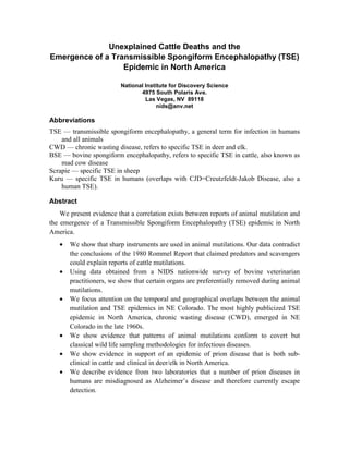
  • 12. National Institute for Discovery Science Argentina wave of mutilations in summer 2002 and their link to infectious disease monitoring will be discussed below. Finally, a large-gage needle was found under the mutilated carcass of an animal near Great Falls Montana and veterinarians reported puncture marks in the jugular of another mutilated animal in Montana (40). The presence of medical hardware at the mutilation scene appears supportive of a sampling operation, rather than cult activity. 5. Specific Organs are Preferentially Removed During Animal Mutilation. A vast body of anecdotal literature exists in the local newspaper archives from 1975-1980 across the nation that documents hundreds, perhaps thousands of animal mutilation reports. Few methodical studies have been conducted, but anecdotal evidence suggests that the eye, ear, tongue, lips, reproductive organs and anus are most commonly reported missing in these animals (1). In order to obtain data on animal mutilations, NIDS conducted a survey among 3849 veterinarian bovine practitioners in the United States (27). Out of the 189 returned questionnaires (see Figure 7), 92 reported mutilations and practitioners who replied to the survey reported 39 cases in which the tongue had been removed, 54 cases documented the removal of an eye, and 70 each for the removal of reproductive organs or rectum. Figure 7. Bovine Practitioner Survey conducted by NIDS indicating the frequency of organs found missing during animal mutilations. 12
  • 13. Unexplained Cattle Deaths & TSE Hence, the NIDS survey suggested the four most common organs removed were rectum, reproductive organs, tongue and eye, thus confirming the anecdotal evidence. 6. Kuru, CJD, BSE and Scrapie -the Prion Diseases Transmissible spongiform encephalopathy (TSE) describes a broad class of neurological diseases that until recently, had been rare and even obscure. D. Carleton Gajdusek’s brilliant investigation of kuru, a mysterious and fatal neurological disease that afflicted over 2000 members of the Fore tribe in New Guinea, together with his discovery of characteristic striking similarities in brain histopathology between kuru and an obscure human disease known as Creutzfeldt-Jakob disease (CJD), earned him the Nobel prize in 1976 (22). Gajdusek and others also linked kuru and CJD with scrapie, an endemic sheep neuropathological condition. Although scrapie had been endemic in British and European sheep for over two centuries, it has only been sporadically documented in the United States since the 1950s. Scrapie became endemic in multiple areas in the U.S. by the mid-1980s, mostly because of the abandonment of a previous USDA policy of slaughtering the entire infected sheep flock. BSE emerged in the United Kingdom in 1985/1986 and as of June 30, 2002, 179,300 cases had been confirmed in British cattle, but the estimated number infected was around 1 million (41). According to USDA officials, BSE has never been detected in the United States, although there is evidence to contradict this assertion (see below). Although Gajdusek maintained kuru was caused by the passage of a “slow virus” as a result of the Fore’s cannibalistic practice of eating their relatives who had succumbed to kuru, biochemical work by Prusiner in the 1980s suggested that TSEs were caused by the aberrant folding of a protein, known as a prion. Prusiner received a Nobel prize in 1997 for his work, although there is still considerable controversy regarding the causative agent of TSEs (see below). In the 1980s, an outbreak of BSE, known as “mad cow disease”, in Great Britain was linked to the practice of feeding rendered cow-parts and other rendered animals back to cows, a process that was later dubbed “high-tech cannibalism” by the media. The common practice of feeding rendered ruminants to other ruminants made herbivore cattle and deer into cannibals and drew parallels with the cannabalism of the Fore tribe. Rhodes describes Gajdusek’s first encounter with kuru in two Fore women in March 1957: “When he first saw the Fore women they could no longer walk. They shivered uncontrollably, not from cold but from brain damage. Their arms and legs pulsed as well with slow, continuous, involuntary tremors-“athetoid movements”, Gajdusek called them. Their speech was blurred. Silly smiles, with grimacing, were prominent. Gadjusek coined the phrase “pathological laughter” for the exaggerated emotionalism of kuru” (22). Gajdusek described the course of kuru: “one month of unsteady gait followed by tremors and athetosis and blurred speech in the second month and in the third month almost complete incapacitation” (22). 13
  • 14. National Institute for Discovery Science Prior to arriving in New Guinea, Gajdusek had traveled extensively in South America, the Middle East, and Central and S.E. Asia, under army funding obtained by Dr. Joe Smadel, Director/Chair of the U.S. Armed Forces Commission on Viral and Rickettsial Diseases at United States Army Walter Reed Hospital. Gajdusek’s purpose was to document epidemic bacterial and viral infectious diseases, with an emphasis on hemorrhagic fever viruses, that were “of military importance” (61). The full title of contract DA-49-007-MD-77 that funded Gajdusek’s travels was: “Field Studies on the Control of Infectious Disease of Military Importance.” Beginning in 1958, Dr Joe Smadel had moved from the United States Army Walter Reed Hospital to the nearby National Institutes of Health (NIH) campus, had set up a sophisticated lab with extensive neuropathology facilities and staff and had begun to receive a steady stream of autopsied kuru brains from the wilds of New Guinea, all sent by Carleton Gajdusek (22). Although the origin of kuru is presently unknown, Gajdusek’s frequent written correspondence with Smadel (61) mentions the acceleration in kuru infection that apparently occurred in the 1940s, although there is evidence from interview testimony with elderly Fore tribes people (62) that the first kuru cases were remembered in the 1920s. The Fore were notoriously vague about time (see for example 63), so attempts to specifically delineate the time when kuru emerged as a full blown epidemic in New Guinea are fraught with error, although Gajdusek appears to estimate the largest leap in the epidemic numbers occurring sometime in the 1940s (61). Nevertheless, it can be unequivocally stated that the importation of dozens of kuru brains and their storage at Fort Detrick, Maryland (63) together with subsequent infectious testing in multiple species (44) at NIH during the 1960s, represented a huge introduction and spread of infectious TSE’s into the United States. Primates (Chimps, Gibbons, Old World monkeys, New World monkeys), guinea pigs, mink, goats, sheep, mice etc were injected with human kuru brain extracts. It is intriguing that the first documented case of CWD appeared in 1967 at a Colorado State University wildlife research facility in Fort Collins, Colorado, within a few years of this intensive cross species kuru testing that originated from experimental facilities at Fort Detrick and Bethesda. 7. Is the Human Food-Chain in United States already compromised? Variant CJD (vCJD) is the only certain example of transmission of animal prion disease to humans. Since 1994, more than 130 people have been diagnosed with vCJD in the UK (4). Recent evidence in transgenic mice (5) suggests that the more common sporadic CJD may also be linked to BSE, in which case animal to human transmission is easier than previously assumed. It has been conclusively shown in Britain and Europe, that BSE emerged in cattle because of the practice of feeding cattle rendered carcasses that included rendered road kill and rendered sheep infected with scrapie. Additional evidence from a team led by Adriano Aguzzi of the University Hospital Switzerland reported a sudden twofold increase in sporadic CJD figures in 2001 and suggested BSE might be to 14
  • 15. Unexplained Cattle Deaths & TSE blame (6). More worryingly, the demonstration of asymptomatic subclinical prion diseases in several mammalian species (7 and references therein) indicates that animals can harbor high levels of prion associated infectious agent without displaying detectable sickness. In this regard, senior author Collinge (7) goes on to say: “The demonstration of animals which can harbor high levels of prion infectivity and detectable levels of PrPSc (infectious prion) without exhibiting any clinical signs of prion disease challenges our understanding of the pathogenic mechanisms involved in these diseases.” BSE according to USDA officials does not and has not existed in the United States. But the recent paradigm-altering laboratory data described by Collinge and his group (7), raise two questions: (a) Is there evidence of transmission to humans of CWD in the United States? And (b) Is there evidence of sub-clinical BSE infection in the United States? In answer to the first question, CJD predominantly occurs in elderly patients. Recent reports describe several cases of CJD in young people in the United States, some of whom are known to have consumed venison (8,9). Perhaps the most definitive answer to the question regarding sub-clinical infection in the United States arose from an unexplained outbreak of a TSE in mink in Michigan in 1985. The mink disease became known as transmissible mink encephalopathy (TME). There had been another outbreak of TME in 1947 in Michigan, killing all animals on a farm and killing 125 mink on a ranch in Minnesota. During the latter epidemic, over the course of a five-month period in 1985, over 4000 mink died of an unexplained TSE. Investigation revealed that the mink had all been fed the infectious agent between June 1 and July 17 1984, since the rancher kept excellent written records. The rancher had fed the mink rendered downer cows with about five percent horsemeat according to an investigation conducted by Dr Richard Marsh, a veterinarian at the University of Wisconsin (11). This outbreak of TME was never explained but the most obvious explanation was a sub-clinical BSE infection in downer cows. Indeed veterinarian Marsh insisted for years that a sub-clinical BSE infection existed in United States cattle. Marsh’s finding was by and large ignored. Nevertheless, his data strongly implied that in the 1980s, while the BSE epidemic was beginning in Britain, an unidentified sub-clinical BSE infection existed in cattle in the United States. Dr Marsh went one step further. He took brains from infected mink and injected them into two calves and 19 months later the calves showed evidence of a form of TSE that was different from the classical symptoms of what we all know as “mad cow” disease or BSE. Marsh’s two calves became lethargic and then fell over. They exhibited symptoms of “downer cow” syndrome. Finally, when Marsh took the brains from the downer calves and injected them back into mink, all of the mink came down with TME, thus confirming that the infectious agent was TME (11). “The real surprise of this experiment is that the clinical signs were quite different from what we’ve seen in Great Britain” Marsh said. “This is what’s changed our perspective on a surveillance of BSE in the United States. We thought BSE in the U.S. would look like BSE in Great Britain-a mad 15
  • 16. National Institute for Discovery Science cow type of disease where the animal would have behavioral changes, become aggressive and look very much like a rabies infection does in cows” (52). In another telling experiment, in 1979 U.S. Department of Agriculture researchers in Mission, Texas inoculated 10 cows with sheep scrapie (50). Three of the 10 animals developed neurological symptoms that were more like “downer cow” symptoms than the classic British “mad cow” BSE. Dr Clarence Gibbs, Acting Chief of the Laboratory of Central Nervous Diseases described the symptoms in the three animals: “progressive difficulty in rising, a stiff-legged gait, incoordination, abnormal tail position, disorientation and terminal recumbency” (50). This experiment refuted the long held assumptions that scrapie was not transmissible to either cattle or humans. Dr. Gibbs bluntly declared “Susceptibility of cattle to scrapie further suggests the possibility that sporadic cases of BSE (mad cow disease) may have occurred in the United States under the clinical picture of the downer cow syndrome….” (50). In yet another confirmatory series of experiments (51), Dr Randall Cutlip from USDA Ames Iowa, repeated the Gibbs experiment, obtained the same results with cows injected with scrapie and described the results as follows: “All calves kept longer than one year became severely lethargic and demonstrated clinical signs of motor neuron dysfunction that was manifest as progressive stiffness, posterior paresis, general weakness and permanent recumbency” (51). This constituted an almost identical litany of symptoms to the earlier Gibbs experiment (50). Why are the similarities in symptoms between the Marsh, Gibbs and Cutlip experiments and the symptoms exhibited by downer cows important? There are two reasons: (a) Every year some 100,000 cows in the United States die of “downer cow” syndrome, a catch-all description that observes the animals died of an undiagnosed ailment, and before 1997 these downer cows were routinely “recycled” via the rendering industry into ruminant feed and (b) Dr. Marsh’s strongly held belief in the possibility that downer cow syndrome has, and is, masking an undetected BSE outbreak in United States cattle. Therefore, even if a small fraction of the annual crop of 100,000 downer cows had BSE, their recycling could have accelerated the spread of the TSE infectious agent through the United States cattle (and captive deer) populations. However, given all this bad news, why has the incidence of CJD (human TSE) apparently remained the same in the United States for the past several decades? CJD is misdiagnosed as Alzheimer’s Disease The official CDC figures suggest that the incidence of CJD in the United States population is extremely low, citing 4751 CJD deaths between 1979-1998 (12). Therefore, according to USDA, the presence of BSE or infectious TSE is not a cause for worry. Is there evidence in humans that prion diseases might be more widespread than previously thought? The answer comes from an intriguing study by one of the top neurodegenerative disease scientists in the world. At Yale University, Dr. Laura Manuelidis examined the post mortem brains of 46 Alzheimer disease patients and found six cases (13%) where the 16
  • 17. Unexplained Cattle Deaths & TSE patients had died of Creutzfeld-Jakob disease (10). The six patients had been misdiagnosed as Alzheimers patients. A second study examined the brains of 54 presumed Alzheimer patients and found 3 cases of CJD (5.5%). The most recent figures for Alzheimer’s in the United States indicate there are 4 million people in the United States who have the disease. These two studies on misdiagnosed CJD imply a range of 200,000-400,000 CJD cases in the United States-an alarming epidemic. Finally, there is no mandatory reporting of CJD cases in most of the states in the U.S., so many CJD cases undoubtedly slip through the cracks. Added to this is the difficulty in correctly diagnosing CJD, as well as the profound reluctance on the part of medical professionals to conduct autopsies on suspected cases of CJD since iatrogenic transfer of CJD from surgical instruments to other people has been documented several times (42). Recently reported research (February 2003) from Italy, showing the detection of significant levels of pathologic prion protein in the olfactory epithelium in sporadic CJD have raised new concerns and questions about the potential infectiousness of CJD (53). In their New England Journal of Medicine paper, the authors warn: “…our data have important implications regarding both the in vivo diagnosis of the condition (CJD) and the risk of infection from living patients” (53). According to Carleton Gajdusek, long considered the father of TSE’s, the infectiousness of Alzheimer’s has not been entirely excluded. In his Nobel prize acceptance speech in 1976 (44), Gajdusek described his experiments involving the injection of brain matter from Alzheimer’s patients into primates. Two primates came down with neurodegenerative plaques, both had been inoculated with brain extracts from patients with familial Alzheimers. These data have never been fully explained. According to Gajdusek: “The diseases transmitted (from Alzheimer’s patients) to primates were clinically and pathologically typical subacute spongiform virus encephalopathies, and did not have pathological features of Alzheimer’s disease in man”. The majority of primates did not come down with disease. These often cited experiments, however, have been criticized (54) since 50% of the “untransmitted” cases used only a single primate for inoculation. More definitive experiments with more primates per innoculum are needed to fully address whether Alzheimer’s is transmissible or not. Other studies have noted an induction of αβ plaques in primates injected with brain homogenates from a patient with Alzheimer’s disease (55). Thus, there is some evidence that Alzheimer’s disease, like CJD, is transmissible. Finally, Han and coworkers at MIT noticed “a striking sequence similarity between NAC, the carboxyl terminus of the beta-amyloid protein, and a region of the scrapie prion protein (PrP) which has been implicated in amyloid formation” (45). Therefore, there are intriguing similarities between the plaques found in the brains of sheep dead from scrapie and the amyloid plaques found in the brains of Alzheimer’s patients. The Remarkable Increase in Alzheimer’s Disease Deaths In United States 1979-2003. Table I, taken from official CDC statistics for Alzheimers Disease (48) illustrates the catastrophic rise of Alzheimer’s Disease in the United States since 1979. 17
  • 18. National Institute for Discovery Science Senile and presenile Alzheimer's organic psychotic Year disease conditions Senility Total 1979 653 1,435 1,229 3,317 1980 1,037 1,644 1,223 3,904 1981 1,473 2,022 1,062 4,557 1982 2,261 2,208 1,178 5,647 1983 4,039 2,624 1,266 7,929 1984 5,846 3,083 1,261 10,190 1985 7,819 3,737 1,343 12,899 1986 8,984 4,296 1,286 14,566 1987 10,877 5,471 1,191 17,539 1988 11,842 6,608 1,411 19,861 1989 12,736 7,967 1,271 21,974 1990 13,391 8,750 1,303 23,444 1991 13,768 9,957 1,412 25,137 Table 1. Alzheimer Annual Mortality Rates 1979-1991 taken from (48). In acknowledging a 20-fold increase in mortality from 1979-1991 from Alzheimer’s Disease, the CDC explains the reasons: “The increasing trend may reflect improvements in diagnosis, awareness of the condition within the medical community, and other unidentified factors rather than substantial changes in the risk of dying from Alzheimer’s Disease” (48). In conversations with several Alzheimer’s groups in the United States, NIDS learned that since Alzheimer’s is still not a mandatory reportable disease, there is a large uncertainty factor in determining the number of cases. There appears to be widespread concurrence in the Alzheimer’s Disease volunteer community that annual rates, as published by the CDC, are probably considerably under-reported. The consensus appears to be that the true figures are unknown. Further, only projections, based on national census data are available, for state-by-state Alzheimer’s figures (see for example 49). Few true epidemiological studies have been conducted to establish whether a particular state, or region within a state, might harbor clusters of Alzheimer’s cases. Regardless of the degree of under-reporting, Table 1 shows the timing of the dramatic rise in Alzheimer’s deaths after 1979 in the United States is coincident with (a) the spread of CWD throughout the wildlife population, (b) the announcements from Dr. Marsh regarding a subclinical infection of BSE in United States cattle and (c) the cessation (around 1980/1981) of the nationwide wave of animal mutilations. Are these three waves of apparently discordant datasets somehow connected? We will discuss the possible connection at the end of the paper. Suffice it to say, it seems difficult to imagine that the extraordinary rise of Alzheimer’s disease since 1979 can simply be explained away by improvements in diagnosis of the disease and by “unidentified factors”. We suggest that one of the unidentified factors in the Alzheimer’s equation may be an unidentified infectious agent within the human food chain. Finally, there is considerable uncertainty 18
  • 19. Unexplained Cattle Deaths & TSE nationally regarding the precise numbers of “early onset” Alzheimer’s Disease in the United States. According to NIDS’s conversations with Alzheimer’s organizations, the conservative estimate for early onset Alzheimer’s varies between 5-10% of total Alzheimer’s cases. The early onset subgroup is defined as those patients who succumbed to symptoms before age 65. Therefore, if the figure of a total of 4 million Alzheimer’s Disease patients is taken as accurate, between 200,000 and 400,000 of those patients showed symptoms before age 65. There are insufficient epidemiological data to answer the question whether the number of “early onset” patients is, or has been, on the rise. 8. A TSE Expert Sounds a Warning. Speaking at the Days of Molecular Medicine conference in La Jolla in March 2002 (65), European prion expert Adriano Aguzzi issued a strong warning against underestimating the dangers of CWD in the United States: “For more than a decade, the U.S. has by-and-large considered mad cows to be an exquisitely European problem. The perceived need to protect U.S. citizens from this alien threat has even prompted the deferral of blood donors from Europe,” he said. “Yet, the threat-from-within posed by CWD needs careful consideration, since the evidence that CWD is less dangerous to humans than BSE is less-than-complete”. Aguzzi went on to point out that CWD is arguably the most mysterious of all prion diseases. “Its horizontal spread among the wild population is exceedingly efficient, and appears to have reached a prevalence unprecedented even by BSE in the UK at its peak. The pathogenesis of CWD, therefore, deserves a vigorous research effort. Europeans also need to think about this problem, and it would be timely and appropriate to increase CWD surveillance in Europe too.” 9. Wisconsin’s CWD Epidemic In contrast to Dr Aguzzi’s sober assessment, on September 13, 2002, James Kazmierczak, a Wisconsin Division of Public Health epidemiologist in an interview with Milwaukee Magazine (21) said: “You could live on a diet of deer brains and never get sick. There is either no, or very low, potential for infection in humans”. The remarkable interview was conducted as a part of an investigation into the growing evidence of widespread CWD infections in Wisconsin deer and in an atmosphere of increasing alarm on the part of hunters. Going into the fall 2002 hunt, Wisconsin state officials estimated there were 1.6 million wild deer, but the number that had tested positive for CWD, as of May 2003 was 210. Wisconsin of course is well known as a national center for Transmissible Mink Encephalopathy. Four out of five known TME outbreaks since 1947 have occurred in Wisconsin. This, combined with the huge density of wild deer in the state prompted Allen Boynton, wildlife biology manager for the Virginia Department of Game and Inland Fisheries to state: “If Wisconsin doesn’t stop CWD, it will move all the way across the 19
  • 20. National Institute for Discovery Science country. This will become a national story” (21). The common practice of feeding herbivore deer back to other deer, especially in the captive deer population is seen as a major route for spreading CWD. In 1995 alone, Wisconsin Department of Natural Resources (DNR) records show that 26,488 road-killed Wisconsin deer were rendered into feed. This feed was used to feed other ruminants, notably cattle and other deer. Since there was nothing remarkable about 1995, this figure can be taken as an annual road-kill average in Wisconsin. If any of the 26,488 animals was infected with CWD, then CWD was passed via the rendering process to both deer and cattle. And, lest there be doubt that CWD can transmit to cattle, a recently published experimental study has unequivocally demonstrated transmission of the CWD agent to cattle by intra-cerebral inoculation (26). NIDS interviewed a prominent and high profile epidemiologist with the Center of Disease Control (CDC) who had investigated three deaths of young people who had consumed venison (8). The epidemiologist informed NIDS that there simply was insufficient evidence to draw a cause and effect relationship between CWD and the three hunters who ate venison. He went on to recommend an in-depth epidemiological study of the hunters in N.E. Colorado who had consumed venison from a known CWD endemic area for many years, but the epidemiologist lamented the lack of political will in the Colorado governor’s office to launch such a study, mainly because of “privacy concerns”. Since sporadic CJD is still not a reportable disease in most states, and because nobody has succeeded in firing up momentum in Colorado to conduct the definitive epidemiological study, even in June 2003, there are no data to either support or negate a direct link between CJD occurrence in humans and the eating of CWD-infected venison. However, it could be simply stated that there is no link because nobody has looked. The situation in the United States with a possible subclinical BSE infection in cattle and an overt CWD epidemic in deer and elk, is reminiscent of the decade-long refusal on the part of the government of the United Kingdom to acknowledge that BSE was capable of jumping the “species barrier” to humans. In January 1996, British Agriculture minister John Gummer appeared on TV to feed his young daughter Cordelia a hamburger to demonstrate that beef was safe. Cordelia refused the hamburger on live TV. The subsequent deaths of over 120 young people from new variant CJD (nvCJD) since 1994 in the UK have long since undermined the existence of the BSE-CJD “species barrier”. The deaths served to brutally highlight the UK government’s stonewalling on BSE. 10. Prion or Virus? Although mostly irrelevant, with one important exception, to the arguments in the present paper, it should be emphasized that there is substantial experimental evidence to indicate that the causative agent of BSE/CJD may in fact contain nucleic acids (13) either in addition to, or even separate from, prions. Many mainstream scientists remain extremely doubtful about an infectious agent (the so-called prion) that can transmit itself without nucleic acid (DNA/RNA). Indeed, during some of the initial observations about kuru, CJD 20
  • 21. Unexplained Cattle Deaths & TSE and BSE experiments failed to show an immune response during infection following transmission. This failure appeared to mitigate against a viral etiology. But recently, these early findings, which appear to demonstrate the “protein only” hypothesis for prion infection, have been shown to be wrong (14). Further, it has been well documented in multiple instances that transmission of the BSE/TSE agent can occur in the absence of detectable prions (17) implying it may be premature to focus on detection of prions only when looking for TSEs. It should also be noted that there was an unprecedented storm of outrage immediately following the announcement that Stanley Prusiner had won the 1997 Nobel Prize for his work on prions. Indeed, renowned TSE researcher Bruce Chesebro made the following statement shortly after Prusiner was awarded his Nobel prize (18): “Although the notion that “protein only” can account for the infectious agent has received considerable publicity as a result of the Nobel prize award to S. Prusiner for the discovery of prions, the fact remains that there are no definitive data on the nature of prions. Prions continue to be vaguely defined, and for the most part this term is used as an operational term for the transmissible agent, but without structural implications”. In the present paper, we take Chesebro’s advice and we use the word prion only as an operational definition of the infectious TSE agent. The putative viral or nucleic acid etiology of BSE and other prion diseases may have important implications for the current widespread testing for CWD in wildlife and for BSE in cattle in the United States. Confusion, controversy or uncertainty regarding the infectious agent strongly increases the likelihood of false negatives in these widespread testing programs. In other words, if the focus in testing remains exclusively on prions, these data suggest that a putative other infectious agent may go undetected. For example, the recent tendency towards live testing for CWD in Unites States deer and elk by sampling tonsils from live animals using immunohistochemical techniques may dramatically increase the likelihood of false negatives. Another example may be the well-publicized failure (as of June 2003) on the part of investigators in Alberta and Saskatchewan to find any other instances of BSE in the herds from which the first confirmed case of mad cow disease appeared in May 2003. In summary, the historical record, especially the work of Dr Richard Marsh suggests (a) there may already be a sub-clinical infection of BSE in the United States cattle herds since the 1980s and (b) a spreading infection from BSE-infected meat and CWD- infected venison may lie undetected in the rapidly increasing Alzheimer’s disease population in the United States. 11. Organs Taken in Mutilations Potentially Contain High Levels of Prions As discussed, both anecdotal evidence and the NIDS survey of bovine practitioners indicate that the most common organs harvested in mutilations are the reproductive organs, anus/large intestine, eye and tongue. Recent experimental data suggests that accumulation of aberrant prions in uterine-placental epithelial cells in the placentome of sheep (28), as 21
  • 22. National Institute for Discovery Science well as in the tongue in hamsters (23) and the eye in humans (24). Further, scrapie is perhaps the most studied of the prion diseases since it has been around the longest. A recent study of the pathogenesis of natural scrapie infection of sheep showed a rapid accumulation of prions in Peyer's patches of the caudal jejunum and ileum, the so-called gut-associated lymphoid tissues (GALT) during the early stages of scrapie infection (29). Thus, recent studies have shown that all of the most commonly removed organs in animal mutilations can harbor high levels of prions. Thus, if animal mutilations were a prion sampling operation, these very organs would preferentially need to be removed for laboratory analysis. 12. Other States Demonstrate a Correlation between Animal Mutilations and Subsequent Outbreaks of CWD. • Utah: NIDS investigation revealed over 20 animal mutilation reports that occurred in an area between Vernal and Roosevelt, Utah between 1985 and 2000. Many of them were investigated by the deputy sheriff of Uinta County (see map). Figure 8. Locations of Animal Mutilations in NE Utah Investigated by NIDS and by NIDS Consultants 1985–1998. 22
  • 23. Unexplained Cattle Deaths & TSE On February 19, 2003 the first case of CWD was reported in a sample from a mule deer buck taken in fall 2002 on Diamond Mountain, just north of Vernal, Utah (47). It is also of interest to note that several anecdotal reports received by NIDS indicate an intense interest in animal mutilation investigations by Carl Whiteside, head of the Colorado Bureau of Investigation in an area between Vernal, Utah and the Colorado border during the late 1970s. Whiteside and his team used a helicopter to move quickly around the area to take samples from mutilated animals in the field. A second case of CWD was reported in May 2003 near Moab, Utah. • Montana: As described, there was an intense wave of both helicopter/UFO sightings and animal mutilations that occurred within a 50-mile radius of Great Falls Montana from 1974-1977 (31). Two mutilations, however, were outliers and were reported a couple of hundred miles southwest of Great Falls. Within 50 miles of these mutilations, the only reported outbreak of CWD in Montana history (as of June 2003) occurred in captive elk in November 1999, in Phillipsburg Montana (58), a mere 50 miles away from the site of the two mutilations. Interestingly, the infected herd of deer were not incinerated but taken to a location north of Great Falls Montana and buried. In summer 2002, less than 100 miles North of this location NIDS received reports of the mutilation of more than 10 animals (35,60). It is also interesting to note that in a recent report (59) five bulls from the herd in Canada from which the BSE-infected animal originated were sent to a feedlot and a ranch Montana. Whether the intensive mutilation wave in the Dupuyer triangle in Montana of summer 2002 had anything to do with a covert search for evidence of BSE infection is not yet known, but the correlations are noteworthy. • New Mexico: One of the highest profile early series of mutilations occurred near Soccorro, New Mexico. Socorro is located at the northern end of White Sands missile range. In late 2002/early 2003 out of 25 mule deer tested from White Sands, four tested positive. A further two tested positive in mid-February 2003, this time outside the White-Sands herd (46). The location at the southern end of the state of New Mexico is very far south from the known epicenter of the CWD epidemic, primarily located in NE Colorado, Nebraska and Wyoming. The finding of CWD near White Sands New Mexico raises questions about how long the CWD has been in the New Mexico wildlife. Since the main locus of animal mutilations in New Mexico occurred near Dulce, approximately three hundred miles north (lying between White Sands and NE Colorado), one has to ask the question whether any CWD has been identified in northern New Mexico? The official answer is no. Regardless, NIDS interviewed several CWD experts, as well as game and wildlife officials from the state of New Mexico, about the unexplained emergence of CWD near the White Sands missile range. All experts pronounced themselves mystified about how an outbreak could have occurred at such a great distance from the 23
  • 24. National Institute for Discovery Science epidemic’s epicenter (NE Colorado). All of the usual possibilities (migration patterns, interstate transport of animals) had been examined and eliminated. • Alberta-Saskatchewan Canada: Because of the recent (May 2003) announcement of a confirmed BSE case in a cow in Alberta, it is worth looking at the links between CWD, BSE and animal mutilations in some detail in Alberta, but especially in Saskatchewan, since the infected animal was imported from that province. Since 1999, about 40 separate outbreaks of CWD in farmed deer and elk have been documented primarily in a region West and NW of Saskatoon and centered around North Battleford, Saskatchewan. The ranch where the BSE positive cow spent considerable time before moving to Alberta was at Baldwinton, a town in NW Saskatchewan that lies in the heart of the CWD endemic area (see arrow on map below). In addition, a further nine cases of CWD in wild deer have been documented in 2002 and 2003 south of that area, primarily in Sask Landing Provincial Park, and the Sandhills area, although one case of CWD in a wild deer was reported in Paradise Hill, that lies in the heart of the CWD epidemic in farmed cervids. The region of NW Saskatchewan, the epicenter of CWD in farmed cervids in Saskatchewan is also the epicenter for about 18 separate cases of animal mutilations investigated since 1994 (see map, Figure 9). A fewer number of cattle mutilations have been reported in the southern area of Saskatchewan that corresponds to where the recent CWD cases have been found in wild deer. Therefore, there is considerable geographical overlap between the locations of the CWD epicenter in Saskatchewan and the locations where animal mutilations were reported since 1994. We hypothesize that the animal mutilations reported in NW Saskatchewan in the past several years may have been a covert prion sampling operation by perpetrators who knew that the TSE infectious agent was spreading from farmed elk/deer in Saskatchewan to wild deer (as has been documented) and thence to cattle. Many of the public pronouncements in the Canadian press in June 2003 regarding the impossibility of the TSE agent jumping the species barrier from cervids to cattle in Saskatchewan, are eerily reminiscent of the same pronouncements made in the United Kingdom in 1995/1996, just prior to people dying from consuming tainted beef. 24
  • 25. Unexplained Cattle Deaths & TSE Figure 9. Geographical overlap between cattle mutilations reports (source: Fern Belzil) and the locations of known CWD outbreaks in Saskatchewan (Source: Canadian Food Inspection Agency map March 2003). The numbered circles are the locations of animal mutilation reports since 1995, stars represent the locations of CWD outbreaks in farmed cervids, and the arrowed spot represents Baldwinton, the location in Saskatchewan from where the BSE positive cow originated. 25
  • 26. National Institute for Discovery Science 13. Monitoring an Occult Epidemic in Argentina On March 13, 2001, an outbreak of foot and mouth disease (FMD) Type A in Argentina was reported to the OIE in the District of Rivadavia, Buenos Aires province. Four additional outbreaks in neighboring provinces were reported in the days following. In August of 2000, serologic evidence of FMD Type A was found in Argentina in areas bordering Paraguay, Brazil, and Uruguay. These cases were linked to illegal imports of cattle. Argentina’s fresh beef exports to the U.S. were suspended in August 2000 due to the serological findings, and reopened in December 2000 with the requirement of additional certification regarding their origin. In response to Argentina's March 13th OIE report, the USDA issued a temporary ban on the importation of all live swine and ruminants, and any fresh swine or ruminant meat (chilled or frozen) and other products of swine and ruminants from Argentina. This ban affects any products processed on or after February 19, 2001. As of March 31, 2001, Argentina had reported a total of 160 FMD outbreaks in 5 provinces (see map). The origin of infection was still under investigation. Figure10. Darkest areas represent affected provinces/departments. Brazil and Uruguay both also reported FMD in the latter half of 2000; Brazil in August 2000 and Uruguay in October 2000. However, while the Argentina outbreaks were caused by type A virus, Brazil and Uruguay both reported outbreaks caused by type O virus. 26
  • 27. Unexplained Cattle Deaths & TSE Unfortunately, convincing evidence emerged that Argentina had hidden the outbreak of FMDV for several months before making the announcement. According to a March 17, 2001 Washington Post article: “Ranchers and beef industry officials said Argentine authorities were aware of the outbreak by the end of last year and, largely in agreement with the meat industry, hid the problem to avoid the panic sweeping Europe over "mad cow” disease. By January, sources said, the government secretly began vaccinating thousands of head of cattle in infected areas in at least three rural provinces” (15). It is noteworthy that (a) Argentina was deceptive in concealing an FMDV epidemic in 2001and (b) a major animal mutilation wave began less than 12 months later in precisely the same provinces that reported the FMDV epidemic (16). According to press reports, up to 400 animals were reported mutilated in Argentina in summer 2002 (16). It is no secret that in 2002, Argentina was desperately negotiating with the International Monetary Fund (IMF) to secure an emergency bailout package for their economy, culminating in January 2003 with an agreement from the IMF to an $11.5 billion loan package (25). The very last thing they needed was a second epidemic, perhaps this time of BSE or CWD. Argentina has consistently and publicly affirmed that the country is “BSE free” and “CWD free”. According to the numerous press reports emanating from Argentina in summer 2002, by far the most common organs harvested from mutilated animals were tongues, eyes, ears, lips and reproductive organs. Only recently, has it become obvious from laboratory research that the tongue (23) and possibly the eye (24) are reservoirs of infectious prions. If we hypothesize that the large Argentina animal mutilation epidemic of summer 2002 was an emergency monitoring program to test for prion diseases in that nation’s cattle, then the apparent overlap between the tissues taken (tongues and eyes) and the required scientific knowledge to focus on these tissues, implies an extremely knowledgeable organization, with access to unpublished data, who performed these field surgeries. One prediction for this hypothesis (see below) is that in the next few years, confirmed cases of BSE/CWD will be detected and or reported in Argentina. Given Argentina’s precarious economic position and their record of dissembling on the FMDV epidemic, there may be a time lapse between detection and reporting. Conclusion, Hypothesis and Predictions The primary conclusion of this paper is that the animal mutilation epidemic of 1970-2003 was and is a monitoring operation for an infectious agent that is spreading through the human food chain (cattle, sheep and wild & farmed deer/elk). In North America (Canada and United States) the infection comprises a full-blown CWD epidemic in deer and elk and a sub-clinical BSE infection in cattle. The infectious agent, unlike all known viruses and bacteria, is almost indestructible and the symptoms in people appear very difficult to diagnose pre-mortem. In short this TSE agent is the perfect stealth killer. If the hypothesis is correct, animal mutilation operations are carried out by a knowledgeable group that is cognizant of the biochemistry and infectious potential of prion 27
  • 28. National Institute for Discovery Science diseases and their fatal spread. How difficult is this knowledge to come by? Beginning in 1958, Dr. Carleton Gajdusek began mailing kuru brains from the wilds of New Guinea to the central neuropathology facility at NIH in Bethesda and Fort Detrick. Hence, these fatal neurodegenerative diseases have been known in the United States, but not highly publicized, since the late 1950s or early 1960s. Only recently, due to the intensive prion research carried in the past two decades has the extent of prion replication become obvious in the eye, tongue, anus/large intestine and reproductive organs (see above) of animals. If these specific tissues are indeed removed during animal mutilation for the purpose of prion monitoring, this implies an intensive knowledge of prion physiology, biochemistry and infectiousness, involving research results not published until relatively recently, on the part of the perpetrators of animal mutilations. The identity of the mutilators remains unknown, but in addition to knowledge of prion infectiousness research, a high level of technology, surgical skill and stealth has been a feature of animal mutilations. It is a recurring theme in law enforcement circles that not a single person has been caught or charged for the crime of animal mutilations in the thirty- year history of the epidemic. Why Leave the Body? This question has plagued investigators ever since the first well-publicized investigations of mutilations began back in the early 1970s. As any reader familiar with the animal mutilation topic will agree, a plethora of hypotheses have sprung up about the perpetrators and their motives for animal mutilations. One of the most quoted hypotheses involves a government operation to monitor radiation or biological warfare testing. But the question “why leave the body?” has never been adequately answered by these hypotheses. The government can just as easily test their own herds, the counter-argument goes, or obtain carcasses from a slaughterhouse if they wish to covertly monitor radiation. Thus, for this and many other reasons, the evidence points away from the government as perpetrators of animal mutilations. Vallee (56) and Smith (57) have suggested intriguing hypotheses that leaving the cow carcass on the ground constitutes a deliberate message. In common with both these authors, we suggest that implicit in the deliberate lack of an attempt to conceal the carcass on the part of the perpetrators of animal mutilation, is a brutal warning. We suggest that attention is being deliberately focused on the mutilated animals. Further, we suggest the warning is that the human food chain is compromised, probably with a prion-associated infectious agent that still remains mostly undetected. 28
  • 29. Unexplained Cattle Deaths & TSE Figure 11. The semiotics of cattle mutilations. Was there a message intended? The eyeball (see foreground in photo) was excised from this animal in Northern California in November 2001 and carefully laid on the ground a few feet away. When found, all the eye-fluid in the excised eyeball was still intact. Figure 12. The sharpness of the cut around the eye appears to convey a message that sharp instruments had been used. Even the hair had been cut (see section 3 of this report). The eye is also a known repository for infectious prions (24). 29
  • 30. National Institute for Discovery Science As discussed above, some of the harrowing consequences of the spread of this TSE infectious agent may lie in a subset of the epidemic of Alzheimer’s disease that is currently ravaging the United States healthcare system. According to CDC estimates (43) there are now 4 million Alzheimer’s patients in the United States, with annual health care costs between $100-500 billion. With the aging population, this cost is projected to soon rise to $1 trillion when 7-8 million have the disease (43). There is also the question of the mysterious early onset Alzheimer’s, currently afflicting about 200,000 Americans. We have presented the evidence that thousands of possible CJD cases may be misdiagnosed as Alzheimer’s and therefore the extent of this CJD epidemic in the United States lies beneath the public’s radar. We hypothesize that the animal mutilators know and have known of the potential damage to humans of this infectious agent in the human food chain. Thus, we hypothesize that animal mutilations serve as both a sampling operation AND a warning. A central implication of this paper is that animal mutilations serve two purposes: as both a covert monitoring operation for the prion infectious agent and as a very graphic public warning, a display that monitoring is being done. The body on the ground, with glaring evidence of highly skilled surgery, serves as a calling card and a warning. From the evidence presented in this paper, we believe the warning is: “A major human food source (beef, elk, deer) is contaminated.” There are several predictions that arise out of the animal mutilation-prion monitoring hypothesis, since the evidence seems to suggest that mutilations will be followed, years or even decades later, by a TSE outbreak: (a) That a rather large outbreak of CWD/TSE will occur in the area around Great Falls Montana in the next several years. (b) Similar large outbreaks will occur in Argentina and in Northern New Mexico in the next few years (the first six cases of CWD were found on and near White Sands missile ground October 2002-February 2003). (c) In the next year or two, there will be an unambiguous link drawn between CWD and sporadic CJD in humans in the United States. (d) Even though the state of California has mandated a ban on importing elk and deer from other states, the highly intense animal mutilation (>30 animals mutilated in 5 years) cluster on and near a ranch in N. California, predicts an outbreak of CWD/TSE in northern California in 5-10 years. (e) In the coming years, as new methods for distinguishing CJD from Alzheimer’s comes on line, there will be a dramatic increase in the incidence of “sporadic” CJD in the population of the United States. 30
  • 31. Unexplained Cattle Deaths & TSE Acknowledgements We thank members of the NIDS science advisory board for reviewing earlier versions of this paper. Other people outside the board also contributed their expertise. They know who they are. NIDS thanks Fern Belzil for providing unpublished data on animal mutilations. References (1) Onet G.E. (1997). Animal Mutilations: What we Know. http://www.nidsci.org/articles/animal1.html (2) The Meeker Herald, Meeker, Colorado, September 6, 1975. (3) Bosque P.J. (2002). Bovine Spongiform Encephalopathy, Chronic Wasting Disease, Scrapie and the Threat to Humans from Prion Disease Epizootics. Current Neurology and Neuroscience Reports. 2: 488-495. (4) http://www.cjd.ed.ac.uk/figures.htm (5) Asante, EA et al. (2002). BSE prions propagate as either variant CJD-like or sporadic CJD- like prion strains in transgenic mice expressing human prion protein EMBO J. 21: 6358-6366 (6) Glatzel M et al (2002). Incidence of Creutzfeldt-Jakob disease in Switzerland. Lancet 360: 139-141. (7) Hill AF & Collinge J (2002). Species-barrier-independent prion replication in apparently resistant species. APMIS. 110: 44-53. (8) Belay ED, et al.(2001). Creutzfeldt-Jakob disease in unusually young patients who consumed venison. Arch Neurol. 58(10): 1673-1678. (9) Peltier et al. (2002). Two cases of early onset sporadic Creutzfeld-Jakob disease in Michigan. Presented at American Academy of Neurology Annual Meeting, Denver Colorado. (10) Manuelidis E.E. and Manuelidis L. (1989), “Suggested links between different types of dementias: Creutzfeld-Jakob disease, Alzheimer disease, and retroviral CNS infections,” ALZHEIMER DISEASE AND ASSOCIATED DISORDERS Vol. 3, Nos. 1-2, pgs. 100-109. (11) Marsh RF et al. (1991). Epidemiological and experimental studies on a new incidence of transmissible mink encephalopathy. Journal Gen. Virol. 72, 589-594. (12) Gibbons RV et al. (2000). Creutzfeld-Jakob Disease in the United States: 1979-1998. JAMA 284: 2322-2323. (13) Manuelidis, L. (1997). Beneath the emperor’s clothes: The body of data in scrapie and CJD. Annales de L’Institut Pasteur. 8, 4, 311-326. (14) Baker CA & Manuelidis L. (2003). Unique inflammatory RNA profiles in microglia in Creutzfeldt-Jakob disease. Proc. Nat. Acad. Sci. 100: 675-679. (15) Faiola, A. (2001). Argentina’s Concealed Outbreak: Meat Exports Banned Months After Livestock Virus was Found. Washington Post. March 17. p A13. (16) Schuessler, J. (2002). Cattle Mutilations in Argentina-2002. Briefing Document. August 2002. (17) Lasmezas C et al. (1997). Transmission of the BSE agent to mice in the absence of detectable prion protein. Science 275: 402-405. (18) Chesebro, B. (1998). BSE and Prions: Uncertainties About the Agent. Science 279: 42-43. (19) Rommel, KM. (1980). Operation Animal Mutilation: Report of the District Attorney First Judicial District State of New Mexico. 31
  • 32. National Institute for Discovery Science (20) http://www.nidsci.org/articles/articles2.html (21) Van de Kamp Nohl, M. (2002). “The Killer Among Us: What state officials aren’t telling you about chronic wasting disease-the politics and blunders behind it’s spread and true dangers”. Milwaukee Magazine. December 2002. (22) Rhodes, R. (1997). Deadly Feasts: The “Prion” Controversy and the Public’s Health. Simon & Schuster. New York. (23) Bartz JC. et al. (2003). Rapid Prion Neuroinvasion following Tongue Infection. Journal Virology. 77(1):583-91 (24) Head MW et al. (2003). Prion protein accumulation in eyes of patients with sporadic and variant Creutzfeldt-Jakob disease. Invest Ophthalmol Vis Sci. 44(1):342-6. (25) ABC News (2003). Massive IMF Loan to see Argentina through Poll. http://abc.net.au/news/newsitems/s769937.htm .January 25, 2003 newscast. (26) Hamir AN. et al (2001). Preliminary findings on the experimental transmission of chronic wasting disease agent of mule deer to cattle. J. Vet. Diagn. Invest. 13: 91-96. (27) NIDS Report. (1997). Results of a Survey Among Bovine Practitioners Concerning Animal Mutilation. http://www.nidsci.org/articles/bovinepractioners.html (28) Tuo, W. et al. (2002). Pregnancy status and fetal prion genetics determine PrPSc accumulation in placentomes of scrapie-infected sheep. Proc Natl Acad Sci U S A 99(9):6310- 5. (29) Van Keulen LJ etal (2002). Early and late pathogenesis of natural scrapie infection in sheep. APMIS 110(1):23-32. (30) Brown LM (2001). Covert Microbiological Experimentation and Livestock Losses. Report submitted to NIDS. (31) NIDS Report (2002). Summary Report on a Wave of UFO/Helicopters and Animal Mutilations in Cascade County, Montana 1974-1977. http://198.63.56.18/pdf/wolverton_report.pdf February 2002. (32) Day G I. Et al. (1980). Capturing and Marking Wild Animals. P 61-88 in Wildlife management Techniques Manual. Sanford D. Shemnitz ed. (33) Gates, CC et al. (1995). Investigation, Control and Epizootiology of Anthrax in a Geographically Isolated, Free-Roaming Population in Northern Canada. Canadian Journal of Veterinary Research 59 (4) 256-264. (34) Kelleher CA et al. (1999). Final Report: Investigation of the Unexplained death of a cow in NE Utah, October 16, 1998. http://www.nidsci.org/articles/ucd_report1.html (35) NIDS Report. (2002). Investigation of a Report of Animal Mutilation in Dupuyer, Montana on June 27, 2001. http://198.63.56.18/pdf/montana_cattlemutilation.pdf (36) Mannaioni G. et al. (1998). Electrophysiological studies on Oxindole, a Neurodepressant Tryptophan Metabolite. Br. J. Pharmacol. 125: 1751-1760. (37) Orcutt JA et al. (1964). Some new compounds exhibiting selective CNS depressant activities. Part 1. Preliminary Observations. Arch. Int. Pharmacodyn. 152: 121-131. (38) NIDS Report (2002). Investigation of a Mutilation Report in Cache County, Utah. http://www.nidsci.org/pdf/cache_county_mutilationreport.pdf (39) Argentinian Veterinarian Pathologist. (2002). Pers. Comm. (40) Donovan R & Wolverton K (1976). Mystery Stalks the Prairie. Pub. THAR Institute, Raynesford Montana. (41) Anderson RM et al. (1996). Transmission dynamics and Epidemiology of BSE in British Cattle. Nature 382: 779-788. 32
  • 33. Unexplained Cattle Deaths & TSE (42) Weber DJ & Rutala WA (2002). Managing the Risk of Nosocomial Transmission of Prion Diseases. Curr. Opin. Infect. Dis. 15: 421-425. (43) Costs of Alzheimer’s Disease. http://www.cdc.gov/diabetes/pubs/costs/figures.htm (44) Gajdusek DC (1976). Unconventional Viruses and the origin and Disappearance of Kuru. Nobel Lecture December 13, 1976. http://www.nobel.se/medicine/laureates/1976/gajdusek- lecture.pdf (45) Han H et al. (1995). The core Alzheimer's peptide NAC forms amyloid fibrils which seed and are seeded by beta-amyloid: is NAC a common trigger or target in neurodegenerative disease? Chem Biol 2(3):163-9 (46) Reed, O (2003). Diseased Deer Outside Range Stuns Game Offical. Albuquerque Journal Feb 15, 2003. http://www.abqtrib.com/archives/news03/021503_news_deer.shtml (47) Knowles S. (2003). Wasting Disease hits Utah. Salt Lake Tribune. Feb 19, 2003. (48) Hoyert DL (1996). Mortality Trends for Alzheimer’s Disease 1979-1991.National Center for Health Statistics. Vital Health Stat 20(28). (49) Alzheimer’s Association (2002). 2000 Population and Alzheimer’s Disease Prevalence Projections. http://search.alz.org/Media/newsreleases/archived/currenttotalschart.htm (50) Gibbs, CJ et al. (1990). Experimental Transmission of Scrapie to Cattle. Lancet 335, 1275. (51) Cutlip RC et al. (1994). Intracerebral Transmission of Scrapie to Cattle. J. Infectious Dis. 169: 814-820. (52) McNair J. (1990). Bypass Protein still Safe. Agri-View. Aug 9, 1990. (53) Zanusso, G. et al. (2003). Detection of Pathologic Prion Protein in the Olfactory Epithelium in Sporadic Creutzfeld-Jakob Disease. New Engl. J. Med. 348: 711-719. (54) Sigurdsson EM et al. (2002). Infectivity of Amyloid Diseases. Trends in Molecular Medicine. 8, 411-413. (55) Baker HF et al. (1994). Induction of β(A4)-amyloid in primates by injection of Alzheimer’s disease brain homogenate. Comparison with transmission of spongiform encephalopathy. Mol. Neurobiol. 8: 25-39. (56) Vallee, J. (1979). Messengers of Deception. And/Or Press Berkeley, California. (57) Smith, FW (1974). Cattle Mutilation: The Unthinkable Truth. Cedaredge, Freedland. (58) (1999) State considers emergency order to act on wasting disease. Billings Gazette 6 November 99. (59) Gransbery, J. (2003). Bulls in BSE herd sold to Montana. Billings Gazette, June 5, 2003. (60) Oyan, K. (2002) Cattle mutilations back: Ranchers, lawmen baffled by crime wave. Great falls Tribune. January 3, 2002 (61) Gajdusek, C (1955-1958). Correspondence on the Discovery and Original Investigations on Kuru Smadel-Gajdusek Correspondence, 1955-1958. DHEW Publication No. (NIH) 76- 1168. Second Printing 1976. (62) Mead S. et al. (2003) Balancing selection at the prion protein gene consistent with prehistoric kurulike epidemics. Science. 25;300 (5619):640-3 (63) Klitzman, R. (1998). The Trembling Mountain. A Personal Account of Kuru, Cannibals and Mad Cow Disease. Plenum. New York NY. (64) Onet, GE and Kelleher, C (1998). An Organophosphorus Insecticide Toxicosis in a Cow. Large Animal Practice, 19, 29-30. (65) Birmingham K. (2002). TSE Threat to USA Increases. Nat Med. 2002 May;8(5):431 33