

Recomendados
Mais conteúdo relacionado
Mais procurados
Mais procurados (20)
Destaque
Destaque (20)
Semelhante a Lecture8 nervous system
Semelhante a Lecture8 nervous system (20)
Mais de Reach Na
Último
Último (20)
Lecture8 nervous system
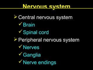
- 1. Nervous system Central nervous system Brain Spinal cord Peripheral nervous system Nerves Ganglia Nerve endings
- 2. Nervous tissue nerve cells NEURONS supporting cells GLIAL CELLS
- 5. Dendrites Multiple processes Receptor processes Usually short Branch profusely forming a dendritic tree Become thinner as they subdivide into branches
- 6. Axons Single processes Effector processes Usually long Don’t branch profusely Ends in terminal arborization – telodendron Have constant diameter
- 7. Types of neurons Sensory (afferent) Motor (efferent) Interneurons
- 8. Types of neurons Bipolar neurons Retina Olfactory mucosa Cochlear and vestibular ganglia Pseudounipolar neurons Sensory neurons Multipolar neurons Motor neurons Interneurons
- 9. Nuclei of glial cells dendrite Axon hillock
- 10. Nissl bodies rough endoplasmic reticulum cisternaes and free ribosomes
- 13. Types of synapses Excitatory Inhibitory
- 14. Types of glial cells Central nervous system Oligodendrocytes Astrocytes Ependymal cells Microglia Peripheral nervous system Schwann cells Satellite cells
- 15. Astrocytes Glial cells of central nervous system 2 types Protoplasmic Fibrous support neurons surround neurons, blood vessels, synapses form a layer on outer surface of the brain and spinal cord control ionic and chemical environment of neurons regulate neuronal activity and metabolism joined to one another by gap junctions forming a continous network
- 19. Microglial cells – Microglia glial cells of central nervous system phagocytic cells – belong to mononuclear phagocytic system macrophages of CNS derive from precursor cells in bone marrow involved in inflammation processes in CNS phagocyte dead neurons and an excess of neurons during embryogenesis
- 20. Ependymal cells glial cells of CNS cuboidal cells line the ventricles of the brain and the central canal of the spinal cord
- 21. Oligodendrocytes glial cells of CNS have only a few small processes myelin – forming cells in CNS
- 23. Cells forming the myelin sheath PNS Schwann cells CNS Oligodendrocytes
- 24. Schwann cell Internode Node of Ranvier
- 27. Peripheral nervous system Myelinated fibers Unmyelinated fibers One Shwann cell→ one axon One Schwann cell→ several axons Axon Shwann cell Shwann cell
- 29. Central nervous system Myelinated fibers Unmyelinated fibers Oligodendrocytes Bare nerve fibers – not imbedded in glial cells
- 30. Peripheral nervous system nerves sensory nerves motor nerves mixed nerves ganglia nerve endings
- 32. Nerve Endoneurium – loose connective tissue Perineurium – a few layers of flattened epithelium – like cells Epineurium – dense irregular connective tissue containing blood vessels
- 41. Nucleolus Satelite cells Nucleus of nerve cell
- 42. Sensory ganglia lie in dorsal roots of spinal cord, outside of CNS contain cell bodies of sensory neurons – pseudounipolar 2 regions central region – nerve fibers peripheral region – cell bodies cell bodies are covered by satellite cells satellite cells – a kind of glial cells of PNS
- 43. Nervous system Somatic nervous system – voluntary functions – control skeletal muscles Autonomic nervous system – involuntary functions – control smooth muscle, secretion of some glands, modulation of cardiac rhythm
- 45. Autonomic nervous system Sympathetic system – presynaptic neurons are located in thoracic and lumbar segments of spinal cord Parasympathetic system – presynaptic neurons have their nuclei in medulla, midbrain, sacral portion of spinal cord
- 46. Nerve endings afferent – sensory receptors efferent
- 47. Sensory receptors exteroceptors proprioceptors interoceptors
- 48. Sensory receptors Nonencapsulated Free nerve endings Merkel endings Encapsulated Meissner corpuscle Pacinian corpuscle Ruffini corpuscle
- 49. Free nerve endings Found in skin and corneal epithelium Terminate in epidermis Devoid of Schwann cells and myelin Respond to touch, heat, cold, pain
- 50. Merkel ending Nerve endings are attached to Merkel cells Merkel cells – modified epidermal cells located in skin Mechanoreceptors – sensitive to touch
- 51. Pacinian corpuscles Encapsulated receptors Ovoid structure resembling a hemisected onion Nerve ending is surrounded by concentric lamellae of flattened cells Found in hypodermis and deed fascia tissues Respond to vibrations and deep pressure
- 53. Meissner’s corpuscles Encapsulated receptors Contain Schwann cells that form irregular, tortuous lamellae Nerve fibers pass between lamellae Found in the papillary layer of hairless skin (lips, fingers, hands, foots) Respond to touch
- 56. Proprioceptors Muscle spindles Golgi tendon organs
- 63. Degeneration and regeneration of peripheral nerve - steps 1. 2. 3. 4. 5. 6. 7. 8. Schwann cells proliferate and bridge the scar. The axon degenerate Anterograde degeneration – (refers to distal segment) – comprise the whole distal segment including axon and myelin sheath Retrograde degeneration – (refers to proximal segment) –extends for a short distance Debris is phagocyted by macrophages. The cell body undergoes chromatolysis. The muscle fiber shows a denervation atrophy. Shwann cell prolipherate within a connective tissue sleeve. The axon grows and penetrate the Schwann cell columns. Schwann cells produce myelin sheath on the regenerated axon.