

Recomendados
Mais conteúdo relacionado
Mais procurados
Mais procurados (20)
Destaque
Destaque (14)
Semelhante a Wound healings
Semelhante a Wound healings (20)
Mais de Siti Julaiha
Mais de Siti Julaiha (12)
Último
Último (20)
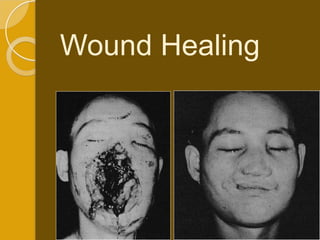
Wound healings
- 2. The Wound Definition Interruption of continuity of tissue resulting from a certain injury especially external physical trauma Wound: a disruption of normal anatomic relations as a result of injury intentional or unintentional.
- 3. Types of traumatic wounds ACCORDING TO SKIN LOSS Closed wound: skin surface intact Without loss of skin ◦ Abrasions: partial division of superficial layer ◦ Contusion: Diffuse extravasation of blood and exudate ecchymotic. ◦ Hematoma: Localized collection in fascial planes – ve signs of inflammation. Opened wound: skin surface interrupted or loss of skin. ◦ Incised ◦ Stab and punctured wound ◦ Lacerated ◦ Crushed and gun shots.
- 4. According to contamination ◦ Clean ◦ Contaminated According to the age of the wound ◦ Early (within 6 hours). ◦ Late ( 6-24h). ◦ delayed (after 24 h).
- 6. WOUND HEALING Natural spontaneous response for restoration of tissue continuity after injury Healing is the interaction of a complex cascade of cellular events that generates ◦ Reconstitution and Resurfacing, ◦ Restoration of the tensile strength of injured skin. Sometimes, tissue has been disrupted so severely that it cannot heal naturally.
- 7. Wound Healing Regardless of causation or tissue type, wound healing presents with identical biochemical and physiologic processes, though wound healing may vary in timing and intensity. Repair Vs Regeneration (speed Vs accuracy) Acute (orderly and timely) Vs Chronic (stalled in inflammatory phase)
- 8. Phases of wound healing: [I] Inflammation (biochemical activation) [II] Proliferation (granulation& cellular activation) [III] Remodeling (maturation &differentiation
- 9. Phases: Inflammatory, Proliferative, & Maturational
- 10. Inflammatory Biochemical -cellular activation Aim: ◦ translation of mechanical injury into biochemical signals. This starts by: ◦ Changes the charge on the surface of collagen molecule. ◦ Platelets aggregation and extravasated plasma contact with the extravascular tissue proteins leads to activation of Hageman's factors (factor XII) and platelets.
- 11. Inflammatory Biochemical -cellular activation Substrate or reactive phase, immediate typically days 1-10 Response to limit and prevent further injury, inflammation, hemostasis, sealing surface, removing necrotic tissue and debris, migration of cells into wound by chemotaxis, cytokines, and growth factors Initial intense local vasoconstriction of arterioles and capillaries followed by vasodilation and vascular permeability
- 12. (signs of inflammation) ◦ Vasodilatation (more persistent) Increase capillary engorgement ,Increase the capillary permeability, and blood flow under effect of histamine and bradykinin, serotonin, prostaglandins from platelets and mast cells flow of the necessary inflammatory cells and factors that fight infection and deriding the wound. ◦ This period is the event responsible for the erythema, edema, and heat observed after tissue injury ◦ Alterations in pH (secondary to tissue and bacterial degradation), The increase fluid tension in the area swelling press of the nerve endings, and tissue hypoxemia at the injury site contribute to the sensation of wound pain
- 13. Defending [migration of inflammatory cells & Chemoattraction to WBCs] ◦ PNL & Lymphocytes invade the wound and fibrin network within 3 hours defending and lysis with their lysosomes. release inflammatory mediators and bactericidal oxygen-free radicals. Lymphocytes also play a role in cellular immunity and antibody production. ◦ O2 is essential for the optimistic results of this defending process.
- 16. 1ry vascular reaction: Activation of clotting factors cascade. Platelet aggregation Clot formation (The scab) which temporarily closes the wound consists mainly of fibrin mesh trapped other blood cells hemostasis. Temporary constricting of small blood vessels (few minutes) temporary blanching.
- 17. Activation of complement system chemotaxis degranulation of mast cells and cytolysis. Platelets: accumulate release alpha granules containing: vasoactive agents chemotactic factors growth factors. Alpha granules (growth factors) initiator proliferative phase by activating the local mesenchymal and epidermal cells.
- 18. Inflammatory Tissue injury & blood vessel damage exposure of subendothelial collagen to platelets and vWF activates the coagulation pathway Plugging: Platelet and fibrin Provisional matrix: platelets, fibrin, and fibronectin Platelet aggregation: Thromboxane (vasoconstrict), thrombin, platelet factor 4
- 19. Platelet s Alpha granules contain: -platelet factor 4: aggregation -Beta-thrombomodulin: binds thrombin -PDGF: chemoattractant -TGF-beta: key component tissue repair Dense granules contain vasoactive substances: adenosine, serotonin, and calcium Other factors released: TXA, Platelet activate factor, Transform. growth factor alpha, Fibroblast growth factor, Beta lysin (antimicrobial), PGE2 and PGI2 (vasodilate) and PGF2 (vasoconstrict).
- 20. Types of growth factors
- 21. Extra Cellular Matrix Mast cell Histamine and platelet serotonin increases capillary vascular permeability Complement factors C5a and leukotreine B4 promote neutrophil chemoattraction As do IL1 and TNF alpha (from endothelial cells & macrophages) Increase chemotactic factors and spillage of intravascular plasma into interstitial fluid aid diapedesis of neutrophils Neutrophils release elastase and proteases, further vasc. dilation and permeability causes inflammation:
- 22. Polymorphonuclear Cells Chemotaxins attract after extravasation Migrate through the ECM by transient interaction with integrins PMNs scavenge, present antigens, provide cytotoxicity-free radicals (H2O2) Migration PMNs stops with wound contamination control usually a few days Persistant contaminant: continuous influx PMN’s and tissue destruction, necrosis, abscess, &
- 23. DEBRIDMENT Macrophages are essential for wound healing. Macrophages (monocytes) enter the wound from the 2nd after wounding and present until the reparative process is complete. Along time macrophages continue ◦ phagocytose & cleaning the wound site of bacteria, debris, F.B and necrotic matter. ◦ producing the activation growth and chemotactic factors similar to those of platelets (complete the function of platelets)…...
- 24. Macrophages Necessary Monocytes migrate & activate: Macrophages Appear when PMN’s disappear 24-48 hr Do the same activities as PMN’s Plus orchestrate release of enzymes (collagenase, elastase), PGE’s, cytokines (IL- 1, TNF alpha, IFN ), growth factors (TGF & PDGF), and fibronectin (scaffold/anchor for fibroblasts) Activate Fibroblasts, endothelial and epithelial cells to form Gran.
- 26. Proliferative Regenerative or Reparative (granulation & cellular activation) starts after 3-5 days takes 5-20 days INCLUDES: Granulation tissue formation Epithelization Contraction Angiogenesis: endothelial cells activate & degrade Basement membrane, migrate, and divide to form more tubules Granulation Tissue: capillary ingrowth, collagen, Macrophages, Fibroblasts, Hyaluroni c acid (GAG)
- 27. Granulation tissue Granulation tissue formation occurs 3- 5 days following injury Includes: Inflammatory cells, Fibroblasts and collagen, ground substance and Vascular and lymphatic proliferation
- 28. Fibroblast The fibroblast is a critical component of granulation tissue. Fibroplasia begins from surrounding mesenchymal cells 3-5 days after injury and may last as long as 14 days. Fibroblasts migrate and proliferate in response to platelets growth factors. Fibroblasts are responsible for the production of collagen, elastin, ground
- 29. Collagen synthesis The collagen fibers which is essential for: ◦ bridging the wound gap ◦ supporting the growing vessels and ◦ wound strength. Fibroblasts Collagen III held together by weak electrostatic forces and is soluble in weak salt solution. It is laid down irregularly and haphazardly then polymerization occurs by cross linked to the collagen molecules Thick strong less soluble collagen [I] become more regular and perpendicular on the plane of wound. The process of collagen synthesis : ◦ Starts on the 3rd day ◦ The peak reaches by the 5-7 days
- 30. This active metabolic process depends mainly on: ◦ Vitamins: B, ascorbic acid ◦ O2 ◦ amino acids ◦ Elements: zinc, iron, copper Collagen formation decreased by: ◦ decrease of vit C. ◦ steroids (high dose). ◦ Protein starvation.
- 31. Proline every 3rd amino- acid and abundant lysine hydroxylation required for x-link Vit C required for normal hydroxylation
- 32. Collagen Type III predominant collagen synthesis days 1-2 Type I days 3-4 Type III replaced by Type I in 3 weeks
- 33. Ground substance ◦ Produced by fibroblasts (water – electrolytes – mucopolysaccharides (proteoglycans) – fibronectins – glycoproteins). Angiogenesis (Vascular and lymphatic proliferation): ◦ The macrophage growth factors stimulates angiogenesis ◦ New capillaries bud from endothelial cells in capillary near the wound edges appear proliferation a new network of capillaries is formed inside the granulation tissues red granulations.
- 34. Epithelization Stats within hours by mitosis of the basal cell layer. The epidermal cells advanced from the edges and creep across the wound surface in a favorable plane dissecting the wound between the living and dead tissue. Migration stops when it meets the opposite advanced epithelium. The new epithelium is thin non-pigmented. Incisional wounds are epithelized within 24-48 hours after injury (distance of less than 1 mm). This epithelial layer provides a seal
- 35. In open wounds: if the wound is moist well oxygenated with viable moist surface and epithelization rapid (few days) and cell migrate over the surface of the wound. However, The process is more slower if the wound dry. The cells burring under the eschar and slowly separating the mobile from the immobile tissue. This explain why the epithelial is more rapid in intact blister than after the blister has been debride and the base of the blister
- 36. In sutured wounds: epithelium may invade the lining of the suture tracks. It usually degenerate with early removal of sutures. However, prolonged sutures ugly punctuate scars. This may be avoided by adhesions taps better cosmoses.
- 37. Epithelialization: Physical Barrier Begins within hours of injury Growth Factors (PDGF, TGF, and EGF) stimulate Mitosis of epithelial cells Migration: dom. factor = epithelial bottleneck (relies on gran. tissue) Epiboly leapfrog like motion until contact inhibition reestablished Early Tensile Strength: blood vessel growth, epithelialization, protein (fibrin) aggregation, later collagen formation
- 38. Wound contraction fibroblasts in the peripheral granulations maturation myofibroblasts centripetal movement of wound edges contraction decrease wound size facilitates closure of a defect. Lag period 2-3 days (with collagen synthesis) with Maximum rapid contraction 3-14 days (The maximal rate of contraction is 0.75 mm/d) It specially occurs at the back of the neck, trunk and face where the skin is loose Contraction must be distinguished from contracture. Contraction is decreased by : x-ray steroids grafting with dermis Burns If prevented slow healing – large fibrous tissue - ugly scar cicatrisation & complications.
- 39. Contraction Vs Contracture Contraction: centripetal movement of the whole thickness of surrounding skin reducing scar Myofibroblasts: special Fibroblasts express smooth muscle and bundles of actin connected through cellular fibronexus to ECM fibronectin, communicate via gap junctions to pull edges of the wound Contracture: the physical constriction or limitation of function as the result of Contraction (scars across joints, mouth, eyelid) Burn/Keloid causing contracture
- 40. Phase of remodeling (maturation &differentiation of scare tissue) ◦ It occurs after 20 days and continue for many months and years or indefinitely. Devascularization Collagen remodeling Cicatrisation
- 41. Maturational Remodeling of wound 3 week-1+year Type I replaces Type III Collagen: net amount doesn’t change after 6 weeks, organization & crosslinking Decreased vascularity, less fibroblasts & hyaluronic acid Peripheral nerves regenerate @ 1mm/day Accelerated Wound Healing: reopening results in quicker healing 2nd time around
- 43. Devascularization: The granulation tissue is gradually replaced by a scare tissue which is relatively acellular and avascular tissue. ◦ pale scare tissue. ◦ The extracellular tissue change its contents. Water is resorbed from the scar.
- 44. Collagen remodeling Collagen remodeling during the maturation phase depends on continued collagen synthesis in the presence of collagen destruction under effect of collagenase. The ratio of the collagen type [I] increase. New collagen is formed in more orderly fashion along the lines of tension in the scare. Facilitating collagen fibers cross-linking and ultimately decreasing scar thickness and increasing wound bursting strength.
- 45. Finally 4-12 w a pale red thick strong scare tend to contract is formed. ◦ Excessive contracture of the scare tissue cicatrisation. [contracture] a pathologic process of excessive fibrosis that limits motion of the underlying tissues and is typically caused by the application of excessive stress to the wound. 12-40 w soft white scare tend to relax. Hyalinization, calcification and even ossification may sometimes occur.
- 46. Closure of Wounds Primary: 1st intention immediately sealed with suturing, skin graft, flap closure (tensile strength) Secondary: Spontaneous involves no active intent to seal wound, gen. For highly contaminated wound, closes by reepithelialization and contraction of the wound (epithelial integrity) Tertiary: delayed primary closure of contaminated wound initially treated to control infection (repeated debridement, abx, wnd vac) then closed by suturing, skin graft, flap design, steri-strip etc.
- 47. Wound Closure
- 48. Summary Inflammatory phase: ◦ A clot forms stop bleeding ◦ Vasodilatation of WBCs ◦ cells of inflammation defending and debridment of injured tissue. Proliferative phase ◦ Epithelization, ◦ Fibroplasia (fibroblasts and collagen), and ◦ Angiogenesis occur during the; additionally, ◦ Granulation tissue forms and ◦ The wound begins to contract. Remodeling (maturation) phase ◦ Collagen forms tight cross-links to other collagen and with protein molecules, ◦ Increasing the tensile strength of the scar.
- 49. The phases of cutaneous wound healing Injury leads to accumulation of platelets and coagulation factors. Coagulation results in fibrin formation and release of PDGF and TGF-band other inflammatory mediators by activated platelets. This leads to more Neutrophil recruitment which signals the beginning of inflammation (24 h). After 48 h macrophages replace neutrophils. Neutrophils and macrophages are responsible for removal of cellular debris and release growth factors to reorganize the cellular matrix. At 72 hours the proliferation phase begins as recruited fibroblasts stimulated by FGF and TFG-b begin to synthesize collagen. Previously formed fibrin forms initial matrix for fibroblasts Collagen cross-linking and reorganization occurs following months after injury in the remodeling phase of repair. Wound contraction follows in large surface wounds and is facilitated by actin-containing fibroblasts (myofibroblasts)
- 51. Wound strength 6 Week = 60% original, 80% final strength 8 Week-1 year ≈ 80% original (Max) Net Collagen = 6 weeks amount stays the same but cont. crosslink increase strength = maturation
- 52. Tensile strength: The work done (force) in breaking a wound per unit area. The bursting strength of a wound is the force required to break a wound regardless of its dimension. Peak tensile strength of a wound occurs approximately 60 days after injury. A healed wound only reaches approximately 80% of the tensile strength of unwounded skin
- 53. the increase of cross linkage between the fibers increase its quality which is reflected in continuing increase in tensile strength. Factors affecting tensile strength: ◦ Factors affecting collagen synthesis specially vit C decrease. ◦ Direction of the w Parallel to the lines of Langer faster the healing and increase the tensile strength In the direction of the pull of the underlying muscle line of creases line scare least visible. ◦ no diff detectable between the wound that are taped and those that are sutured .
- 54. Complications of wound healing 1. Bleeding - shock - anemia. 2. Injury of Imp. structures. 3. Infection: 4. Dehiscence (bursting wound). 5. Implantation or epidermoid cyst 6. Keloid formation 7. Pigmentation tattooing 8. Painful scare local or reformed neuroma. 9. Cicatrization : burns deformity stricture and stenosis in tubes. 10.Neoplasia: sq cell ca. on scare tissue. 11.F.b. retained
- 55. Impediments to Wound Healing Bacteria>105/cm2 : Decreased O2 content, collagen lysis, prolonged inflammation Devitalized Tissue & Foreign Body: Retards Granulation Tissue formation and healing Cytotoxic drugs: 5FU, MTX, Cyclosporine, FK-506 can impair wound healing. D- Penicillamine- inhibit collagen x-linking Chemotherapy: no effect after 14 days Radiation: Collagen synthesis abnormal, fibrosis of vessel
- 56. More Impediments Diabetes: impedes the early phase response Malnurishment: Albumin<3.0, Vit-C Smoking: vasoconstriction, atherosclerosis, carboxyhemo globin, decreased O2 delivery Steroids: inhibit macrophages, PMNs, Fibroblast collagen synthesis, cytokines, and decreased wound tensile strength -Vit A (25,000 IU QD) counteracts effect of steroids DENERVATION has NO EFFECT on Wound Healing
- 57. Dehiscence (bursting wound) PF: ◦ Infection. ◦ Weak scare due to continuous strain (coughing vomiting) or stretch Decrease the bursting strength. ◦ Rapid absorbed catgut. ◦ Poor surgical technique. ◦ General conditions poor wound healing ◦ Decrease nutrition (Proteins) and vitamins (vit c)
- 58. Keloid formation & hypertrophic scars Unknown etiology P.F. ◦ Sex: females ◦ Race: black ◦ Repeated trauma ◦ TB patients – burns ◦ Irritation of FB, hair, keratin ◦ Age: In young, thin skin 1st year of life. And very old ◦ Common Site: Neck over the sternum. Wounds that cross skin tension lines or wounds that are located on the ear lobes or presternal and deltoid areas.
- 59. Keloids: Beyond the Borders Excess Deposition of Collagen Causes Scar Growth Beyond the Border of the Original wound Tx: XRT, steroids, silicone sheeting, pressure, excise. often Autosomal Dominant, Refractory to Darker Pigment, Often Tx & not preventable above clavicle but not always
- 60. Difference: Keloid grow beyond the wound borders It does not tend to resolve spontaneously. ◦ Hypertrophic scars stay within the limit of the original wound and do tend to regress spontaneously. It can form as late as a year after injury ◦ whereas Hypertrophic scars are generally seen soon after tissue injury, if the active scare continue more than 6 month it is considered true keloid which
- 61. Hypertrophic Scar: confined within Excess collagen deposit causing raised scar remains within the original wound confines Darker pigmented skin & flexor surfaces of upper torso Often occurs in burns or wounds that take a long time to heal, sometimes preventable Can regress spontaneously Tx: steroids, silicone, pressure garments
- 62. Histologically: Keloid also contain a greater amount of type III collagen than a mature scar, which suggests a failure in scar maturation. The collagen is loose disorganized wavy pattern of irregularly shaped fibers with a lower content of collagen cross-links compared to normal skin. keloid and hypertrophic scars have rich blood supply, high
- 63. TTT: The recurrence rate of these abnormal scars is high. Conservative management includes: Intralesional injection of triamcinolone. pressure, Laser, and radiotherapy. Excision & grafting :only if no response to conservative management.
- 64. Maggots
- 65. Langer’s Lines Lines lie perpendicular to underlying muscle fibers, as fibers contract wound edges are reapproximated as apposed to gapping caused parallel wound edges (plus camouflage)
- 66. Cachexia, anorexia Cancer Altered host metabolism. Protein catabolism Abnormal inflammatory cell response Impaired healing (decreased chemotaxis and phagocyte function) Risk of infection
- 69. Intraoperative surgical factors Length & Direction The best cosmetic results may be achieved when incisions are made parallel to the direction of the tissue fibers. Tissue handling, Hemostasis, Maintain ing moisture Materials of closure.
- 70. Diseases Assoc With Abnormal Wound Healing Osteogenesis Imperfecta: Type I Collagen defect Ehler-Danlos syndrome: Collagen disorder, 10 types Marfan Syndrome: fibrillin defect (collagen) Epidermolysis Bullosa: Excess fibroblasts Tx: phenytoin Scurvy: Vit C req. for proline hydroxylation Pyoderma gangrenosum: proud flesh
- 71. Mechanisms of regeneration, wound healing and repair
- 72. Repair of tissue damage can be broadly separated into two processes Regeneration ◦ Restitution of lost tissue Tissue with high proliferative capacity = labile tissue (e.g hematopoietic cells, epithelial cells of skin and gastrointestinal tract regenerate from stem cells) Quiescent tissues = stable tissue which normally have low levels of replication, however can undergo rapid cell division when stimulate (e.g pancreas, kidney, parenchymal cells of liver, ; mesenchymal cells as lymphocytes, fibroblasts, smooth muscle , endothelial cells) Healing ◦ may restore original structures but results in collagen deposition and scar formation in tissue where scaffold is disrupted
- 73. Regeneration requires Presence of stem cells for renewal or tissue cells that are capable to divide in response to growth factors Intact tissue scaffold Most of the processes that are referred to as “regeneration “ in mammalian organs are actually compensatory growth processes that involve cell hypertrophy and hyperplasia (e.g liver regeneration)
- 74. Stem cells They are undifferentiated cells that do not yet have a specific function. They can replicate for a long period of time and give rise to differentiated cells. In every cell division one cell retains its self renewing capacity while the other cell can undergo differentiation (“asymmetric replication”)
- 75. Two types of stem cells embryonic stem cells ◦ derived from the inner cell mass of a blastocyst from in vitro fertilized eggs ◦ are pluripotent and can generate all tissues
- 76. adult (somatic) stem cells ◦ they are present in small numbers in various tissues of the adult body ◦ are typically programmed to form different cell types of their own tissue and are therefore multipotent ◦ in tissues with high turn over (hematopoietc system, epithelial lining of the gut and skin) they are instrumental in renewal ◦ although present in a variety of permanent non- dividing tissues they are not very active
- 77. Bone marrow contains two different types of adult stem cells: the hematopoietic stem cell and the bone marrow stromal cell
- 78. Potential plasticity of hematopoietic stem cells
- 79. Hematopoietic stem cell transplantation: an established treatment option for hematological disorders and cancers Hematopoietic stem cells can be retrieved from the peripheral blood or the bone marrow and identified by the expression of the CD34 marker 2X10⁶ HSC/KG body weight of recipient are needed for a successful autologous HSC transplantation Under steady state conditions the number of CD34+ cells in peripheral blood is 1-5/mm3 Mobilization procedures of CD34+ stem cells into the peripheral blood can be accomplished by administration of G-CSF or GM-CSF to the donor and can increase the HSC count in the peripheral blood 50 fold
- 80. Repair by Healing (Scarring) Healing is a fibro-proliferative responses that “patches” rather than restores tissue and involves the following processes ◦ Induction of an inflammatory response to remove dead and damaged tissue ◦ Proliferation of parenchymal and connective tissue cells ◦ Angiogenesis (blood vessel formation) and formation of granulation tissue ◦ Synthesis of ECM proteins and collagen deposition ◦ Tissue remodeling ◦ Wound contraction ◦ Acquisition of wound strength It usually leads to scar formation and does not lead to complete
- 81. Angiogenesis = growth of new blood vessels Angiogenesis occurs in the healthy body for healing wounds and for restoring blood flow after tissue injury Healthy angiogenesis is tightly controlled by a serious of “on” and “off switches (Angiogenic growth factors versus angiogenesis inhibitors) In many serious diseases the body loses control over angiogenesis and angiogenesis-related diseases occur when new blood vessels grow excessively or
- 83. Angiogenesis / Neovascularization is critical to chronic inflammation and fibrosis,tumor growth and vascularization of ischemic tissue Sprouting
- 84. VEGF and Angiopoietins are the most important angiogenic factors
- 85. Role of extracellular matrix in wound healing and scar formation Extracellular matrix (ECM) is formed by specific secreted macromolecules that form a network on which cells grow and migrate along ECM is secreted locally and forms a significant proportion of the tissue volume ECM sequesters ◦ water that provides turgor to soft tissues ◦ and minerals that provides rigidity to skeletal muscles ◦ Forms a reservoir for growth factors ECM proteins assemble into two general organizations ◦ Interstitial matrix (present between cells) ◦ Basement membrane [BM] (produced by epithelial and mesenchymal cells and is closely associated with the cell surface)
- 86. Three groups of macromolecules constitute the ECM Fibrous structural proteins ◦ Collagen ◦ Fibrillins Adhesive glycoproteins ◦ Cadherin ◦ Integrins ◦ Immunoglobulin family ◦ Selectins Proteoglycans and Hyaluronic Acid
- 87. Fibrous structural proteins ◦ Collagens Collagens are the most abundant proteins 27 different types Type I,II, III, V and XI are the most abundant (interstitial or fibrillar collagens) Provide tensile strength of tissue Fibrillar collagen requires hydroxylation of proline and lysine in procollagen which is dependent on Vitamin C Type IV is the main component of Basemant membrane and forms sheets) ◦ Elastins and Fibrillins Provide tissue with the ability to recoil Elastins are found in large vessels, uterus, skin and ligaments Fibrillins form a scaffolding for the deposition of elastins Marfan syndrome is an inherited autosomal dominant defect in fibrillin synthesis. Without the structural support provided by fibrillin, many tissues are weakened, which can have severe consequences, for example, ruptures in the walls of major
- 89. Proteoglycans and hyaluronic acid ◦ Proteoglycans (mucoproteins) are formed of glucosaminoglycans (GAGs) covalently attached to core proteins and are highly negatively charged Biophysical functions due to ability to fill space, bind and organize water molecules and repel negatively charges molecules They are ideal lubricating fluids in the joint due to high viscosity and low compressibility Biochemical functions are mediated by specific binding of GAGs to other macromolecules e.g Antithrombin III (AT III) binds tightly to heparin and heparan sulfates and inactivates factor II, IXa and XIa thus controlling blood coagulation Proteoglycans (such as Syndecan) act as reservoirs for growth factors secreted into the ECM by binding the latter.
- 90. TGF- functions as a central regulator of tissue repair and negatively regulates both acquired and adaptive immunity Lack of the TGF gene in mice results in excessive tissue inflammation and autoimmunity resulting in death of the animals, however increased activity leads to excessive scar formation and loss of organ function
- 91. Mechanisms of fibrosis TH-2 cytokines IL-4 and IL-13 lead to “alternative” activation of macrophages Nicole Meissner-Pearson
- 92. Mechanisms of Fibrosis: a result of chronic inflammation and repair Fibrosis: excessive accumulation of extracellular-matrix components such as collagen that is produced by local fibroblasts leading to a permanent fibrotic scar Macrophages and fibroblasts are the main effector cells involved in the pathogenesis of fibrosis Pro-fibrotic mediators such as TGF-b and IL-13 amplify this process The degradation of collagen is controlled by Matrix- Metallo-proteinases (MMPs) and are activated by IFN-g Therefore the net increase of collagen within a wound is controlled by the balance of these opposing mechanisms Although severe acute injuries can cause marked tissue remodeling. Fibrosis that is associated with chronic injury (repetitive) is unique in that the adaptive immune response is thought to have an important role
- 94. TH-2 cytokines IL-4 and IL-13 lead to “alternative” activation of macrophages Macrophages differentiate into at least two functionally distinct populations depending on whether they are exposed to TH-1 or TH-2 cytokines TH-1 cytokine activate NOS2 in classically activated macrophages whereas TH-2 cytokines IL-4 and IL-13 preferentially stimulate Arginase-1 (ARG1) leading to an alternative activation pathway ARG1 promotes the generation of polyamines and L- proline via metabolism of L-arginine to L-ornithine and activation of ODC and OAT Polyamines are crucial for cell growth and L-proline is a substrate for collagen synthesis
- 96. A balance between TH-2 and TH-1 cytokines is necessary to promote healing but inhibit excessive fibrotic tissue remodeling Therapeutics that modulate this balance may be beneficial in patients suffering from fibrotic diseases. Drugs that directly inhibit TGF-b1 and IL-13 might prove the safest and most effective approach
- 97. Fibrotic tissue remodeling can result in loss of organ function Fibrotic changes can occur in various vascular diseases including ◦ Cardiac diseases ◦ Peripheral vascular diseases They affect as well main organ systems like ◦ Skin ◦ Lung ◦ Liver ◦ Kidney
- 98. Artificial Skin The term “artificial skin” has been used to describe a cell-free membrane comprising a highly porous graft copolymer of type I collagen and chondroitin 6-sulfate which degrades at a specific rate in the wound and regenerates the dermis in dermis-free wounds in animal models and patients
- 99. History of Skin Replacement Thirty years ago, burn surgeons determined that badly burned skin should be removed as quickly as possible, followed by immediate and permanent replacement of the lost skin. Doctors are able to take a postage stamp-sized piece of skin from the patient and grow the skin under special tissue culture conditions. • From this small piece of skin, technicians can grow enough skin to cover nearly the entire body in just 3 weeks • Although attempts to cover wounds and treat severe burns is cited as far back as 1500 B.C., it has only been in the past few centuries that a significant number of solutions have emerged. • The bulk of these solutions involve using skin grafts from humans (allografts) or animals (xenografts), or using membranes fabricated from natural or synthetic polymers.
- 100. History of Artificial Skin The first synthetic skin was invented by John F. Burke, V. Yannas, at the Massachusetts Institute of Technology (MIT) In 1979 Burke and Yannas used their artificial skin on their first patient, a woman whose burns covered over half her body Integra is the first FDA approved tissue engineered product for burn and reconstructive surgery. It was Patented on August 14, 1990
- 101. Why is it Needed? When skin is damaged or lost due to severe injury or burns, bacteria and other microorganisms have easy access to warm, nutrient-rich body fluids. To treat a severe burn, surgeons first remove the burned skin and then quickly cover the underlying tissue, usually with a combination of laboratory-grown skin cells and artificial skin.
- 102. A Literary Review Wayne R. Fischer ME 597 Introduction to Solid Biomechanics Boise State University May 8th, 2003
- 103. The best material for wound closure is the patient’s own skin; however autografting has several disadvantages (Schulz, 2000): 1. The donor site is a new wound. 2. Scarring and pigmentation changes occur. 3. Dermis is not replaced. 4. Donor site is a potential site for infection. 5. Donor site is not unlimited. 6. Extensive burns makes it impossible.
- 104. Cadaver Skin: Allograft as a Temporary Skin Substitute The annual national requirement for cadaver skin is estimated to be only 3000 m2. Yet only 14% to 19% of human skin needed is being recovered.
- 105. Xenografts • Xenografts, particularly porcine skin grafts, are commercially available and are an effective means of short-term wound closure (Yannas, 1980). • A Xenograft is normally removed on the third or fourth day of use before extensive adhesion onto the wound bed sets in, thereby necessitating its traumatic excision prior to drying and sloughing off (Yannas, 1980).
- 106. Observations from designing dermal replacements (Schulz, 2000) The thicker the dermal layer of a split-thickness skin graft, the less the graft contracts. Partial-thickness wounds with superficial dermal loss heal with less hypertrophic scarring. Full-thickness skin grafts contract minimally. The length of illness in burn cases is essentially restricted to the length of time the burn wound is open. Full-thickness dermal injuries heal by contraction and hypertonic scarring, producing subepithelial scar tissue that is nothing like the original dermis.
- 107. Research topics of Dr. Yannas at Dept. of Engineering, MIT 1. Study the mechanical behavior of artificial skin as a function of processing variables. 2. Study the surface tension of artificial skin. 3. Study the stress relaxation rate of artificial skin in standardized solutions of tissue enzymes. 4. Study the design of novel processes for the inexpensive and reproducible fabrication of artificial skin. 5. Study the pore structure of artificial skin by scanning electron microscopy. 6. Study the moisture permeability of artificial skin.
- 108. Synthetic Polymers (Yannas, 1980) The use of synthetic polymers has not so far led to the solution of the problem of a skin substitute. A high incidence of infection and a relatively low capacity for inducing vascularisation and epithelialisation are frequently reported. However, useful insights into the requirements for a satisfactory skin replacement have been discovered through the use of synthetic polymers.
- 109. Schematic Representation of Specific Mechanical Problems that Should Not Arise (Yannas, 1985).
- 110. Specific Physiochemical and Mechanical Problems to Overcome (Yannas, 1985). a) Skin graft does not displace air pockets efficiently from graft-woundbed interface. c) Shear stress causes buckling of graft, rupture of graft woundbed bond and formation of air pockets. e) Excessively high moisture flux rate through graft causes dehydration and development of shrinkage stresses at edges and peeling.
- 111. Specific Physiochemical and Mechanical Problems to Overcome (Yannas, 1985). b) Flexural rigidity of graft is excessive; graft does not deform sufficiently under its own weight to make contact with depressions in woundbed surface, thus air pockets form. d) Peeling force lifts graft away from woundbed. f) Very low moisture flux causes fluid accumulation at graft- woundbed interface and peeling.
- 112. Literary Review Up to 1990’s (Beele, 2002) Antiquity: Indian description of using autologous soft tissue flaps. Greeks used dressings for skin wounds. Renaissance: Amboise-Pare provide wound healing foundation. 1850’s: Reverdin and Thiersch use autologous skin grafts. 1914: Kreibich was the first person to cultivate keratinocytes in vitro. 1948: Medawar autotransplanted keratinocytes. 1960’s: Yannas and Burke begin their work using materials science mechanics. 1975: Rheinwald & Green describe a technique to cultivate human keratinocytes. 1980’s: Yannas and Burke describe a bilaminate collagen-glycosaminoglycan matrix with a silicon surface. After take of the matix. The silicon surface is removed and can be replaced with autologous cultured epidermal cells. 1981: Bell constructs the first living skin equivalent with collagen fibroblast gel with keratinocytes cultured on top of contracted gel. 1983: Helton used cultured allografts in burn patients 1985: Boyce and Ham introduce an alternative culturing method.
- 113. Further Research (Buras, 1989) The actual biological elements and events being critically tested in mechanical studies are only guessed at, and analysis can rarely go beyond the science of mechanics. There are promising possibilities: Pulsed ultrasound techniques may soon provide accurate imaging of skin structures as well as measurements of blood flow in the skin. The multifrequency shear wave method may be able to resolve mechanical properties of the epidermal tissues discretely.
- 114. Some References And we’re up and walking again! Beele, H. Artificial skin: Past, present and future. The International Journal of Artificial Organs. 25(3): 163-173, 2002. Jones, I., Currie, L., Martin, R. A guide to biological skin substitutes. British Journal of Plastic Surgery. 55: 185- 193, 2002. Schulz III, J.T., Tompkins, R.G., Burke, J.F. Artificial Skin. Annu. Rev. Med. 51: 231-244, 2000. Yannas, I.V. Artificial Skin and Dermal Equivalents. In The Biomedical Engineering Handbook, ed. J. D. Bronzino, pp. 2025-2038. Boca Raton: CRC Press, 1995.
- 115. Literary Review: 1990’s to Present (Chart in British 2002 Journal of Plastic Surgery)
- 116. Types of Skin Replacements Epicel skin replacement technology ◦ Introduced by Genzyme Biosurgery in 1987. ◦ Physicians isolate individual cells from a postage-stamp-sized biopsy of skin. ◦ Grow the cells for about 2 to 3 weeks and allow them to form individual sheets of tissue. ◦ Then surgeons transplant these sheets of tissue to the burnt area where these sheets fuse over time with the burnt area.
- 117. Types of Skin Replacements Integra Dermal Regeneration Template® ◦ Semi -synthetic approach to skin regeneration ◦ Researchers develop a bi-layer membrane system called the Dermal Regeneration Template ◦ The first and only FDA approved tissue engineered product for burn and reconstructive surgery ◦ Dermal replacement layer is constructed of a porous, biodegradable matrix of cross-linked bovine tendon collagen and the glycos- aminoglycan chondroitin 6-sulfate.
- 118. ◦ Second layer acts as a temporary replacement (Epidermal ) – made from silicone polymer Following completion of the dermal layer physicians replace the temporary epidermal with an epidermal auto-graft. Skin Graft ◦ taking cells from a non-burned epidermal layer of skin, growing them into large sheets of cells in a laboratory
- 119. How does it work? The burn surgeon drapes a sheet of Integra ® over the wounded area for 2 to 4 weeks. Allows the victim’s cells to grow a new dermis on top of matrix of the Integra ®. Then the surgeon removes the top layer of the Integra® and applies a very thin sheet of the victim’s own epithelial cells. Over time, a normal epidermis (except for the Skin replacement. scientists at Integra Using a bilayer membrane system, absence of hair follicles) is LifeSciences help repair skin lesions and burns.
- 120. General Design Properties Essential Design Properties ◦ "The dermal replacement should provide both the information necessary to control the inflammatory and contractile processes and also the information necessary to evoke ordered recreation of autologous tissue in the form of a neodermis" (Schulz, 2000). ◦ "The initial replacement material should provide immediate physiologic wound closure and be eliminated once it has provided sufficient information for neodermis reconstitution of " (Schulz). ◦ It should protect the wound by providing a barrier to the outside (Beele, 2002) ◦ It should control water evaporation and protein and electrolyte loss and It should limit excessive heat loss (Beele) ◦ It should decrease pain and allow early mobilization and It should provide an environment for accelerated wound
- 121. More General Design Properties Physical Characteristics ◦ It should be easy to manipulate the product, i.e. easy to place and dress the skin substitute effectively (Beele) ◦ It should improve the cosmetic appearance of the scar (Beele) Availability ◦ It should be readily available off the shelf and custom made. Cost ◦ Cost should not preclude the use of the device.
- 122. Advantages -Disadvantages of Temporary Skin Substitutes
- 123. Advantages and Disadvantages of Permanent Skin Substitutes
- 125. Cultured skin cells , polyglycolic fabric , collage gels and glycosaminoglycans are incorporated Rapid Keratinocyte cultures are obtained by growing the same on a feeder layer of irradiated fibroblasts. Also , neonatal fibroblasts are being used (Paed Medical Center , Munster –
- 126. The graft is a bilayer membrane. In this approach, the top layer , a Silicone Layer incorporates the features of moisture control. While the bottom layer delivers the performance of sealing the skin breach and preventing scarring. The top layer is removed after a period of about 10–15 days. Following removal of the top layer, the epidermal cover is provided by covering with a thin epidermal graft.
- 127. Design Parameters The ratio of the time constant of biodegradation to the time constant for normal healing of a skin incision ideally must be unity Adequate Moisture Flux Dermis Regeneration Template - porous matrix seeded with cells, induces synthesis of a new dermis, simultaneously synthesis of a new epidermis occurs by migration of epithelial cell. The depth of tissue loss must be known as epithelial cells cannot migrate if loss of tissue is high.
- 128. Limitation 3-4 weeks are required for the cell expansion in the graft Certain limitations while implanting the graft Air pocketing between graft and underlying skin Shear stresses can cause buckling of the skin while grafting Excess or low Moisture flux may cause lifting of the skin or edema underneath No appendages of skin Take rates are less.
- 129. Advantages Less Scarring Reduction in the nutritional requirements may be observed while using skin substitutes Can be used even in extensive burns Does not initiate an inflammatory or foreign body response
- 131. Biological skin substitutes The epidermis injuries are healed by regeneration of the epidermis The migration of keratinocytes from the periphery of the wound and the proliferation would lead to the total healing without scars However if dermis is injured recovery is harder since the dermis cannot regenerate In order to shorten the healing process or abolish the side effects, skin substitutes should be used temporarily or permanently.
- 132. Desired properties for skin substitutes Adhere to the substrate Be durable and sufficiently elastic to tolerate some deformation Allow evaporative water loss at the rate typical of the external layer Have optimal water permeability to prevent either desiccation of the wound or fluid accumulation under the covering
- 133. Categories of skin substitutes Skin substitutes for wound closure Skin substitutes for wound cover Wound closure requires a material to restore the epidermal barrier function and become incorporated into the healing wound Wound cover necessitates a material which relies in the in growth of granulation for adhesion. They are used in superficial burns
- 134. Skin substitutes for wound cover Biobrane Transcyte Cultured allogenic keratinocytes Apligraf ( graft skin ) Dermagraft
- 135. Biobrane It is a bilaminate membrane of nylon mesh bonded to a thin layer of slicone which is semipermeable The nylon mesh is coated with peptides to aid adherence Used temporarily for freshly excised full thickness wounds
- 136. Transcyte The difference between trancyte and biobrane is the seeding the neonatal fibroblast on to the collagen coated nylon membrane Since nylon is not biodegradable, it cannot be used as a dermal substitute The removal process is more successful because of less bleeding
- 138. Cultured allogenic keratinocytes The survival period of allogenic cells is one week The healing with allogenic cells can be enhanced with cytokines and growth factors by the cells They are used as dressing for chronic wounds
- 139. Apligraf ( graft skin ) Composed of two layers. Inner layer- gel type 1 bovine collagen with living neonatal fibroblast Outer layer – neonatal allogenic keratinocytes Used to treat chronic ulcers
- 140. Dermagraft Cyropreserved human fibroblast derived dermal substitute on polyglactin-910 mesh scaffold Enhances healing by stimulating the ingrowth of firbovascular tissue from the wound bed Used in chronic lesions
- 141. Skin substitutes for wound closure Alloderm Integra Cultured autologous keratinocytes Composite epidermal skin substitutes
- 142. Alloderm Derived from human cadaveric skin in which the epidermis is removed and cellular components of the dermis are extracted and preserved to avoid specific immune response After application, the wound bed is repopulated, revascularised and incorporated into the tissue
- 144. Integra Produced by Burke and Yannas, producer of GAG(gylcosamine) sponges. Widely accepted skin substitute Integration of the GAG sponge with a silicone layer Pore size-70-200micrometer,to allow the migration of patient’s own endothelial cells and fibroblasts Pore size small- delay or the prevention of biointegration is observed Pore size large- insufficient attachment are for invading host cells Advantage-improved elasticity Disadvantage- cost
- 145. Cultured autologous keratinocytes Grown in vitro conditions as confluent sheets Since they are fragile, they require separation from tissue culture substrate by using proteolytic enzyme before they are applied to the wound bed Cultured autologous delivery systems Fibrin-glue suspension Fibrin glue sheets Upside down membrane delivery systems Sprayed cell suspension
- 146. Composite epidermal-dermal skin substitute Healing quality can be enhanced by combining the cultured keratinocytes with a dermal matrix Keratinocytes should be maintained on a biomaterial Epidermis binds-to the biomaterial and receives adequate nutrition, the epidermal barrier is replaced. COS Approximately €700 for 125 cm 2 T (Journal of Pediatric Orthopaedics 14(5):381-384, September 2005)
- 147. Recent Advances
- 148. Artificial Skin From Hair Roots Fraunhofer - Gesellschaft (2008, January 4). Growing Artificial Skin From Hair Roots Euroderm and the Fraunhofer Institute for Cell Therapy and Immunology in Leipzig have been granted approval to produce artificial skin from patients’ own cells. Few hairs off the back of the patient’s head are pulled Adult stem cells from the roots are extracted,
- 149. ICX-SKN - Mimicking nature Paul Kemp and colleagues at British biotech company Intercytex Fully and consistently integrates into the human body No need for further grafting
- 151. FILM Skin – For robots, Artificial Limbs Flexible, Integrated, Lightweight, Multifunctiona l skin Oak Ridge National Laboratory's Nanomaterials Synthesis and Properties Group Carbon Nanotubes are being used The material can be designed to behave as both a temperature and pressure sensor, as a flexible electrical conductor, or as part of a polymer material with mechanical and thermal properties similar to those of human skin.
- 152. Skin cells genetically engineered to be resistant to bacteria Scientists at the Cincinnati Shriners Hospital for Children have engineered bacteria resistant skin cells. Due to delay in angiogenesis, the skin is vulnerable to bacteria as there are no circulating macrophages. Hence incorporating anti-bacterial factors like Human Beta Defensin 4, will help void bacteria at an initial stage
- 154. HOW IT WORK
- 163. In the Near Future
- 164. Self Healing Artificial Skin http://www.mvac.uiuc.edu Microvascular Autonomic Composites Initiative (µVAC) is creating materials with a microvascular network, capable of pumping self-healing polymers to repair sites of skin breech Skin capable of healing, even though only to a certain degree, could prove incredibly useful for the robotics industry.
- 165. The surface layer acts as a catalyst for the healing agent, causing it to Microvascular network polymerize upon contact embedded in the substrate layer carrying the healing agent Residue healing agent repairing cracks on the surface of the µVAC material.
- 166. Microvascular Autonomic Composites Initiative (µVAC) is for Robotic Skin….. Imagine the same with our Artificial Skin Skin that regenerates when breeched accidentally or intentionally
- 167. Questions for Future Research How does the skin transform and grow naturally on a biochemical and physiologic level? How can these natural transformations be combined with concepts from materials science and biomechanics in order to develop and design a cost effective and viable skin substitute? Which designs already incorporate natural growth components with concepts from materials science and biomechanics? How can these designs be enhanced or re-deigned using the concepts within the domain of materials science and biomechanics?
- 168. Overview- Integumentary Development Development of Skin Skin ◦ largest organ ◦ protective layer ◦ 2 embryonic origins ◦ epidermis surface ectoderm ◦ dermis mesoderm
- 169. A totally new epidermis is present every 25 to 45 days. Melanocytes create melanin, the substance that gives our skin color. These cells are found deep in the epidermis layer. Accumulations of melanin are packaged in melanosomes (membrane-bound granules). These granules form a pigment shield against UV radiation for the keratinocyte nuclei.
- 170. Embryology of skin Ectoderm forms the surface epidermis and the associated glands. Mesoderm forms the underlying connective tissue of dermis and hypodermis
- 172. Skin tissue culture. Kultur jaringan kulit . Keratinocyte cells is the most upper part of skin, epidermis.
- 173. For burns grafting The keratinocyte cell is the building block of the top layer of skin, the epidermis. Burns are traditionally treated by taking a thin layer of skin from an undamaged area (donor site) and placing it onto the burn wound. In severely burnt patients, the donor sites are few and the potential to re-harvest the area is limited. Skin allografts (harvested from one person and grafted onto another) can be used as temporary dressings, but are rejected by the body within a short time.
- 174. The Tissue Culture laboratory grows keratinocyte cells into epithelial grafts for burn patients in hospitals. From a small piece (2 x 2cm) of the patient’s own skin, it can be can grew enough epithelial grafts to cover a whole person in 3 weeks. The individual grafts are typically 10 x 7cm in size and are multi-layered, very much like normal epidermis. At the base of the graft is the basal cell layer. As the cells move through each layer of the skin, they become increasingly differentiated. Once the epithelial graft is placed on the patient and exposed to air, the top layer takes on the protective role of the skin by becoming
- 175. Normal epidermis
- 176. Cultured epithelium (H&E stain, magnification approx. x350)
- 177. Detached cultured epithelial graft
- 178. For chronic skin ulcer treatment It has been established a keratinocyte cell line from neonatal foreskin. These cells are free of contamination by HIV, Hepatitis B & C and CMV and have been used to produce cultured epithelial allografts for the successful treatment of chronic leg ulcers. Cryo-preserved allografts are available as biological dressings for immediate use on request.
- 179. One of medium culture for skin growing in the lab. medium culture consist of insulin, epidermal growth factor (EGF), hydrocortisone, Penicillin /streptomycin and fungizone.
Notas do Editor
- Ditutupluka: permukaankulitutuhTanpahilangnyakulitLecet: pembagiansebagianlapisandangkalMemar: Diffuse ekstravasasidarah dan eksudatecchymotic.Hematoma: Localized koleksidalampesawatfasia - vetanda-tandaperadangan.Dibukaluka: permukaankulittergangguatauhilangnyakulit.BertakukTusuk dan lukatertusukRobekHancur dan tembakansenjata.
- AlamresponspontanuntukpemulihankelangsunganjaringansetelahcederaPenyembuhanadalahinteraksidarikaskadekompleksperistiwaseluler yang menghasilkanPemulihan dan Resurfacing,Pemulihankekuatantarikkulit yang terluka.Kadang-kadang, jaringantelahterganggubegituparahsehinggatidakdapatmenyembuhkansecaraalami.
- Tujuan:terjemahandaricederamekanismenjadisinyalbiokimia.Hal inidimulaidengan:Perubahanmuatanpadapermukaanmolekulkolagen.Trombositagregasi dan menghubungi plasma extravasateddengan protein jaringanekstravaskulermenyebabkanaktivasifaktor Hageman's (faktor XII) dan platelet.
- Substratataufasereaktif, segera biasanya 1-10 hariResponuntukmembatasi dan mencegahcederalebihlanjut, peradangan, hemostasis, penyegelanpermukaan, membuangjaringannekrotik dan puing-puing, migrasiselkelukaolehchemotaxis, sitokin, dan faktorpertumbuhanvasokonstriksilokalawalintensarteriola dan kapilerdiikutiolehvasodilatasi dan permeabilitasvaskular
- Vasodilatasi (lebihpersisten) Meningkatkan engorgement kapiler, Peningkatanpermeabilitaskapiler, dan darahmengalirdibawahpengaruhhistamin dan bradikinin, serotonin, prostaglandin dari platelet dan sel mast alirandarisel-selinflamasi yang diperlukan dan faktor-faktor yang melawaninfeksi dan mencaciluka.Periodeiniadalahperistiwabertanggungjawabatas edema, eritema, dan panasdiamatisetelahcederajaringanPerubahanpada pH (sekunderuntukjaringan dan degradasibakteri), Keteganganmeningkatcairandidaerahtersebut pembengkakantekan dariujungsaraf, dan hipoksemiajaringandilokasicederaberkontribusisensasi rasa sakitluka
- PNL & Limfositmenyerangjaringanluka dan fibrin dalamwaktu 3 jam membela dan lisisdenganlisosommereka.pelepasan mediator inflamasi dan radikalbebasoksigenbakterisida.Limfositjugamemainkanperandalamimunitasselular dan produksiantibodi.O2 adalahpentinguntukhasiloptimisprosesmembela.
- Aktivasikaskadefaktorpembekuan.Agregasi plateletPembentukanbekuan (kudis The) yang sementaramenutuplukaterutamaterdiridari fibrin mesh terjebakseldarahlainnya hemostasis.Sementarakonstriksipembuluhdarahkecil (beberapamenit) blansingsementara.
- Statistikdalamhitungan jam oleh mitosis lapisansel basal.Sel-sel epidermis majudaritepi dan merayapdiseluruhpermukaanlukadalampesawatmenguntungkanbedahlukaantarajaringanhidup dan mati. Migrasiberhentiketikamemenuhiepitelmajuberlawanan.Epitelbarutipis non-pigmen.lukainsisionaladalahepithelizeddalamwaktu 24-48 jam setelahluka (jarakkurangdari 1 mm). Lapisanepitelmenyediakansegelantaraluka yang mendasari dan lingkungan.
- Dalamlukaterbuka: jikalukalembabberoksigenbaikdenganpermukaanlembab yang layak dan epitelisasicepat (beberapahari) dan selbermigrasidiataspermukaanluka.Namun, prosesinilebihlambatjikalukakering. Sel burring bawaheschar dan perlahanmemisahkan mobile darijaringanbergerak.Hal inimenjelaskanmengapaepitellebihcepatmelepuhutuhdaripadasetelahmelepuhtelahdebride dan dasarmelepuhdiizinkanuntukkering.
- Dalamlukadijahit: epiteldapatmenyeranglapisandari trek jahit. Biasanyamerosotdenganpenghapusanawaljahitan. Namun, jahitanbekasluka lama menekankanjelek. Inidapatdihindaridengankeranadhesi cosmoses lebihbaik.
- fibroblas di perifer granulasi pematangan myofibroblasts. gerakan sentripetal dari tepi luka kontraksi penurunanukuran luka memfasilitasi penutupan cacat.Lag periode 2-3 hari (dengan sintesis kolagen) dengan kontraksi Maksimum cepat 3-14 hari (Tingkat maksimal kontraksi adalah 0,75 mm / d)Hal ini khususnya terjadi di belakang batang, leher dan wajah di mana kulit yang longgarKontraksi harus dibedakan dari contracture.Kontraksi berkurang oleh:sinar Xsteroidgrafting dengan dermisBurns Jika dicegah memperlambat penyembuhan - jaringan fibrosa besar - bekas luka jelek cicatrisation & komplikasi.
- Devascularization:Jaringan granulasi secara bertahap digantikan oleh jaringan SCARE yang jaringan relatif aselular dan avaskular. jaringan pucat.Jaringan ekstraseluler mengubah isinya. Air diresorpsi dari bekas luka.
- Kolagenrenovasiselamafasepematangantergantungpadasintesiskolagenlanjutandihadapanpenghancurankolagendibawahpengaruhkolagenase.Rasiojeniskolagen [I] meningkat. Kolagenbarudibentukdilebihteratursepanjanggarisketegangan SCARE.seratkolagenMemfasilitasi cross-linking dan akhirnyapenurunanketebalanbekasluka dan meningkatkanketahananretak.
- Primer: 1 niat segera disegel dengan penjahitan, graft kulit, flap penutupan (kekuatan tarik)Sekunder: spontan tidak melibatkan maksud aktif untuk menutup luka, gen. untuk sangat terkontaminasi luka, menutup oleh reepithelialization dan kontraksi luka. (Integritas epitel)Tersier: penutupan primer tertunda yang terkontaminasi luka awalnya dirawat untuk mengendalikan infeksi (diulang debridemen, Abx, VAC WND) kemudian ditutup dengan penjahitan, skin graft, flap desain, steri-strip dll
- FasepenyembuhanlukakulitCederamenyebabkanakumulasitrombosit dan faktorkoagulasi.Koagulasihasildalampembentukan fibrin dan pelepasan PDGF dan TGF-band mediator inflamasilainnyadengan platelet diaktifkan. Hal inimenyebabkanrekrutmenNeutrofillebih yang merupakantandaawalperadangan (24 jam).Setelah 48 jam gantimakrofagneutrofil. Neutrofil dan makrofagbertanggungjawabuntukpemindahanreruntuhanselular dan faktorpertumbuhanrilisuntukmereorganisasimatriksselular.Pada 72 jam faseproliferasidimulaisebagaidirekrutfibroblasdistimulasioleh FGF dan TFG-b mulaimensintesiskolagen.Sebelumnyaterbentuk fibrin bentukawalmatriksuntuk fibroblast Kolagen cross-linking dan reorganisasiterjadibulanberikutnyasetelahcederadalamtahaprenovasiperbaikan. kontraksi Luka berikutdalamlukapermukaanbesar dan difasilitasiolehaktin yang mengandungfibroblas (myofibroblasts)
- Pekerjaan yang dilakukan (gaya) dalammemecahkanluka per satuanluas.Kekuatanledakanlukaadalahgaya yang dibutuhkanuntukmemecahkanlukaterlepasdaridimensinya.Puncakkekuatantariklukaterjadisekitar 60 harisetelahcedera.Luka sembuhhanyamencapaisekitar 80% darikekuatantarikkulit unwounded
- peningkatan hubungan silang antara serat peningkatan kualitas yang tercermin dalam melanjutkan peningkatan kekuatan tarik.Faktor-faktor yang mempengaruhi kekuatan tarik:Faktor-faktor yang mempengaruhi sintesis kolagen khusus vit C menurun.Arah wSejajar dengan garis-garis Langer penyembuhan lebih cepat dan meningkatkan kekuatan tarik Dalam arah tarikan garis otot yang mendasari lipatan garis menakut-nakuti paling terlihat.tidak ada diff terdeteksi antara luka yang direkam dan mereka yang dijahit.
- HambatanuntukPenyembuhan LukaBakteri> 105/cm2: O2 konten Penurunan, lisis kolagen, radang berkepanjanganDevitalized Tissue & Asing Body: menghambat pembentukan jaringan granulasi dan penyembuhanobat sitotoksik: 5FU, MTX, Siklosporin, FK-506 dapat mengganggu penyembuhan luka. D-Penisilamin-menghambat kolagen x-linkingKemoterapi: tidak berlaku setelah 14 hariRadiasi: sintesis kolagen abnormal, fibrosis kapal
- Diabetes: menghambattahapawal responMalnurishment: Albumin <3.0, Vit-CMerokok: vasokonstriksi, aterosklerosis, carboxyhemoglobin, penurunanpengiriman O2Steroid: menghambatmakrofag, PMNs, fibroblast kolagensintesis, sitokin, luka dan penurunankekuatantarik -Vit A (IU 25.000 QD) melawanefekdari steroid Denervasitelah NO PENGARUH padaPenyembuhan Luka
- Kelahiran kembaliRestitusi jaringan hilangJaringan dengan kapasitas proliferasi tinggi jaringan = labil (misalnya sel hematopoietik, sel-sel epitel kulit dan saluran pencernaan regenerasi dari sel batang)Diam jaringan = jaringan stabil yang biasanya memiliki tingkat rendah replikasi, namun dapat mengalami pembelahan sel cepat ketika merangsang (misalnya pankreas, ginjal, sel-sel parenkim hati,; sel mesenchymal sebagai limfosit, fibroblast, otot polos, sel endotel)Penyembuhandapat mengembalikan struktur asli tapi hasil dalam deposisi kolagen dan pembentukan parutdalam jaringan di mana perancah tergangguatau kerusakan terjadi pada jaringan tidak membagi = tetap (misalnya sistem saraf pusat, otot skeletal dan jantung)
- Kehadiran sel-sel induk untuk perpanjanganatau sel-sel jaringan yang mampu membagi dalam menanggapi faktor pertumbuhanUtuh jaringan perancahSebagian besar proses yang disebut sebagai "regenerasi" pada organ mamalia sebenarnya proses pertumbuhan kompensasiyang melibatkan hipertrofi dan hiperplasia sel (misalnya regenerasi hati)
- Mereka adalah sel terdiferensiasi yang belum memiliki fungsi yang spesifik.Mereka dapat mereplikasi untuk jangka waktu yang panjang dan menimbulkan sel-sel dibedakan.Pada setiap pembelahan sel satu sel mempertahankan kapasitas itu sendiri memperbaharui sementara sel lainnya dapat mengalami diferensiasi (â € œasymmetric replicationâ €)
- merekahadirdalamjumlahkecildiberbagaijaringantubuhorangdewasabiasanyadiprogramuntukmembentukberbagaijenisseljaringanmerekasendiri dan olehkarenaitumultipotendalamjaringandengangilirannyatinggiselama (sistemhematopoietc, lapisanepitelusus dan kulit) merekaadalah instrumental dalampembaharuanmeskipunhadirdalamberbagaipermanen non-membagijaringanmerekatidaksangataktif
- Sumsum tulang berisi dua jenis sel induk dewasa: sel hematopoietik batang dan sel sumsum tulang stroma
- Penyembuhan adalah tanggapan FIBRO-proliferasi bahwa "patch" daripada jaringan mengembalikan dan melibatkan proses-proses berikutInduksi respon inflamasi untuk menghilangkan jaringan yang mati dan rusakProliferasi sel jaringan parenkim dan ikatAngiogenesis (pembentukan pembuluh darah) dan pembentukan jaringan granulasiSintesis protein ECM dan deposisi kolagenJaringan remodelingLuka kontraksiAkuisisi kekuatan lukaBiasanya menyebabkan pembentukan parut dan tidak mengarah untuk menyelesaikan restitusi jaringan terluka
- Angiogenesis terjadidalamtubuh yang sehatuntukpenyembuhanluka dan untukmemulihkanalirandarahsetelahcederajaringanangiogenesis Sehatsecaraketatdikontrololehserius "on" dan "switch off (faktorpertumbuhanangiogenik versus inhibitor angiogenesis)Dalambanyakpenyakitseriustubuhkehilangankontrolatas angiogenesis dan penyakit yang berkaitandengan angiogenesis terjadiketikapembuluhdarahbarutumbuhsecaraberlebihanataukurang
- Matriksekstraseluler (ECM) dibentukolehmakromolekuldisekresispesifik yang membentukjaringandimanasel-seltumbuh dan bermigrasibersamaECM disekresisecaralokal dan membentuksebuahproporsi yang signifikandari volume jaringanECM disekapair yang menyediakanturgorpadajaringanlunakdan mineral yang memberikankekakuanpadaototrangkaMembentuk reservoir untukfaktorpertumbuhanECM protein merakitmenjadiduaorganisasiumumMatriksinterstisial (sekarangantarsel)Basement membran [BM] (diproduksiolehsel epithelial dan mesenchymal dan eratterkaitdenganpermukaansel)
- Proteoglikan dan asamhyaluronicProteoglikan (mucoproteins) dibentukdariglucosaminoglycan (GAG) kovalenmenempelpada protein inti dan sangatbermuatannegatiffungsiBiofisikkarenakemampuannyauntukmengisiruang, mengikat dan mengaturmolekul air dan mengusirnegatifbiayamolekulMerekaadalahcairanpelumaspadasendi yang ideal karenaviskositastinggi dan kompresibilitasrendah fungsibiokimiadimediasioleh GAG pengikatanspesifikuntukmakromolekul lainmisalnyaantithrombin III (AT III) mengikateratsulfat heparin dan heparan dan menginaktivasifaktor II, IXa dan Xia demikianpembekuandarahmengendalikanProteoglikan (sepertiSyndecan) bertindaksebagai reservoir untukfaktorpertumbuhandisekresike ECM dengancaramengikatkedua.
- Fibrosis:berlebihan akumulasi komponen matriks ekstraseluler-seperti kolagen yang dihasilkan oleh fibroblas lokal yang mengarah ke bekas luka fibrosis permanenMakrofag dan fibroblas merupakan sel efektor utama yang terlibat dalam patogenesis fibrosisPro-mediator fibrosis seperti TGF-b dan IL-13 memperkuat proses iniKerusakan kolagen dikendalikan oleh Matrix--proteinase Metallo (MMPs) dan diaktifkan oleh IFN-gOleh karena itu peningkatan bersih kolagen dalam luka dikendalikan oleh keseimbangan mekanisme ini berlawananMeskipun cedera akut berat dapat menyebabkan ditandai remodeling jaringan.Fibrosis yang berhubungan dengan cedera kronis (berulang) adalah unik dalam bahwa respon imun adaptif diduga memiliki peran penting
- Makrofag berdiferensiasi menjadi paling sedikit dua populasi fungsional yang berbeda tergantung pada apakah mereka terkena TH-1 atau TH-2 sitokinTH-1 sitokin mengaktifkan NOS2 di klasik makrofag diaktifkan sedangkan TH-2 sitokin IL-4 dan IL-13 preferentially merangsang Arginase-1 (ARG1) yang mengarah ke jalur alternatif aktivasiARG1 mempromosikan generasi Poliamina dan L-prolin melalui metabolisme L-arginin untuk L-ornithine dan aktivasi ODC dan OATPoliamina sangat penting untuk pertumbuhan sel dan L-prolin merupakan substrat untuk sintesis kolagen