SlideShare uma empresa Scribd logo
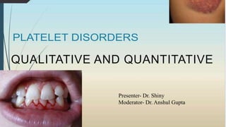
Presented by- Dr. Shiny
QUALITATIVE AND QUANTITATIVE
Presenter- Dr. Shiny
Moderator- Dr. Anshul Gupta
THROMBOCYTOPENIA
refers to decrease in the number of platelets in peripheral blood (<1.5 lacs/cmm).
may result from four main mechanisms-
Increased peripheral destruction of platelets
Decreased production of platelets in bone marrow
Dilutional thrombocytopenia
Sequestration in enlarged spleen.
Platelet counts between 50,000 and 20,000/cmm usually cause mild spontaneous bleeding.
Platelet count below 20,000/cmm is associated with risk of severe haemorrhage.
IDIOPATHIC THROMBOCYTOPENIC PURPURA
(ITP)
In ITP, autoantibodies or immune complexes bind to platelets and cause their premature
peripheral destruction.
Megakaryocytes are normal or increased in bone marrow.
 ITP occurs in two forms—acute and chronic.
Acute ITP is a short-lasting illness which occurs in children following viral infection or
vaccination.
Chronic ITP is an disorder of insidious onset with multiple remissions and relapses,
occurs predominantly in adult women, and is not preceded by infection or associated
with any underlying disease.
PATHOGENESIS
Acute ITP:
In acute ITP, immune complexes of viral antigens and host anti-viral antibodies
bind to Fc receptors on platelets that leads to immune destruction of platelets by
macrophages in spleen.
Alternatively, antiviral antibodies may cross-react with platelet antigens.
•Chronic ITP: In chronic ITP, autoantibodies are predominantly IgG.
•These antibodies are directed against specific platelet glycoproteins GpIIb/IIIa or
GpIb/IX in majority of patients. Thus, in addition to causing destruction of platelets,
these autoantibodies also induce platelet dysfunction by blocking GpIIb/IIIa
receptors.
•Antibody-coated platelets are recognised by Fc receptors on macrophages and
destroyed mainly in spleen.
CLINICAL FEATURES- ACUTE ITP
•Incidence of ITP increases with age.
•Clinical presentation varies from severe thrombocytopenia and bleeding to
asymptomatic mild thrombocytopenia detected incidentally.
•Acute ITP predominantly affects children between 2-6 yrs of age with sex ratio 1:1.
•The disease often follows viral respiratory infection or vaccination after an interval of 2
to 3 weeks.
•There is increased incidence during winter and spring.
•The disease starts suddenly with mucocutaneous membrane bleeding in the
form of purpuric spots and ecchymoses (especially on legs), bleeding from
gums, nose, gastrointestinal tract and hematuria.
•Intracranial hemorrhage though rare can be fatal.
•Spleen tip may be palpable but significant splenomegaly is unusual.
•The disease is self-limited and spontaneous complete remissions usually
occur within 2 to 6 weeks in more than 80% of patients.
CLINICAL FEATURES- CHRONIC
ITP
Chronic ITP occurs in young adults- more common in females (3F:1M).
There is an insidious onset of superficial bleeding from skin and mucous membrane;
menorrhagia in females.
Chronic bleeding can cause iron deficiency anaemia.
History of preceding viral infection or any underlying disease is lacking.
Spleen is not palpable in chronic ITP and in the presence of splenomegaly alternative
diagnosis should be considered.
Some patients have asymptomatic thrombocytopenia and are discovered incidentally
during routine blood counts.
New Terminology Proposed By
International Working Group (2009)
• The term immune thrombocytopenia should be used in place of immune
thrombocytopenic purpura as bleeding is absent in majority of patients
• Primary ITP is defined as an autoimmune disorder with isolated thrombocytopenia
(platelet count <100,000/cmm) without any underlying cause.
• Secondary ITP refers to all other forms of immune thrombocytopenia except
primary ITP, e.g. drug-induced; associated with Helicobacter pylori, Hep. C virus, or HIV
• Phases of ITP:
1. Newly diagnosed: First 3 months after diagnosis
2. Persistent ITP: 3 to 12 months from diagnosis
3. Chronic ITP: >12 months
EXAMINATION OF PERIPHERAL
BLOOD:
•Blood loss may lead to anemia.
•In children, lymphocytes and eosinophils are frequently increased.
•In acute ITP, platelets are markedly reduced (<20,000/cmm) while in chronic ITP
platelet count is variable (usually moderately low, i.e. around 50,000/cmm).
•Morphologically, platelets are frequently large(megathrombocytes).
•In chronic ITP, bleeding manifestations are frequently mild as compared to the degree of
thrombocytopenia; this is due to the presence of large, giant platelets in circulation,
which are functionally hyperactive.
•The number of large platelets is proportional to megakaryocyte number in marrow.
BONE MARROW EXAMINATION:
•In bone marrow, megakaryocytes are normal or increased in number and
frequently show morphological changes such as hypogranularity of
cytoplasm, vacuolisation, lack of platelet budding, nuclear non-lobulation or
hypolobulation and dense nuclear chromatin.
•These morphologic abnormalities are seen in any condition associated with
accelerated platelet destruction and are not specific for ITP.
•If clinical features, CBC, and blood smear are indicative of ITP, bone marrow
examination is not necessary for diagnosis of ITP.
Coagulation profile: Prolonged BT and deficient clot retraction are the
usual abnormalities.
Tests for blood coagulation are normal.
Platelet antibodies: Levels of platelet-associated immunoglobulins are raised in
majority (more than 90%) of patients with ITP.
This test, however, is neither sufficiently sensitive nor specific for ITP.
Therefore, it is not necessary for diagnosis.
DIFFERENTIAL DIAGNOSIS
Diagnosis of ITP is one of exclusion since there are no specific clinical or lab features.
In neonates and small children- maternal ITP, FNAIT, and inherited thrombocytopenia
 Drug-induced thrombocytopaenia, post transfusion isoimmune purpura by history.
Isolated thrombocytopenia in SLE.
Evans’ syndrome (immune hemolytic anemia+thrombocytopenia)
Autoimmune thrombocytopenia- lymphoma, chronic lymphocytic leukaemia
HIV is emerging as a common cause of thrombocytopenia
Hereditary thrombocytopenia (Bernard-Soulier syndrome, Wiskott-Aldrich syndrome).
DIAGNOSIS
Diagnosis of ITP is based on combination of following features:
• Mucocutaneous type of bleeding with abrupt onset (acute ITP) or insidious onset
(chronic ITP)
• No other abnormality on physical examination with patient otherwise being normal.
• Presence of isolated thrombocytopenia with no other abnormality on CBC.
• Bone marrow examination is usually normal.
• Exclusion of other causes of thrombocytopenia.
TREATMENT OF ITP
• Steroids: Prednisolone, Dexamethasone, Methyleprednisolone.
• Intravenous immunoglobulin (IVIG): The mechanism of action is probably
blockade of the reticuloendothelial system, preventing phagocytosis of platelets-
The dose is 0.5 to 1 g/kg daily for 2 or 3 days, repeated every 10 to 21 days as needed.
The platelet count usually begins to rise within 2 to 4 days
• Other immunosuppressive drugs: mycophenolate,azathioprine (imuran)
• Rituximab (Mabthera)
• Thrombopoiesis-stimulating agents
• Recombinant FVIIa
Second-line Management
• Splenectomy: traditional second-line treatment for many years. It remains the
most effective treatment with the highest rate of complete and durable remissions.
• Thrombopoiesis-stimulating agents: support the Platelet count as long as they
are continued, but do not induce remissions. Romiplostin, Eltrombopag.
NEONATAL ALLOIMMUNE
THROMBOCYTOPAENIA
When the foetal platelets possessing paternally derived antigens lacking in the mother
enter maternal circulation during gestation or delivery, formation of alloantibodies is
stimulated.
These maternal antibodies cross the placenta and cause destruction of foetal platelets.
Firstborn babies are also frequently affected.
The most common platelet antigen against which antibodies form is HPA-1a (PlAl).
The condition is self-limited and usually resolves by 3 weeks (maximum 3 months)
after delivery.
There is a risk of intracranial haemorrhage due to trauma during vaginal delivery.
In severe cases purpura and hemorrhages are evident at birth or manifest within a few hours.
Alloimmune neonatal thrombocytopaenia should be distinguished from other causes of
thrombocytopenia as described earlier.
POST-TRANSFUSION PURPURA
In this very rare but life-threatening disorder, sudden onset of thrombocytopenia and
bleeding occurs about 1 week to 10 days following blood transfusion in some adult
multiparous women.
In all cases, donor platelets possess HPA-1a antigen while this antigen is lacking on
patient’s platelets.
Patients are probably sensitized previously during pregnancy by foetal platelets having
HPA-1a antigen.
The condition is treated by intravenous gamma globulin and plasmapheresis.
THROMBOTIC
MICROANGIOPATHIES
Thrombotic microangiopathies are characterised by microvascular
thrombosis, microangiopathic haemolytic anaemia (MAHA), and
thrombocytopaenia.
Include-
1. Thrombotic thrombocytopenic purpura (TTP)
2. Haemolytic uraemic syndrome (HUS).
THROMBOTIC THROMBOCYTOPENIC
PURPURA
This uncommon disorder is characterized by formation of hyaline microthrombi
in microcirculation of various organs due to aggregation of platelets.
In idiopathic TTP, autoantibodies against A disintegrin and metalloprotease
with thrombospondin type 1 motif 13 lead to deficiency of ADAMTS13 and
accumulation of ultra-large vWF multimers that bind large number of platelets.
In familial TTP (Upshaw-Sohulman syndrome) ADAMTS13 deficiency results
from mutations in ADAMTS13 gene
The disorder mainly affects young adults and is slightly more common in females.
The pentad of manifestations includes:
(i) Microangiopathic haemolytic anaemia: Haemolysis of red cells results from
their passage across fibrin strands of microthrombi in circulation. Clinically
patients have pallor and frequently icterus.
PBF shows presence of fragmented and nucleated red cells and reticulocytosis
(ii) Bleeding manifestations secondary to severe thrombocytopaenia such as
petechiae, ecchymoses, epistaxis and gastrointestinal/genitourinary bleeding.
Coagulation studies (PT, APTT) are normal in most patients;
(iii) Fluctuating neurologic dysfunction such as altered level of consciousness,
seizures, coma
(iv) Renal abnormalities: proteinuria, haematuria, azotaemia;
(v) Fever.
Most patients respond to transfusions of fresh frozen plasma or to plasmapheresis.
HAEMOLYTIC UREMIC
SYNDROME
characterised by triad of acute renal failure, thrombocytopenia, and microangiopathic
haemolytic anaemia.
Classified into two types: typical and atypical.
Typical HUS occurs predominantly in children <5 years of age and is associated with
Shiga toxin-producing Escherichia coli O157:H7; it is characterised by a prodrome of
diarrhoeal illness followed by MAHA, thrombocytopaenia, and renal failure.
Atypical HUS has similar clinical features but is not preceded by diarrhoeal prodrome.
Thrombocytopenia Due To Increased
Platelet Sequestration Or Pooling
Normally about 30% of total platelets in the body are sequestered in the spleen.
In conditions associated with enlargement of spleen, splenic platelet pool expands
and may reach up to 90% in some cases.
Due to compensatory increase in platelet production in bone marrow,
thrombocytopenia is usually mild.
PSEUDOTHROMBOCYTOPE
NIA
When platelet counts are determined by electronic cell counters on blood
samples collected in EDTA, sometimes a falsely low result may be obtained.
Examination of a parallel peripheral blood smear made from EDTA
anticoagulated blood, however, reveals large clumps of platelets and platelets
rosetting around neutrophils.
The platelet clumping results from presence of EDTA-dependent antiplatelet
antibody in some patients.
EDTA alters the conformation of GpIIb/IIIa complex and exposes neoantigen.
The antibody reacts with this cryptic antigen and causes platelet clumping only in vitro.
These antibodies do not have any clinical significance.
Incorrect diagnosis of thrombocytopenia can be avoided by simultaneous examination of
peripheral blood film along with determination of direct platelet count.
THROMBOCYTOSIS
•This refers to increase in the platelet count above normal (> 4 lac/cmm).
•Primary Thrombocytosis- Thrombocytosis due to myeloproliferative disorders
•It can be usually distinguished from reactive (secondary) thrombocytosis by
the presence in the former of leucocytosis and immature white cells and
nucleated red cells in peripheral blood, defective platelet function (deficient
epinephrine-induced platelet aggregation) and splenomegaly.
Thrombocytosis in essential thrombocythemia is associated with
thromboembolic and bleeding manifestations.
In reactive thrombocytosis, platelet count is modestly elevated and has no
clinical significance.
However, persistent thrombocytosis following splenectomy for chronic
haemolytic anaemia may result in increased risk of thromboembolic
complications if haemolysis is not completely corrected.
DISORDERS OF PLATELET
FUNCTION
Disorders of platelet function are classified into two broad categories:
inherited and acquired
INHERITED DISORDERS OF PLATELET
FUNCTION-
BERNARD-SOULIER SYNDROME
•rare autosomal recessive congenital bleeding disorder.
•In this disease, adhesion of platelets to subendothelium is defective due to
congenital absence of glycoprotein Ib receptor complex (which consists of
GpIb, V and IX) on platelet surface.
•This receptor is essential for binding of platelets to subendothelium via vWF
•It is caused by defects in GP9,GP1BB and GP1BA genes.
•The haemorrhagic manifestations usually begin in infancy or early childhood.
•They are of moderate to marked degree and consist of purpuric spots, easy or
spontaneous bruising, and mucosal bleeding.
Characteristic laboratory abnormalities include-
giant platelets on peripheral blood smear
mild to moderate thrombocytopenia not proportional to the severity of bleeding,
abnormal platelet function studies (in the form of prolonged bleeding time, impaired
platelet aggregation with ristocetin not corrected by addition of normal plasma,
normal platelet aggregation with other agonists), and absence of GpIb, V, and IX.
No satisfactory form of therapy is available.
Severe haemorrhagic episodes are managed by platelet transfusions.
Repeated platelet transfusions can induce alloimmunisation and formation of
antibodies against glycoproteins that are absent.
GLANZMANN’S
THROMBASTHENIA
•In this very rare autosomal recessive bleeding disorder, platelet aggregation is
deficient due to absence of GpIIb/IIIa recepter complex on platelets.
•The disorder results from mutations in genes ITGB3 (located on 17q21.32) or
ITGA2B (located on 17q21.32).
•Normally upon activation of platelets, GpIIb/IIIa receptors become exposed
on platelet surface and serve as binding sites for fibrinogen. Fibrinogen
molecules form bridges between adjacent platelets during aggregation.
In Glanzmann’s thrombasthenia, the absence of fibrinogen binding due to lack of
receptors is responsible for deficient aggregation.
On PBF, platelets appear small and discrete (i.e. they are not in clumps due to lack of
aggregation)
platelet count is normal
platelet function studies are abnormal (in the form of prolonged bleeding time, poor
clot retraction, platelet aggregation is absent with ADP, epinephrine, collagen, and
arachidonic acid and normal with ristocetin), and lack of GpIIb/IIIa complex.
No effective form of therapy is available. Severe haemorrhages are treated by platelet
transfusions.
STORAGE POOL
DEFICIENCY
Deficiency of intracellular granules of platelets is referred to as storage pool
deficiency.
It may involve dense granules, alpha granules, or both.
Dense granule storage pool deficiency: This is the most common type of
hereditary platelet function disorder. Patients usually present with mild
mucocutaneous bleeding. Electron microscopy reveals absence of dense
granules. Intraplatelet levels of ADP, serotonin, and calcium are diminished.
Ristocetin-induced aggregation is normal.
Alpha granule storage pool deficiency (Gray platelet syndrome): In this condition,
which has been described in only a few patients, alpha granules and their contents
are diminished or absent.
These patients have a mild bleeding diathesis.
Platelets are mildly decreased in number, are large in size, and appear pale-grey on
stained blood smears.
Defective platelet aggregation has also been described.
Reticulin fibres are increased in bone marrow.
ACQUIRED DISORDERS OF PLATELET
FUNCTION
Drugs- Aspirin- results in inability of the platelets to synthesise thromboxane A2 and
failure of platelet secretion. This forms the basis of use of aspirin as anti-platelet
drug in practice.
Other drugs- NSAIDS, penicillin, cephalosporins, local anaesthetics, dipyridamole,
dextran, and heparin.
Myeloproliferative neoplasms
Paraproteinaemias- coat the platelet surface and inhibit adhesion and aggregation
Uraemia- prolongation of bleeding time and impaired platelet aggregation
REFERENCES
1. Essentials of Hematology 3rd edition, Kawthalkar
2. Textbook of Hematology, 4th edition, Tejindar Singh
3. https://www.slideshare.net/pediatricsmgmcri/platelet-disoders
4. https://www.slideshare.net/PrincessAlenAguilar/platelet-disorders-36549484
Platelet disorders- qualitative and quantitative

Mais conteúdo relacionado

Semelhante a Platelet disorders- qualitative and quantitative

CME thrombocytopenia
CME thrombocytopeniaCME thrombocytopenia
CME thrombocytopenia
Alaa Habbal
 
medicine.Bleeding disorders.(dr.sabir) (new powerpoint)
medicine.Bleeding disorders.(dr.sabir) (new powerpoint)medicine.Bleeding disorders.(dr.sabir) (new powerpoint)
medicine.Bleeding disorders.(dr.sabir) (new powerpoint)
student
 
Medicine.Bleeding disorders.(dr.sabir)
Medicine.Bleeding disorders.(dr.sabir)Medicine.Bleeding disorders.(dr.sabir)
Medicine.Bleeding disorders.(dr.sabir)
student
 

Semelhante a Platelet disorders- qualitative and quantitative (20)

ITP by dr. Mohib Ali
ITP by dr. Mohib AliITP by dr. Mohib Ali
ITP by dr. Mohib Ali
 
Hemolytic uremic syndrome
Hemolytic uremic syndrome Hemolytic uremic syndrome
Hemolytic uremic syndrome
 
plateletdisoders-160916040647.pptx
plateletdisoders-160916040647.pptxplateletdisoders-160916040647.pptx
plateletdisoders-160916040647.pptx
 
CME thrombocytopenia
CME thrombocytopeniaCME thrombocytopenia
CME thrombocytopenia
 
Platelet disorders
Platelet disordersPlatelet disorders
Platelet disorders
 
thrombo done1.ppt
thrombo done1.pptthrombo done1.ppt
thrombo done1.ppt
 
medicine.Bleeding disorders.(dr.sabir) (new powerpoint)
medicine.Bleeding disorders.(dr.sabir) (new powerpoint)medicine.Bleeding disorders.(dr.sabir) (new powerpoint)
medicine.Bleeding disorders.(dr.sabir) (new powerpoint)
 
Medicine 5th year, 10th lecture (Dr. Sabir)
Medicine 5th year, 10th lecture (Dr. Sabir)Medicine 5th year, 10th lecture (Dr. Sabir)
Medicine 5th year, 10th lecture (Dr. Sabir)
 
thrombo done2.ppt
thrombo done2.pptthrombo done2.ppt
thrombo done2.ppt
 
IDIOPATHIC THROMBOCYTOPENIC PURPURA (ITP)
IDIOPATHIC THROMBOCYTOPENIC PURPURA  (ITP)IDIOPATHIC THROMBOCYTOPENIC PURPURA  (ITP)
IDIOPATHIC THROMBOCYTOPENIC PURPURA (ITP)
 
Approach to thrombocytopenia
Approach to thrombocytopeniaApproach to thrombocytopenia
Approach to thrombocytopenia
 
Pediatric thrombocytopenia
Pediatric thrombocytopeniaPediatric thrombocytopenia
Pediatric thrombocytopenia
 
Itp
ItpItp
Itp
 
Immune Thrombocytopenia (ITP)
Immune Thrombocytopenia (ITP)Immune Thrombocytopenia (ITP)
Immune Thrombocytopenia (ITP)
 
idiopathic thrombocytopinic purpura
idiopathic thrombocytopinic purpuraidiopathic thrombocytopinic purpura
idiopathic thrombocytopinic purpura
 
Disseminated Intravascular Coagulation
Disseminated Intravascular CoagulationDisseminated Intravascular Coagulation
Disseminated Intravascular Coagulation
 
Medicine.Bleeding disorders.(dr.sabir)
Medicine.Bleeding disorders.(dr.sabir)Medicine.Bleeding disorders.(dr.sabir)
Medicine.Bleeding disorders.(dr.sabir)
 
Idiopathic thrombocytopenic purpura.pptx
Idiopathic thrombocytopenic purpura.pptxIdiopathic thrombocytopenic purpura.pptx
Idiopathic thrombocytopenic purpura.pptx
 
Thrombocytopenia
ThrombocytopeniaThrombocytopenia
Thrombocytopenia
 
Bleeding disorders
Bleeding disordersBleeding disorders
Bleeding disorders
 

Mais de DrShinyKajal

Mais de DrShinyKajal (20)

An Octogenarian Patient Of Intracapsular Neck Of Femur Fracture With Pre-oper...
An Octogenarian Patient Of Intracapsular Neck Of Femur Fracture With Pre-oper...An Octogenarian Patient Of Intracapsular Neck Of Femur Fracture With Pre-oper...
An Octogenarian Patient Of Intracapsular Neck Of Femur Fracture With Pre-oper...
 
A case presentation of Exchange Transfusion in new born infant with Neonatal ...
A case presentation of Exchange Transfusion in new born infant with Neonatal ...A case presentation of Exchange Transfusion in new born infant with Neonatal ...
A case presentation of Exchange Transfusion in new born infant with Neonatal ...
 
A Case presentation of Massive Transfusion in post LSCS PPH patient
A Case presentation of Massive Transfusion in post LSCS PPH patientA Case presentation of Massive Transfusion in post LSCS PPH patient
A Case presentation of Massive Transfusion in post LSCS PPH patient
 
case presentation on diagnosis of beta thalassemia major
case presentation on diagnosis of beta thalassemia majorcase presentation on diagnosis of beta thalassemia major
case presentation on diagnosis of beta thalassemia major
 
Troubleshooting in Transfusion transmissible infection TTI laboratory
Troubleshooting in Transfusion transmissible infection TTI laboratoryTroubleshooting in Transfusion transmissible infection TTI laboratory
Troubleshooting in Transfusion transmissible infection TTI laboratory
 
Case presentation on HEMOTHERAPY IN SHOCK- hypovolemia and hemorrhagic shock
Case presentation on HEMOTHERAPY IN SHOCK- hypovolemia and hemorrhagic shockCase presentation on HEMOTHERAPY IN SHOCK- hypovolemia and hemorrhagic shock
Case presentation on HEMOTHERAPY IN SHOCK- hypovolemia and hemorrhagic shock
 
Case presentation onHeterozygous variant of Hemoglobin E
Case presentation onHeterozygous variant of  Hemoglobin ECase presentation onHeterozygous variant of  Hemoglobin E
Case presentation onHeterozygous variant of Hemoglobin E
 
Case presentation on Antibody screening- how to solve 3 cell and 11 cell panel?
Case presentation on Antibody screening- how to solve 3 cell and 11 cell panel?Case presentation on Antibody screening- how to solve 3 cell and 11 cell panel?
Case presentation on Antibody screening- how to solve 3 cell and 11 cell panel?
 
Tissue Banking and Umbilical Cord Blood Banking
Tissue Banking and Umbilical Cord Blood BankingTissue Banking and Umbilical Cord Blood Banking
Tissue Banking and Umbilical Cord Blood Banking
 
Transfusion support in Surgery- elective surgery, cardiac surgery, MSBOS, Tra...
Transfusion support in Surgery- elective surgery, cardiac surgery, MSBOS, Tra...Transfusion support in Surgery- elective surgery, cardiac surgery, MSBOS, Tra...
Transfusion support in Surgery- elective surgery, cardiac surgery, MSBOS, Tra...
 
vin willebrand factor, disease and Hemophilia
vin willebrand factor, disease and Hemophiliavin willebrand factor, disease and Hemophilia
vin willebrand factor, disease and Hemophilia
 
Granulocyte antigens & antibodies and their role in transfusion medicinepptx
Granulocyte antigens & antibodies and their role in transfusion medicinepptxGranulocyte antigens & antibodies and their role in transfusion medicinepptx
Granulocyte antigens & antibodies and their role in transfusion medicinepptx
 
Adsorption and Elution- heal elution ether elution Autoadsorption alloadsorpt...
Adsorption and Elution- heal elution ether elution Autoadsorption alloadsorpt...Adsorption and Elution- heal elution ether elution Autoadsorption alloadsorpt...
Adsorption and Elution- heal elution ether elution Autoadsorption alloadsorpt...
 
NAT nucleic acid amplification technology Testing- Reverse transcription-poly...
NAT nucleic acid amplification technology Testing- Reverse transcription-poly...NAT nucleic acid amplification technology Testing- Reverse transcription-poly...
NAT nucleic acid amplification technology Testing- Reverse transcription-poly...
 
New Emerging Pathogens in blood and blood components transfusion
New Emerging Pathogens in blood and blood components transfusionNew Emerging Pathogens in blood and blood components transfusion
New Emerging Pathogens in blood and blood components transfusion
 
pathogen inactivation of cellular components.pptx
pathogen inactivation of cellular components.pptxpathogen inactivation of cellular components.pptx
pathogen inactivation of cellular components.pptx
 
1. Bio preservation of Red Cell Components 2. CULTURED RBCs 3. solvent plasma
1. Bio preservation of Red Cell Components 2. CULTURED RBCs 3. solvent plasma1. Bio preservation of Red Cell Components 2. CULTURED RBCs 3. solvent plasma
1. Bio preservation of Red Cell Components 2. CULTURED RBCs 3. solvent plasma
 
Storage of Blood Components- equipments, effects of improper storage, transpo...
Storage of Blood Components- equipments, effects of improper storage, transpo...Storage of Blood Components- equipments, effects of improper storage, transpo...
Storage of Blood Components- equipments, effects of improper storage, transpo...
 
Peripheral blood stem cell transplantation- sources mobilization preservation...
Peripheral blood stem cell transplantation- sources mobilization preservation...Peripheral blood stem cell transplantation- sources mobilization preservation...
Peripheral blood stem cell transplantation- sources mobilization preservation...
 
DONOR RECRUITMENT AND RETENTION STRATEGIES.pptx
DONOR RECRUITMENT AND RETENTION STRATEGIES.pptxDONOR RECRUITMENT AND RETENTION STRATEGIES.pptx
DONOR RECRUITMENT AND RETENTION STRATEGIES.pptx
 

Último

Circulation through Special Regions -characteristics and regulation
Circulation through Special Regions -characteristics and regulationCirculation through Special Regions -characteristics and regulation
Circulation through Special Regions -characteristics and regulation
MedicoseAcademics
 

Último (20)

Is preeclampsia and spontaneous preterm delivery associate with vascular and ...
Is preeclampsia and spontaneous preterm delivery associate with vascular and ...Is preeclampsia and spontaneous preterm delivery associate with vascular and ...
Is preeclampsia and spontaneous preterm delivery associate with vascular and ...
 
DIGITAL RADIOGRAPHY-SABBU KHATOON .pptx
DIGITAL RADIOGRAPHY-SABBU KHATOON  .pptxDIGITAL RADIOGRAPHY-SABBU KHATOON  .pptx
DIGITAL RADIOGRAPHY-SABBU KHATOON .pptx
 
Antiplatelets in IHD, Dose Duration, DAPT vs SAPT
Antiplatelets in IHD, Dose Duration, DAPT vs SAPTAntiplatelets in IHD, Dose Duration, DAPT vs SAPT
Antiplatelets in IHD, Dose Duration, DAPT vs SAPT
 
In-service education (Nursing Mangement)
In-service education (Nursing Mangement)In-service education (Nursing Mangement)
In-service education (Nursing Mangement)
 
Relationship between vascular system disfunction, neurofluid flow and Alzheim...
Relationship between vascular system disfunction, neurofluid flow and Alzheim...Relationship between vascular system disfunction, neurofluid flow and Alzheim...
Relationship between vascular system disfunction, neurofluid flow and Alzheim...
 
Scientificity and feasibility study of non-invasive central arterial pressure...
Scientificity and feasibility study of non-invasive central arterial pressure...Scientificity and feasibility study of non-invasive central arterial pressure...
Scientificity and feasibility study of non-invasive central arterial pressure...
 
Effects of vaping e-cigarettes on arterial health
Effects of vaping e-cigarettes on arterial healthEffects of vaping e-cigarettes on arterial health
Effects of vaping e-cigarettes on arterial health
 
Final CAPNOCYTOPHAGA INFECTION by Gauri Gawande.pptx
Final CAPNOCYTOPHAGA INFECTION by Gauri Gawande.pptxFinal CAPNOCYTOPHAGA INFECTION by Gauri Gawande.pptx
Final CAPNOCYTOPHAGA INFECTION by Gauri Gawande.pptx
 
Cervical screening – taking care of your health flipchart (Vietnamese)
Cervical screening – taking care of your health flipchart (Vietnamese)Cervical screening – taking care of your health flipchart (Vietnamese)
Cervical screening – taking care of your health flipchart (Vietnamese)
 
CURRENT HEALTH PROBLEMS AND ITS SOLUTION BY AYURVEDA.pptx
CURRENT HEALTH PROBLEMS AND ITS SOLUTION BY AYURVEDA.pptxCURRENT HEALTH PROBLEMS AND ITS SOLUTION BY AYURVEDA.pptx
CURRENT HEALTH PROBLEMS AND ITS SOLUTION BY AYURVEDA.pptx
 
𝕔ALL #𝕘IRLS Service in Jaipur %(8901183002)# Jaipur #𝕔ALL #𝕘IRLS
𝕔ALL #𝕘IRLS Service in Jaipur %(8901183002)#  Jaipur #𝕔ALL #𝕘IRLS𝕔ALL #𝕘IRLS Service in Jaipur %(8901183002)#  Jaipur #𝕔ALL #𝕘IRLS
𝕔ALL #𝕘IRLS Service in Jaipur %(8901183002)# Jaipur #𝕔ALL #𝕘IRLS
 
ANATOMY OF THE LOWER URINARY TRACT AND MALE [Autosaved] [Autosaved].pptx
ANATOMY OF THE LOWER URINARY TRACT AND MALE [Autosaved] [Autosaved].pptxANATOMY OF THE LOWER URINARY TRACT AND MALE [Autosaved] [Autosaved].pptx
ANATOMY OF THE LOWER URINARY TRACT AND MALE [Autosaved] [Autosaved].pptx
 
Aptopadesha Pramana / Pariksha: The Verbal Testimony
Aptopadesha Pramana / Pariksha: The Verbal TestimonyAptopadesha Pramana / Pariksha: The Verbal Testimony
Aptopadesha Pramana / Pariksha: The Verbal Testimony
 
Gauri Gawande(9) Constipation Final.pptx
Gauri Gawande(9) Constipation Final.pptxGauri Gawande(9) Constipation Final.pptx
Gauri Gawande(9) Constipation Final.pptx
 
Compare home pulse pressure components collected directly from home
Compare home pulse pressure components collected directly from homeCompare home pulse pressure components collected directly from home
Compare home pulse pressure components collected directly from home
 
A thorough review of supernormal conduction.pptx
A thorough review of supernormal conduction.pptxA thorough review of supernormal conduction.pptx
A thorough review of supernormal conduction.pptx
 
Why invest into infodemic management in health emergencies
Why invest into infodemic management in health emergenciesWhy invest into infodemic management in health emergencies
Why invest into infodemic management in health emergencies
 
Vaccines: A Powerful and Cost-Effective Tool Protecting Americans Against Dis...
Vaccines: A Powerful and Cost-Effective Tool Protecting Americans Against Dis...Vaccines: A Powerful and Cost-Effective Tool Protecting Americans Against Dis...
Vaccines: A Powerful and Cost-Effective Tool Protecting Americans Against Dis...
 
Circulation through Special Regions -characteristics and regulation
Circulation through Special Regions -characteristics and regulationCirculation through Special Regions -characteristics and regulation
Circulation through Special Regions -characteristics and regulation
 
Young at heart: Cardiovascular health stations to empower healthy lifestyle b...
Young at heart: Cardiovascular health stations to empower healthy lifestyle b...Young at heart: Cardiovascular health stations to empower healthy lifestyle b...
Young at heart: Cardiovascular health stations to empower healthy lifestyle b...
 

Platelet disorders- qualitative and quantitative

  • 1. Presented by- Dr. Shiny QUALITATIVE AND QUANTITATIVE Presenter- Dr. Shiny Moderator- Dr. Anshul Gupta
  • 2.
  • 3.
  • 4.
  • 5.
  • 6.
  • 7. THROMBOCYTOPENIA refers to decrease in the number of platelets in peripheral blood (<1.5 lacs/cmm). may result from four main mechanisms- Increased peripheral destruction of platelets Decreased production of platelets in bone marrow Dilutional thrombocytopenia Sequestration in enlarged spleen. Platelet counts between 50,000 and 20,000/cmm usually cause mild spontaneous bleeding. Platelet count below 20,000/cmm is associated with risk of severe haemorrhage.
  • 8.
  • 9.
  • 10.
  • 11. IDIOPATHIC THROMBOCYTOPENIC PURPURA (ITP) In ITP, autoantibodies or immune complexes bind to platelets and cause their premature peripheral destruction. Megakaryocytes are normal or increased in bone marrow.  ITP occurs in two forms—acute and chronic. Acute ITP is a short-lasting illness which occurs in children following viral infection or vaccination. Chronic ITP is an disorder of insidious onset with multiple remissions and relapses, occurs predominantly in adult women, and is not preceded by infection or associated with any underlying disease.
  • 12.
  • 13. PATHOGENESIS Acute ITP: In acute ITP, immune complexes of viral antigens and host anti-viral antibodies bind to Fc receptors on platelets that leads to immune destruction of platelets by macrophages in spleen. Alternatively, antiviral antibodies may cross-react with platelet antigens.
  • 14. •Chronic ITP: In chronic ITP, autoantibodies are predominantly IgG. •These antibodies are directed against specific platelet glycoproteins GpIIb/IIIa or GpIb/IX in majority of patients. Thus, in addition to causing destruction of platelets, these autoantibodies also induce platelet dysfunction by blocking GpIIb/IIIa receptors. •Antibody-coated platelets are recognised by Fc receptors on macrophages and destroyed mainly in spleen.
  • 15.
  • 16. CLINICAL FEATURES- ACUTE ITP •Incidence of ITP increases with age. •Clinical presentation varies from severe thrombocytopenia and bleeding to asymptomatic mild thrombocytopenia detected incidentally. •Acute ITP predominantly affects children between 2-6 yrs of age with sex ratio 1:1. •The disease often follows viral respiratory infection or vaccination after an interval of 2 to 3 weeks. •There is increased incidence during winter and spring.
  • 17. •The disease starts suddenly with mucocutaneous membrane bleeding in the form of purpuric spots and ecchymoses (especially on legs), bleeding from gums, nose, gastrointestinal tract and hematuria. •Intracranial hemorrhage though rare can be fatal. •Spleen tip may be palpable but significant splenomegaly is unusual. •The disease is self-limited and spontaneous complete remissions usually occur within 2 to 6 weeks in more than 80% of patients.
  • 18. CLINICAL FEATURES- CHRONIC ITP Chronic ITP occurs in young adults- more common in females (3F:1M). There is an insidious onset of superficial bleeding from skin and mucous membrane; menorrhagia in females. Chronic bleeding can cause iron deficiency anaemia. History of preceding viral infection or any underlying disease is lacking. Spleen is not palpable in chronic ITP and in the presence of splenomegaly alternative diagnosis should be considered. Some patients have asymptomatic thrombocytopenia and are discovered incidentally during routine blood counts.
  • 19. New Terminology Proposed By International Working Group (2009) • The term immune thrombocytopenia should be used in place of immune thrombocytopenic purpura as bleeding is absent in majority of patients • Primary ITP is defined as an autoimmune disorder with isolated thrombocytopenia (platelet count <100,000/cmm) without any underlying cause. • Secondary ITP refers to all other forms of immune thrombocytopenia except primary ITP, e.g. drug-induced; associated with Helicobacter pylori, Hep. C virus, or HIV • Phases of ITP: 1. Newly diagnosed: First 3 months after diagnosis 2. Persistent ITP: 3 to 12 months from diagnosis 3. Chronic ITP: >12 months
  • 20. EXAMINATION OF PERIPHERAL BLOOD: •Blood loss may lead to anemia. •In children, lymphocytes and eosinophils are frequently increased. •In acute ITP, platelets are markedly reduced (<20,000/cmm) while in chronic ITP platelet count is variable (usually moderately low, i.e. around 50,000/cmm). •Morphologically, platelets are frequently large(megathrombocytes). •In chronic ITP, bleeding manifestations are frequently mild as compared to the degree of thrombocytopenia; this is due to the presence of large, giant platelets in circulation, which are functionally hyperactive. •The number of large platelets is proportional to megakaryocyte number in marrow.
  • 21.
  • 22. BONE MARROW EXAMINATION: •In bone marrow, megakaryocytes are normal or increased in number and frequently show morphological changes such as hypogranularity of cytoplasm, vacuolisation, lack of platelet budding, nuclear non-lobulation or hypolobulation and dense nuclear chromatin. •These morphologic abnormalities are seen in any condition associated with accelerated platelet destruction and are not specific for ITP. •If clinical features, CBC, and blood smear are indicative of ITP, bone marrow examination is not necessary for diagnosis of ITP.
  • 23. Coagulation profile: Prolonged BT and deficient clot retraction are the usual abnormalities. Tests for blood coagulation are normal. Platelet antibodies: Levels of platelet-associated immunoglobulins are raised in majority (more than 90%) of patients with ITP. This test, however, is neither sufficiently sensitive nor specific for ITP. Therefore, it is not necessary for diagnosis.
  • 24.
  • 25. DIFFERENTIAL DIAGNOSIS Diagnosis of ITP is one of exclusion since there are no specific clinical or lab features. In neonates and small children- maternal ITP, FNAIT, and inherited thrombocytopenia  Drug-induced thrombocytopaenia, post transfusion isoimmune purpura by history. Isolated thrombocytopenia in SLE. Evans’ syndrome (immune hemolytic anemia+thrombocytopenia) Autoimmune thrombocytopenia- lymphoma, chronic lymphocytic leukaemia HIV is emerging as a common cause of thrombocytopenia Hereditary thrombocytopenia (Bernard-Soulier syndrome, Wiskott-Aldrich syndrome).
  • 26. DIAGNOSIS Diagnosis of ITP is based on combination of following features: • Mucocutaneous type of bleeding with abrupt onset (acute ITP) or insidious onset (chronic ITP) • No other abnormality on physical examination with patient otherwise being normal. • Presence of isolated thrombocytopenia with no other abnormality on CBC. • Bone marrow examination is usually normal. • Exclusion of other causes of thrombocytopenia.
  • 27.
  • 28. TREATMENT OF ITP • Steroids: Prednisolone, Dexamethasone, Methyleprednisolone. • Intravenous immunoglobulin (IVIG): The mechanism of action is probably blockade of the reticuloendothelial system, preventing phagocytosis of platelets- The dose is 0.5 to 1 g/kg daily for 2 or 3 days, repeated every 10 to 21 days as needed. The platelet count usually begins to rise within 2 to 4 days
  • 29. • Other immunosuppressive drugs: mycophenolate,azathioprine (imuran) • Rituximab (Mabthera) • Thrombopoiesis-stimulating agents • Recombinant FVIIa Second-line Management • Splenectomy: traditional second-line treatment for many years. It remains the most effective treatment with the highest rate of complete and durable remissions. • Thrombopoiesis-stimulating agents: support the Platelet count as long as they are continued, but do not induce remissions. Romiplostin, Eltrombopag.
  • 30. NEONATAL ALLOIMMUNE THROMBOCYTOPAENIA When the foetal platelets possessing paternally derived antigens lacking in the mother enter maternal circulation during gestation or delivery, formation of alloantibodies is stimulated. These maternal antibodies cross the placenta and cause destruction of foetal platelets. Firstborn babies are also frequently affected. The most common platelet antigen against which antibodies form is HPA-1a (PlAl). The condition is self-limited and usually resolves by 3 weeks (maximum 3 months) after delivery.
  • 31. There is a risk of intracranial haemorrhage due to trauma during vaginal delivery. In severe cases purpura and hemorrhages are evident at birth or manifest within a few hours. Alloimmune neonatal thrombocytopaenia should be distinguished from other causes of thrombocytopenia as described earlier.
  • 32. POST-TRANSFUSION PURPURA In this very rare but life-threatening disorder, sudden onset of thrombocytopenia and bleeding occurs about 1 week to 10 days following blood transfusion in some adult multiparous women. In all cases, donor platelets possess HPA-1a antigen while this antigen is lacking on patient’s platelets. Patients are probably sensitized previously during pregnancy by foetal platelets having HPA-1a antigen. The condition is treated by intravenous gamma globulin and plasmapheresis.
  • 33. THROMBOTIC MICROANGIOPATHIES Thrombotic microangiopathies are characterised by microvascular thrombosis, microangiopathic haemolytic anaemia (MAHA), and thrombocytopaenia. Include- 1. Thrombotic thrombocytopenic purpura (TTP) 2. Haemolytic uraemic syndrome (HUS).
  • 34. THROMBOTIC THROMBOCYTOPENIC PURPURA This uncommon disorder is characterized by formation of hyaline microthrombi in microcirculation of various organs due to aggregation of platelets. In idiopathic TTP, autoantibodies against A disintegrin and metalloprotease with thrombospondin type 1 motif 13 lead to deficiency of ADAMTS13 and accumulation of ultra-large vWF multimers that bind large number of platelets. In familial TTP (Upshaw-Sohulman syndrome) ADAMTS13 deficiency results from mutations in ADAMTS13 gene
  • 35. The disorder mainly affects young adults and is slightly more common in females. The pentad of manifestations includes: (i) Microangiopathic haemolytic anaemia: Haemolysis of red cells results from their passage across fibrin strands of microthrombi in circulation. Clinically patients have pallor and frequently icterus. PBF shows presence of fragmented and nucleated red cells and reticulocytosis (ii) Bleeding manifestations secondary to severe thrombocytopaenia such as petechiae, ecchymoses, epistaxis and gastrointestinal/genitourinary bleeding.
  • 36. Coagulation studies (PT, APTT) are normal in most patients; (iii) Fluctuating neurologic dysfunction such as altered level of consciousness, seizures, coma (iv) Renal abnormalities: proteinuria, haematuria, azotaemia; (v) Fever. Most patients respond to transfusions of fresh frozen plasma or to plasmapheresis.
  • 37.
  • 38. HAEMOLYTIC UREMIC SYNDROME characterised by triad of acute renal failure, thrombocytopenia, and microangiopathic haemolytic anaemia. Classified into two types: typical and atypical. Typical HUS occurs predominantly in children <5 years of age and is associated with Shiga toxin-producing Escherichia coli O157:H7; it is characterised by a prodrome of diarrhoeal illness followed by MAHA, thrombocytopaenia, and renal failure. Atypical HUS has similar clinical features but is not preceded by diarrhoeal prodrome.
  • 39.
  • 40. Thrombocytopenia Due To Increased Platelet Sequestration Or Pooling Normally about 30% of total platelets in the body are sequestered in the spleen. In conditions associated with enlargement of spleen, splenic platelet pool expands and may reach up to 90% in some cases. Due to compensatory increase in platelet production in bone marrow, thrombocytopenia is usually mild.
  • 41. PSEUDOTHROMBOCYTOPE NIA When platelet counts are determined by electronic cell counters on blood samples collected in EDTA, sometimes a falsely low result may be obtained. Examination of a parallel peripheral blood smear made from EDTA anticoagulated blood, however, reveals large clumps of platelets and platelets rosetting around neutrophils. The platelet clumping results from presence of EDTA-dependent antiplatelet antibody in some patients.
  • 42. EDTA alters the conformation of GpIIb/IIIa complex and exposes neoantigen. The antibody reacts with this cryptic antigen and causes platelet clumping only in vitro. These antibodies do not have any clinical significance. Incorrect diagnosis of thrombocytopenia can be avoided by simultaneous examination of peripheral blood film along with determination of direct platelet count.
  • 43.
  • 44.
  • 45. THROMBOCYTOSIS •This refers to increase in the platelet count above normal (> 4 lac/cmm). •Primary Thrombocytosis- Thrombocytosis due to myeloproliferative disorders •It can be usually distinguished from reactive (secondary) thrombocytosis by the presence in the former of leucocytosis and immature white cells and nucleated red cells in peripheral blood, defective platelet function (deficient epinephrine-induced platelet aggregation) and splenomegaly.
  • 46. Thrombocytosis in essential thrombocythemia is associated with thromboembolic and bleeding manifestations. In reactive thrombocytosis, platelet count is modestly elevated and has no clinical significance. However, persistent thrombocytosis following splenectomy for chronic haemolytic anaemia may result in increased risk of thromboembolic complications if haemolysis is not completely corrected.
  • 47.
  • 48.
  • 49. DISORDERS OF PLATELET FUNCTION Disorders of platelet function are classified into two broad categories: inherited and acquired
  • 50.
  • 51. INHERITED DISORDERS OF PLATELET FUNCTION- BERNARD-SOULIER SYNDROME •rare autosomal recessive congenital bleeding disorder. •In this disease, adhesion of platelets to subendothelium is defective due to congenital absence of glycoprotein Ib receptor complex (which consists of GpIb, V and IX) on platelet surface. •This receptor is essential for binding of platelets to subendothelium via vWF •It is caused by defects in GP9,GP1BB and GP1BA genes.
  • 52. •The haemorrhagic manifestations usually begin in infancy or early childhood. •They are of moderate to marked degree and consist of purpuric spots, easy or spontaneous bruising, and mucosal bleeding. Characteristic laboratory abnormalities include- giant platelets on peripheral blood smear mild to moderate thrombocytopenia not proportional to the severity of bleeding, abnormal platelet function studies (in the form of prolonged bleeding time, impaired platelet aggregation with ristocetin not corrected by addition of normal plasma, normal platelet aggregation with other agonists), and absence of GpIb, V, and IX.
  • 53. No satisfactory form of therapy is available. Severe haemorrhagic episodes are managed by platelet transfusions. Repeated platelet transfusions can induce alloimmunisation and formation of antibodies against glycoproteins that are absent.
  • 54.
  • 55. GLANZMANN’S THROMBASTHENIA •In this very rare autosomal recessive bleeding disorder, platelet aggregation is deficient due to absence of GpIIb/IIIa recepter complex on platelets. •The disorder results from mutations in genes ITGB3 (located on 17q21.32) or ITGA2B (located on 17q21.32). •Normally upon activation of platelets, GpIIb/IIIa receptors become exposed on platelet surface and serve as binding sites for fibrinogen. Fibrinogen molecules form bridges between adjacent platelets during aggregation.
  • 56. In Glanzmann’s thrombasthenia, the absence of fibrinogen binding due to lack of receptors is responsible for deficient aggregation. On PBF, platelets appear small and discrete (i.e. they are not in clumps due to lack of aggregation) platelet count is normal platelet function studies are abnormal (in the form of prolonged bleeding time, poor clot retraction, platelet aggregation is absent with ADP, epinephrine, collagen, and arachidonic acid and normal with ristocetin), and lack of GpIIb/IIIa complex. No effective form of therapy is available. Severe haemorrhages are treated by platelet transfusions.
  • 57. STORAGE POOL DEFICIENCY Deficiency of intracellular granules of platelets is referred to as storage pool deficiency. It may involve dense granules, alpha granules, or both. Dense granule storage pool deficiency: This is the most common type of hereditary platelet function disorder. Patients usually present with mild mucocutaneous bleeding. Electron microscopy reveals absence of dense granules. Intraplatelet levels of ADP, serotonin, and calcium are diminished. Ristocetin-induced aggregation is normal.
  • 58. Alpha granule storage pool deficiency (Gray platelet syndrome): In this condition, which has been described in only a few patients, alpha granules and their contents are diminished or absent. These patients have a mild bleeding diathesis. Platelets are mildly decreased in number, are large in size, and appear pale-grey on stained blood smears. Defective platelet aggregation has also been described. Reticulin fibres are increased in bone marrow.
  • 59. ACQUIRED DISORDERS OF PLATELET FUNCTION Drugs- Aspirin- results in inability of the platelets to synthesise thromboxane A2 and failure of platelet secretion. This forms the basis of use of aspirin as anti-platelet drug in practice. Other drugs- NSAIDS, penicillin, cephalosporins, local anaesthetics, dipyridamole, dextran, and heparin. Myeloproliferative neoplasms Paraproteinaemias- coat the platelet surface and inhibit adhesion and aggregation Uraemia- prolongation of bleeding time and impaired platelet aggregation
  • 60.
  • 61.
  • 62. REFERENCES 1. Essentials of Hematology 3rd edition, Kawthalkar 2. Textbook of Hematology, 4th edition, Tejindar Singh 3. https://www.slideshare.net/pediatricsmgmcri/platelet-disoders 4. https://www.slideshare.net/PrincessAlenAguilar/platelet-disorders-36549484