PATHway to Decoding the Impact of Cancer Immunotherapy: Latest Advances in Biomarker Testing and Pathologic Response Assessment
•
0 gostou•4 visualizações

Co-Chairs Kurt A. Schalper, MD, PhD, and Vamsidhar Velcheti, MD, prepared useful Practice Aids pertaining to solid tumor for this CME/MOC activity titled “PATHway to Decoding the Impact of Cancer Immunotherapy: Latest Advances in Biomarker Testing and Pathologic Response Assessment.” For the full presentation, downloadable Practice Aids, and complete CME/MOC information, and to apply for credit, please visit us at https://bit.ly/3SsmiMV. CME/MOC credit will be available until February 14, 2025.

Recomendados
Recomendados
Mais conteúdo relacionado
Semelhante a PATHway to Decoding the Impact of Cancer Immunotherapy: Latest Advances in Biomarker Testing and Pathologic Response Assessment
Semelhante a PATHway to Decoding the Impact of Cancer Immunotherapy: Latest Advances in Biomarker Testing and Pathologic Response Assessment (20)
Mais de PVI, PeerView Institute for Medical Education
Mais de PVI, PeerView Institute for Medical Education (20)
Último
Último (20)
PATHway to Decoding the Impact of Cancer Immunotherapy: Latest Advances in Biomarker Testing and Pathologic Response Assessment
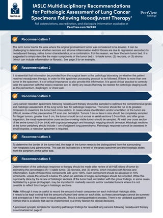
- 1. IASLC Multidisciplinary Recommendations for Pathologic Assessment of Lung Cancer Specimens Following Neoadjuvant Therapy1 Full abbreviations, accreditation, and disclosure information available at PeerView.com/GZR40 Recommendation 1 The term tumor bed is the area where the original pretreatment tumor was considered to be located. It can be challenging to determine whether necrosis and stromal inflammation and/or fibrosis are due to regression secondary to neoadjuvant therapy, native tumor characteristics, or a combination. For this reason, we favor the term tumor bed. It is suggested to simply describe the major components of the tumor bed: (1) viable tumor, (2) necrosis, or (3) stroma (which can include inflammation or fibrosis). See page 3 for an example. Recommendation 2 It is essential that information be provided from the surgical team to the pathology laboratory on whether the patient received neoadjuvant therapy, in order for this specimen processing protocol to be followed. If there is more than one tumor in the specimen, it is of critical importance to also provide this information. It is good clinical practice to correctly label the specimen with the lobe(s) resected and to clarify any issues that may be needed for pathologic staging such as the pericardium, diaphragm, or chest wall. Recommendation 3 Lung cancer resection specimens following neoadjuvant therapy should be sampled to optimize the comprehensive gross and histologic assessment of the lung tumor bed for pathologic response. The tumor should be cut in its greatest dimension to maximize the tumor bed cross section. In cases where identification and/or orientation of the tumor are difficult, review of the preoperative CT scan can be helpful. Tumors 3 cm or less in size should be completely sampled. For larger tumors, greater than 3 cm, the tumor should be cut across in serial sections 0.5-cm thick, and after gross inspection, the most representative cross section showing viable tumor should be sampled. At least one cross section of the entire tumor (0.5-cm thick) with a gross photograph and histologic mapping should be made. Histologic sections at the tumor periphery should include 1 cm of adjacent lung parenchyma. Pathologic response cannot be assessed in small biopsies; a resection specimen is required. Recommendation 4 To determine the border of the tumor bed, the edge of the tumor needs to be distinguished from the surrounding non-neoplastic lung parenchyma. This can be facilitated by a review of the gross specimen and the histologic slides from the periphery of the tumor bed. Recommendation 5 Determination of the pathologic response to therapy should be made after review of all H&E slides of tumor by estimating the percentages of (1) viable tumor, (2) necrosis, and (3) stroma, which includes both fibrosis and inflammation. Each of these three components add up to 100%. Each component should be assessed in 10% increments, unless the amount is below 5% when an estimate of single percentages should be recorded. While this is primarily done by the review of histologic sections of the tumor bed, correlation with the gross findings, in some cases facilitated by a gross photograph, may be important in markedly necrotic and/or cavitated tumors where it is not possible to reflect this change in histologic sections. Note: Although it may be useful to record the amount of each component on each individual histologic slide, it needs to be kept in mind that the amount of tumor bed varies on each slide, so these percentages cannot be summed and averaged as if they were in equal amounts. This is a semiquantitative process. There is no validated quantitative method that is available that can be implemented in a timely fashion for clinical decisions. A proposed synoptic template for reporting pathologic findings for resected lung cancers following neoadjuvant therapy is summarized on page 2.
- 2. Recommended Synoptic Template for Recording Lung Cancers Following Neoadjuvant Therapy This page has been designed for your use. Please feel free to print and use it in your practice. Primary tumor Type of neoadjuvant therapy • No known presurgical therapy: _____ • Type of neoadjuvant therapy – Chemotherapy: ____________________ – Radiotherapy: ____________________ – Immunotherapy (please specify): ____________________ – TKI (please specify): ____________________ – Other (please specify): ____________________ Treatment effect in primary tumor • Percentage of viable tumor (record in 10% increments except below 10%; then record single digits between 1%-5%): _____ • No residual viable tumor identified: _____ • Percentage of necrosis: _____ • Percentage of stroma (includes fibrosis and inflammation): _____ Grade of inflammation (choose the appropriate grade) _____ Mild _____ Moderate _____ Marked Method (choose what was used for evaluation) • Correlation was made with a gross photograph of tumor cut surface: Yes _____ No _____ • Evaluation was aided by use of tumor mapping to match a gross photograph to histologic sections: Yes _____ No _____ • Evaluation was aided by radiologic pathologic correlation: Yes _____ No _____ Treatment effect in lymph node metastases • Total number of lymph node stations examined: _____ • Total number of lymph nodes examined: _____ • No carcinoma present: _____ • Total number of lymph nodes with metastatic carcinoma: _____ • Lymph node stations involved by tumor with treatment-related changes: _____ • Lymph node stations with treatment-related changes without viable tumor: _____ • Largest tumor focus: mm at station number: _____ • Extracapsular extension present: _____ • No extracapsular extension (comment): __________________________________________________________________ Comments: ___________________________________________________________________________________________ _____________________________________________________________________________________________________ _____________________________________________________________________________________________________ _____________________________________________________________________________________________________
- 3. IASLC Multidisciplinary Recommendations for Pathologic Assessment of Lung Cancer Specimens Following Neoadjuvant Therapy1 Full abbreviations, accreditation, and disclosure information available at PeerView.com/GZR40 Histologic Components of the Tumor Bed (A) Schematic image showing how percentage compositions are assigned. The tumor bed is divided into viable tumor area, necrosis, and stroma. Stroma includes inflammation and fibrosis. (B) A representative hematoxylin-and-eosin stained slide image (left) and a corresponding color illustration of the distribution of the components (right). The blue, red, and green areas represent viable tumor, necrosis, and stroma, respectively. Tumor bed = X + Y + Z = 100% Viable tumor (X%) Necrosis (Y%) Stroma (Z%) Viable tumor = 50% Necrosis = 40% Stroma = 10% A B Tumor bed
- 4. IASLC Multidisciplinary Recommendations for Pathologic Assessment of Lung Cancer Specimens Following Neoadjuvant Therapy1 Full abbreviations, accreditation, and disclosure information available at PeerView.com/GZR40 Recommendation 6 Definition of major pathologic response (MPR) MPR is defined as the reduction of viable tumor to the amount beneath an established clinically significant cutoff based on prior evidence according to the individual histologic type of lung cancer and a specific therapy. The historical definition of MPR for all histologic types of lung cancer is ≤10% of viable tumor with no viable tumor required for pathologic complete response (pCR). MPR is calculated as the estimated size of viable tumor divided by the size of the tumor bed. For the moment, this is the cutoff being used in multiple active clinical trials. However, recent data suggests the MPR in the conventional chemotherapy setting may differ according to histologic type: ie, adenocarcinoma versus squamous cell carcinoma. If after review of histologic sections, the percentage of viable tumor is near the cutoff for MPR, additional histologic sections should be submitted. The pathology report should record the total number of blocks of tumor bed that were examined even if the blocks did not consist entirely of tumor bed but also included some uninvolved lung. For colloid adenocarcinomas, where tumor cells are only focal, the mucin pools should be included in the percentage of viable tumor. However, if there are only areas of extracellular mucin without any apparent viable tumor cells within the mucin, we suggest regarding this as stroma. Further study is needed to address this point. MPR can also be classified for the lung primary in the setting where the lung primary shows little or no viable tumor, but lymph nodes show viable metastatic carcinoma (ypT0, N1, 2 or 3). However, the prognostic and therapeutic implications of this clinical setting are not known. Recommendation 7 Definition of pathologic complete response (pCR) pCR is defined as the lack of any viable tumor cells on review of H&E slides after complete evaluation of a resected lung cancer specimen including all sampled regional lymph nodes. Such tumors would be staged as ypT0N0, according to the 8th edition AJCC and UICC staging systems. Note If no tumor is seen in the initial sections and tissue from the tumor bed remains, additional histologic sections should be made. The number of additional sections should be whatever seems reasonable in the individual setting depending on the size of the tumor bed and the capacity of the individual pathology laboratory. If the histologic changes in the initial sections obtained do not show findings that fit for the effects of therapy, the possibility that the wrong area was sampled should be considered. In such cases the gross specimen may need to be re-evaluated using radiologic pathologic correlation and if additional lesions are identified, these should be sampled. The pathology report should record the total number of blocks of tumor bed that were examined even if the entire block did not consist of tumor bed. The identification of incidental lesions of squamous cell carcinoma in situ, atypical adenomatous hyperplasia, adenocarcinoma in situ, or minimally invasive adenocarcinoma in the surrounding lung parenchyma that are clearly separate from the main tumor for which neoadjuvant therapy was administered does not disqualify a case for classification as MPR or CPR. This proposal is based on clinical judgement, as currently no clinical data exist to make a specific recommendation. In the setting of multiple tumors where a second invasive predominant lung carcinoma is present that was regarded preoperatively to be an intrapulmonary metastasis but determined to be a second synchronous primary after clinical, radiologic, pathologic, and/or molecular assessment, it is questionable whether the terms MPR or CPR should be used if the main tumor otherwise meets the above criteria. No data exists currently to address this question.
- 5. IASLC Multidisciplinary Recommendations for Pathologic Assessment of Lung Cancer Specimens Following Neoadjuvant Therapy1 Full abbreviations, accreditation, and disclosure information available at PeerView.com/GZR40 Recommendation 8 In the absence of more systemic data regarding the evaluation of tumors following immunotherapy and molecular targeted therapy, the same approach to pathologic assessment of resected lung cancers in the neoadjuvant setting in evaluating the percentage of viable tumor, necrosis, and stroma should be used, regardless of the type of neoadjuvant therapy administered whether it was radiation, chemotherapy, targeted therapy, immunotherapy, chemoradiation, chemoimmunotherapy, or chemotherapy-targeted therapy. There also may be different features that can be addressed depending on the type of therapy such as immune cell infiltrates in patients who received immunotherapy. Recommendation 9 In most cases, the lymph nodes are small enough to completely sample, but if there is a very large metastasis or tumor bed (>2 cm), the lymph node can be bisected and the central slice through the tumor can be submitted in designated cassettes. This should also be done during grossing of intraoperative frozen sections of lymph nodes. Depending on individual laboratory resources, more extensive or even complete sampling can also be done. Then, the same approach can be used for histologic evaluation that is used for the resected lung cancer, reporting percent viable tumor, necrosis, and stroma. CPR in a lymph node can be recognized if there is a well-defined scar and/or area of tumor necrosis in the absence of identifiable viable tumor cells. Recommendation 10 The following recommendations are made for T-factor staging of neoadjuvant lung cancer resection specimens. Tumor size If the viable tumor forms a discrete mass where the size can be measured with a ruler either grossly or microscopically (where it can be measured on a single H&E slide), this is the preferred approach (see figure). However, if the viable tumor cannot be measured with a ruler because of either grossly indistinct borders, multiple foci interspersed among necrosis and/or stroma, or it is present on multiple slides, the viable invasive tumor size should be estimated using the following formula. Viable invasive tumor size (cm) = tumor bed size x percentage of viable invasive tumor Estimating invasive size by adjusting for lepidic component In tumors that have a component of lepidic growth, tumor size estimation should use the principles introduced in the 8th Edition TNM classification that record both size and invasive sizes, but only use the invasive size for T-factor determination. Thus, in the neoadjuvant setting, viable tumor–size estimation for such cases may need two adjustments: one for the invasive size excluding the lepidic component and a second for the percentage of viable tumor, as outlined above. However, the clinical implications of this adjustment for the lepidic component are not known in the neoadjuvant setting. T3: Multiple tumors considered to represent intrapulmonary metastases If there is more than one tumor within a lobe, the pathologic response or percentage of viable tumor should be reported for each tumor, unless the number of intrapulmonary metastases are too numerous to count. Recommendation 11 Ongoing neoadjuvant studies with targeted therapies and immunotherapies in resectable NSCLC represent a unique source of information, and the International Association for the Study of Lung Cancer strongly recommends and promotes the design and implementation of an international database to collect uniformly clinical and pathologic information with the ultimate goal of fostering collaboration and facilitating the identification of surrogate end points of long-term survival. 1. Travis WD et al. J Thorac Oncol. 2020;15:709-740.
- 6. Pan-Tumor Pathologic Scoring of Response to Neoadjuvant Immunotherapy Full abbreviations, accreditation, and disclosure information available at PeerView.com/GZR40 Need and Rationale • Immune checkpoint inhibitor (ICI) therapy is rapidly transitioning from advanced- to early-stage cancers, with several new FDA approvals in the neoadjuvant setting and many active neoadjuvant ICI clinical trials underway, creating a need for a standardized, universal scoring system for pathologic response that encompasses features characteristic of immunotherapy and spans different tumor types The definitive surgical resection specimen of a treated tumor provides a unique window at an early timepoint to assess treatment effect, and evidence is accumulating in multiple tumor types that percentage of residual viable tumor (% RVT) calculated on H&E slides in surgical pathology specimens can be used as a surrogate marker of long-term survival Long-term patient outcomes can be predicted in weeks to months when the definitive surgical pathology specimen is received Pathologic findings can help guide further therapy decisions Terms and Definitions1 % RVT = percentage of residual viable tumor assessed on post-treatment specimen Viable tumor = surface area of viable tumor after therapy Tumor bed = surface area of tumor before therapy • pCR = pathologic complete response, 0% residual viable tumor • MPR = major pathologic response, ≤10% residual viable tumor • pPR = partial pathologic response, >10% and ≤50% residual viable tumora Commonly Studied Thresholds of Pathologic Response • The surface area of the tumor bed is determined using features that are common across tissues and tumor types Immune-Related Pathologic Response Criteria (irPRC) Adjuvant immunotherapy Neoadjuvant immunotherapy Specimen analyzed by pathologists Healthy cell T cell Tumor cell Artery Immunotherapy a Preliminary definition used in melanoma (not for all tumor types).
- 7. Pan-Tumor Pathologic Scoring of Response to Neoadjuvant Immunotherapy Full abbreviations, accreditation, and disclosure information available at PeerView.com/GZR40 Benefits of a Standardized Pan-Tumor irPRC Scoring System • Allows for direct comparisons of relative therapeutic efficacy across different tumor types • Provides a scoring system for tumor types not previously treated in the neoadjuvant setting • Avoids different scoring systems arising within clinical trials that are subsequently difficult to implement by pathologists clinically (similar to PD-L1 IHC) Identifying Features of Tumor Regression and Calculating % RVT2 Proposed reproducible, quantitative irPRC: % RVT is assessed by dividing the total surface area of RVT (circled in blue below) by the total tumor bed area × 100; the total tumor bed area (circled in green below) is composed of the regression bed area + RVT area + areas of necrosis % RVT = Total tumor bed area Total area involved by viable tumor × 100 Recognition of regression influences the score: patterns of immune-mediated regression Pan-tumor irPRC leverages the pathologist’s ability to distinguish between intratumoral stroma and regressed tumor Plasma cells Cholesterol clefts Foamy macrophages Neovascularization Proliferative fibrosis Tumor-infiltrating lymphocytes Tertiary lymphoid structure Granulomas
- 8. Pan-Tumor Pathologic Scoring of Response to Neoadjuvant Immunotherapy Full abbreviations, accreditation, and disclosure information available at PeerView.com/GZR40 IASLC vs Pan-Tumor irPRC: Common Ground2-4 IASLC vs Pan-Tumor irPRC: Key Differences2-4 IASLC and Pan-Tumor irPRC Grossing • Submit complete cross section or entire tumor, including tumor interface with adjacent normal lung • Photo with block mapping can be helpful RVT reporting recommendations • If RVT ≥10%, report in 10% increments Tumor bed definition • Total area originally involved by tumor • Excludes other diagnosable processes such as pneumonia, downstream obstructive changes, and other associated nonspecific “reactive” processes Lymph nodes • Recommend quantitative scoring of lymph nodes using the same approach as in the primary tumor IASLC irPRC Tumor bed components (= 100%) % tumor + % necrosis + % stroma % tumor (including intratumoral stroma that is not regression) + % necrosis + % regression % tumor Area of tumor epithelium only Area of tumor epithelium and intratumoral stroma % stroma Area of inflammation and fibrosis (including intratumoral stroma) N/A % regression/regression bed N/A Region(s) of the radiographically and grossly identifiable “tumor mass” that have been replaced by a wound healing response (proliferative fibrosis with neovascularization) and evidence of immune activation and tumor cell death Development and validation Development: neoadjuvant chemotherapy Validation: ongoing Development: neoadjuvant anti–PD-1 Validation: chemo alone and chemo + IO 1. Krishnamoorthy M et al. J Natl Cancer Inst. 2021;113:823-832. 2. Cottrell TR et al. Ann Oncol. 2018;29:1853-1860. 3. Travis WD et al. J Thorac Oncol. 2020;15:709-740. 4. Analysis provided courtesy of Janis Taube, MD, MSc.
- 9. Principles and Guideline Recommendations for PD-L1 Assessment Full abbreviations, accreditation, and disclosure information available at PeerView.com/GZR40 a Images courtesy of Sanjay Mukhopadhyay, MD. b Images courtesy of Kurt A. Schalper, MD, PhD. Principles of PD-L1 Assessment by IHC • Use a validated IHC assay and proper controls (eg, cell lines, tonsil, placenta) • Sample should meet preanalytical requirements (eg, fixation, thin section, storage) • Consider tumor/assay-specific differences (eg, pattern, scoring system, cutpoints) • PD-L1 detected in tumor and stromal cells (eg, >100 cells, only tumor score reliable) • Membranous staining in tumor cells; can be cytoplasmic in immune cells (incomplete) • The intensity of staining is not considered in the scoring (eg, any intensity counts) • Exclude areas of dysplasia, noninvasive disease, necrosis, and nontumor tissue • PD-L1 staining can be heterogeneous (eg, integrate scores) Tumor and Immune/Stromal Cells Can Express PD-L1a Non–Small Cell Lung Carcinoma Gastric Adenocarcinoma PD-L1 Staining Can Be Heterogeneousb
- 10. Principles and Guideline Recommendations for PD-L1 Assessment Full abbreviations, accreditation, and disclosure information available at PeerView.com/GZR40 1. https://www.cap.org/protocols-and-guidelines/upcoming-cap-guidelines/pd-l1-testing-of-patients-with-lung-cancer-for-immunooncology-therapies. Draft Recommendation Statements 1. In patients with advanced NSCLC, clinicians should use a validated PD-L1 IHC expression assay, in conjunction with other targetable genomic biomarker assays where appropriate, to optimize selection for treatment with immune checkpoint inhibitors 2. Clinicians should test for PD-L1 expression using the best available specimen (note: laboratories should ensure appropriate validation has been performed on all specimen types and fixatives) 3. When feasible, laboratorians should use clinically validated PD-L1 IHC assays as intended 4. Laboratories that choose to use laboratory-developed tests for PD-L1 expression should validate according to the requirements of their accrediting body 5. Laboratorians should report PD-L1 IHC results using a percent expression score 6. Clinicians should not use TMB alone to select patients with advanced NSCLC for immune checkpoint inhibitors based on insufficient evidence in this population CAP Guidelines: Testing of Patients With Lung Cancer for Immunotherapies1
- 11. MSI/MMR in Immunotherapy Biomarker Testing Full abbreviations, accreditation, and disclosure information available at PeerView.com/GZR40 PCR + Capillary Electrophoresis Immunohistochemistry MLH1 MSH2 MSH6 PMS2 • No specific DNA requirement • Only tumor biopsy/tissue • 4 x 5-micron sections • Any tumor cell content • Widely available • Sensitivity: 85% • >20- to 30-ng DNA • Tumor and nontumor sample • 4 to 10 x 5-micron sections • >25% to 30% tumor cells • Not widely available • Sensitivity: 93% How to Test for MSI or dMMR: Immunohistochemistry and PCR + Capillary Electrophoresisa MSI IHC Requirements and Performance PCR Test Requirements/Performance a Images courtesy of Kurt A. Schalper, MD, PhD.
- 12. MSI/MMR in Immunotherapy Biomarker Testing Full abbreviations, accreditation, and disclosure information available at PeerView.com/GZR40 How to Test for MSI or dMMR: Next-Generation Sequencing1 • Tumor only required • Obtained as part of some NGS panels that may already be performed as part of standard of care (not a separate test) • May have improved performance for NGS detection for certain tumor types (non-CRC) • Sensitivity: >95% MSI NGS Requirements/Performance
- 13. MSI/MMR in Immunotherapy Biomarker Testing Full abbreviations, accreditation, and disclosure information available at PeerView.com/GZR40 1. Papke DJ et al. Mod Pathol. 2018;31:1882-1890. 2. https://documents.cap.org/documents/mmr-msi-summary-recommendations.pdf. 3. Bartley AN et al. Arch Pathol Lab Med. 2022;146:1194-1210. 4. Vikas P et al. J Clin Oncol. 2023;JCO2202462. CAP Guidelines: MMR and MSI Testing for Immune Checkpoint Inhibitor Therapy2-4 Strength of Recommendation Guideline Statement Strong 1. For patients with CRC being considered for immune checkpoint inhibitor therapy, pathologists should use MMR-IHC and/or MSI by PCR for the detection of DNA MMR defects. Although MMR-IHC or MSI by PCR is preferred, pathologists may use a validated MSI by NGS assay for the detection of DNA MMR defects. Note: MSI by NGS assay must be validated against MMR-IHC or MSI by PCR and must show equivalency. Strong 2. For patients with gastroesophageal and small bowel cancer being considered for immune checkpoint inhibitor therapy, pathologists should use MMR-IHC and/or MSI by PCR over MSI by NGS for the detection of DNA MMR defects. Note: This recommendation does not include esophageal squamous cell carcinoma. Strong 3. For patients with endometrial cancer being considered for immune checkpoint inhibitor therapy, pathologists should use MMR-IHC over MSI by PCR or NGS for the detection of DNA MMR defects. Conditional 4. For patients with cancer types other than CRC, GEA, small bowel, and endometrial being considered for immune checkpoint inhibitor therapy, pathologists should test for DNA MMR, although the optimal approach for the detection of MMR defects has not been established. Note: Assays must be adequately validated for the specific cancer type being tested with careful consideration of performance characteristics of MMR-IHC and MSI by NGS or PCR for the detection of DNA MMR defects. Strong 4. For all cancer patients being considered for immune checkpoint inhibitor therapy based upon defective MMR, pathologists should not use TMB as a surrogate for the detection of DNA MMR defects. If a tumor is identified as TMB high, pathologists may perform IHC and/or MSI by PCR to determine if high TMB is secondary to MMR deficiency. Strong 5. For cancer patients being considered for immune checkpoint inhibitor therapy, if an MMR deficiency consistent with Lynch syndrome is identified in the tumor, pathologists should communicate this finding with the treating physician.