SlideShare uma empresa Scribd logo
1 de 24
Baixar para ler offline
REVIEW ARTICLE
                                                                                                                 Martin J. London, MD
                                                                                                              William C. Oliver, Jr, MD
                                                                                                                Gregory A. Nuttall, MD
                                                                                                                         Section Editors


                              Anesthesia and Adult Congenital Heart Disease
                                   Pierre-Guy Chassot, MD,* and Dominique A. Bettex, MD†


C     ONGENITAL HEART DISEASES (CHDs) are common
      pathologies because they occur in 0.5% to 1% of births;
among them, complex malformations are less frequent (0.15%
                                                                                 correct cyanosis and relieve pulmonary hypertension;
                                                                                 however, they have significant long-term complica-
                                                                                 tions, particularly when the anatomic right ventricle
of births).1 The major advances made over the past 30 years in                   (RV) must function as the systemic ventricle.
congenital cardiac surgery have resulted in an increased num-                 3. Nonoperated patients: some malformations are diag-
ber of children born with heart disease who enjoy long-term                      nosed only in adulthood because they are benign (eg,
survival; 85% of these babies are expected to reach adulthood.2                  ASD and bicuspid aortic valve) or because they pro-
The 15-year survival rate is 80% and 95% for complex and                         duce a progressive mismatch between systemic and
simple CHD, respectively; half of the patients with complex                      pulmonary circulation and ventricular failure (eg, Eb-
physiology are over 25 years old.3 Moreover, this population is                  stein anomaly and congenitally corrected transposition
growing at a rate of 5% each year.2 Any anesthesiologist might                   of great arteries). Spontaneous development of collat-
therefore encounter one of these patients in his/her daily prac-                 eral circulation may obviate a restricted flow, like
tice of cardiac or noncardiac surgery. His/her role is pivotal in                aortopulmonary collaterals in the cases of tight pulmo-
the management of these cases, particularly during noncardiac                    nary stenosis. Finally, some patients are immigrants
surgery and obstetrics when his/her understanding of the patho-                  from countries without access to corrective cardiac
physiology should lead the decisions of the operative team.4                     surgery.
   In adulthood, patients with CHD can be allotted to 3 different             Five different factors have an independent prognostic value
categories:                                                                for perioperative complications: pulmonary hypertension, cya-
   1. Patients with previous complete correction of their                  nosis, reoperation, arrhythmias, and ventricular dysfunction.
      defect: even complete anatomic correction can leave                  Grown-up patients with CHD may additionally develop pathol-
      hemodynamic sequelae, which are proportional to the                  ogies that are characteristic of adulthood, such as arterial hy-
      remodeling of the cardiac chambers and to the age at                 pertension, diabetes,and coronary artery disease. These issues
      which the operation was performed—the earlier the                    become more prominent because this population is growing
      surgery ( 3 years), the less important the sequelae.                 older.
      Nevertheless, only ductus arteriosus, ostium secundum                   After an overview of the anatomic nomenclature of CHD,
      atrial septal defect (ASD), and isolated ventricular sep-            this article describes the hemodynamic peculiarities of the main
      tal defect (VSD) may be corrected without after effects              congenital pathologies as they appear in adults, along with the
      if surgery is in the first years of life.5                            pathophysiology of their correction or palliation. Finally, some
   2. Patients with partial or palliative surgery: the hemody-             recommendations will be provided concerning their manage-
      namic behavior of these patients is frequently complex               ment by the anesthesiologist.
      and far different from normal physiology (eg, after a
      Fontan procedure). These “physiologic” repairs might                                  ANATOMIC NOMENCLATURE

                                                                              The extreme diversity of CHDs calls for structural classifi-
   From the *Department of Anaesthesiology, University Hospital of         cation. The basic concept of this classification is a segmental
Lausanne, Lausanne, Switzerland; and †Department of Cardiac An-            approach6,7; the heart is considered in terms of 3 segments
aesthesia, Institute for Anaesthesiology, University Hospital of Zürich,   (atria, ventricles, and arterial trunks) connected via 2 junctions
Zürich, Switzerland.                                                       (atrioventricular and ventriculoarterial) (Fig 1). The definition
   Address reprint requests to Pierre-Guy Chassot, MD, Department of       of the segments is based on their intrinsic morphology because
Anaesthesiology, University Hospital of Lausanne, BH-10, CHUV,
                                                                           the usual criteria of size and position are not relevant in
CH-1011 Lausanne, Switzerland. E-mail: pchassot@chuv.hospvd.ch
   © 2006 Elsevier Inc. All rights reserved.
                                                                           congenital diseases. In the sequential analysis, 5 criteria are
   1053-0770/06/2003-0025$32.00/0                                          used to determine the anatomic structures of the heart:8
   doi:10.1053/j.jvca.2005.12.016                                             1. Situs: it can be solitus (normal) or inversus; because it
   Key words: congenital heart disease, adult anesthesia, intraopera-             is concordant with the abdominal status, the situs is
tive transesophageal echocardiography                                             mainly defined by the position of the right atrium (RA).

414                                                    Journal of Cardiothoracic and Vascular Anesthesia, Vol 20, No 3 (June), 2006: pp 414-437
ADULT CONGENITAL HEART DISEASE                                                                                                 415


  Fig 1. Segmental analysis. The
heart is divided into 3 segments
connected through 2 junctions.
The atrioventricular valves belong
to the ventricular segment.
(Adapted with permission from
Chassot and Bettex.116) (Color ver-
sion of figure is available online.)


   2. Concordance or discordance of successive segments:             2. The dimension of the defect: a shunt of small size
      instead of following each other normally (concor-                 (restrictive shunt) allows the passage of a small volume
      dance), the different anatomic segments can be in an              of blood but generates a high-pressure gradient be-
      inappropriate relative position (discordance).                    tween cavities or vessels; a large shunt does not impede
   3. Segmental connections: the junctional parts between               the blood flow, resulting in an absence of significant
      segments are the atrioventricular canal between atria             gradient and a large volume of blood.
      and ventricles and the infundibulum (or conus arterio-         3. The enlargement of the receiving chambers: isolated
      sus) between ventricles and arterial trunks.                      defects situated upstream of the atrioventricular (AV)
   4. Anatomic markers of each cardiac chamber: left ven-               valves (ASD, anomalous pulmonary venous return)
      tricle is defined by 2 papillary muscles, a mitral valve           cause right-sided chamber dilatation, whereas lesions
      and a partially fibrous outlet; the right ventricle is             downstream of the AV junctions (VSD, ductus arteri-
      defined by 3 papillary muscles, one of which is inserted           osus) induce early-on left-sided chamber dilatation; in
      on the septum; a tricuspid valve, of which the septal             both cases, the pulmonary artery is dilated and its flow
      insertion is more apical than the mitral annulus; and a           increased.
      completely muscular outlet.                                    The importance of a shunt is judged by the ratio between the
   5. Associated abnormalities: dysmorphic chamber struc-         pulmonary flow and the systemic flow: Qp/Qs. It is calculated
      tures, obstructive lesions, or septal defects.              by catheterization or by echocardiography. It is the ratio of the
   The anatomic descriptions in this review are mainly based on   flow or cardiac output measured in the pulmonary artery and in
transesophageal echocardiography (TEE) because it corre-          the aorta (or left ventricular outflow tract). An L-to-R shunt,
sponds to the usual approach of the anesthesiologist in the       which brings arterialized blood into the pulmonary circulation,
operating room.                                                   can also be calculated by the ratio of the O2 saturations as
   Shunts may be located at the level of an ASD or a VSD.         follows: Qp/Qs        (SaO2     SvO2)/(SpvO2       SpaO2), where
They may also be situated at the level of the central veins       SaO2 is arterial saturation (radial or femoral artery), SvO2 is
(anomalous pulmonary venous connections) or of the great          central venous saturation (mixed venous blood), SpvO2 is pul-
arteries (ductus arteriosus, aortopulmonary fistula, and coro-     monary venous saturation, and SpaO2 is pulmonary artery
nary fistula). A shunt is defined by 3 characteristics.             saturation (enriched by the shunt). Because SpvO2         SaO2 if
   1. The direction of the flow: left-to-right (L-to-R), right-    the pulmonary gas exchange is normal, then Qp/Qs (SaO2
      to-left (R-to-L), or bidirectional. R-to-L shunts induce    SvO2)/(SaO2 SpaO2).
      cyanosis. The flow through a shunt increases if the             In the following example, the mixed venous blood (SvO2
      upstream pressure is increased or the downstream pres-      74%) is enriched by an L-to-R VSD shunt to a pulmonary
      sure decreased; in case of systemic arterial vasodila-      artery value of 90%: Qp/Qs         (98%     74%)/(98%       90%)
      tion, the flow of an L-to-R shunt declines, whereas the      and Qp/Qs 24/8 3:1. In this example, the ratio of pulmo-
      flow of an R-to-L shunt increases. To diminish a cya-        nary-to-systemic flow is 3:1. In case of an R-to-L shunt, the
      notic shunt, the pulmonary artery resistances and the       pulmonary flow is decreased; a ratio of 0.5:1, for example,
      RV afterload must be decreased or the systemic arterial     indicates that the pulmonary flow is half the volume of sys-
      and left ventricle (LV) afterload increased.                temic flow.
416                                                                                                                    CHASSOT AND BETTEX


  Fig 2. The LSVC. (A) In basal view, the LSVC (arrow) appears as a supplementary vessel between the left upper pulmonary vein (LUPV) and
left atrial appendage (LAA). (B) In 4-chamber view of the right cavities, the coronary sinus (CS) looks very enlarged; the microbubbles injected
in a left forearm vein appear in the coronary sinus.


                   PATHOLOGIC APPROACH                                       After correction of caval or pulmonary venous return, the
   Usually, congenital heart defects are classified according to           flow must present a normal biphasic systolic-diastolic pattern
their impact on pulmonary blood flow, leading to cyanotic or               on TEE imaging, with a maximal velocity below 1 m/s. A
noncyanotic pathologies. In this review, however, the authors             continuous nonphasic pattern and a peak velocity of 2 m/s are
will consider the different pathologies following the segmental           indicative of restriction.10
approach previously mentioned. Each description will be com-                 The ASD represents 7% of all CHD and 30% of the adult
pleted by comments concerning their surgical corrections, their           CHD population; it is the second most common congenital
anesthesia management, and their intraoperative TEE imaging.              defect in adulthood after aortic bicuspid valve.11 The interatrial
   Persistence of a left superior vena cava (LSVC) is found in            septum may present 4 types of defects: ostium secundum
0.5% of the general population and up to 10% of patients with             (defect at the level of the fossa ovalis, representing 75% of the
CHD.9 In 90% of the cases, the LSVC drains into the coronary              cases), ostium primum (defect in the AV septum, 15% of the
sinus, which is strikingly enlarged on TEE images (Fig 2). The            cases), sinus venosus (frequently associated with an abnormal
right superior vena cava can be normal, absent, or hypoplastic.           caval or pulmonary venous connection, 10%), and coronary
It is a harmless anomaly, except under the following situations:          sinus defect (unroofing of the coronary sinus into the left atrium
   1. In case of central vein placement, there is an increased            [LA], rare) (Fig 3). The volume overload secondary to the
       risk of thrombosis of the coronary sinus; catheters                L-to-R shunt is proportional to the dimensions of the defect; it
       should be removed as soon as possible. Pulmonary                   induces a dilation of the RA, a dilated hypertrophy of the RV,
       artery catheters are contraindicated.                              and an increase in size of the PA (Fig 4); the diameter of the PA
   2. During cardiopulmonary bypass, the LSVC and coro-                   is larger than the diameter of the aorta, and the flow velocity is
       nary sinus flow must be drained by the venous cannu-                increased in the pulmonary artery and veins. The normal ratio
       lation.                                                            of 0.6 between right- and left-heart dimensions and velocities
   3. If retrograde cardioplegia is used, the solution will               may be more than doubled. The diagnosis is made at echocar-
       escape into the LSVC, without protecting the heart.                diography; there is an echo dropout area in the interatrial
   4. In case of radiofrequency ablation, pacemaker, or                   septum and a bright continuous systolic-diastolic flow from the
       transjugular portosystemic shunt implantation, the                 LA to RA (Fig 5).
       technique must avoid damaging the coronary sinus.                     The L-to-R flow presents a typical biphasic cyclic pattern
   Only partial anomalous pulmonary venous drainage is en-                (Fig 5). Variations are related to the cardiac cycle and to
countered in adults; 1 or 2 pulmonary veins drain into the                respiration. One peak of flow occurs during late systole and
systemic venous return. It is frequently associated with a sinus          early diastole (synchronous with “v” wave), and one peak
venosus ASD. The most common form is the anomalous drain-                 during the atrial contraction (synchronous with “a” wave). A
age of the right upper pulmonary vein into the RA or into the             short period of R-to-L shunt can usually be recorded during
base of the SVC. The right cavities are dilated because of                early systole and mid-diastole.12,13 This flow pattern is consis-
volume overload.                                                          tent with the instantaneous cyclic pressure differences between
ADULT CONGENITAL HEART DISEASE                                                                                                 417


                                   Superior sinus
                                   venosus defect                                                              Foramen
                                                                                                               ovale (PFO)


                                   Fossa ovalis                                                                Atrio-ventricular
                                   defect:                                                                     septal defect:
                                   ostium secundum                                                             ostium primum

                                                                                                                Tricuspid
  Fig 3. ASDs. The view of the                                                                                  valve
interatrial septum is taken from
the right atrium; the different    Inferior sinus
types of ASDs are depicted.        venosus defect                                                              Coronary
(Adapted with permission from
Chassot and Bettex.116)                                                                                        sinus defect


the left and right atria14 (Fig 6). The most important shunt       ventricles, by systemic and pulmonary venous capacitance, and
reversal is observed in protosystole when the mitral annulus       by AV regurgitation.
descent abruptly increases the LA volume and therefore de-           The R-to-L component of shunt flow is increased during
creases its pressure.15 The pressure gradient between atria is     sudden intrathoracic pressure drops (increased venous return to
modified by changes in the compliance and afterload of the          RA and decreased venous return to LA) like during spontane-


   Fig 4. ASDs. (A) Schematic drawing of the shunt, with RA and RV enlargement. (B) Ostium primum defect (arrow), contiguous to the
tricuspid annulus; the RV is enlarged. (C) Ostium secundum defect (arrow) in the middle part of the septum; the RV is enlarged.
418                                                                                                                 CHASSOT AND BETTEX


  Fig 5. ASD flow. (A) Color flow through an ASD (left-to-right shunt). (B) Pulse wave Doppler spectral display of the ASD flow; although the
flow is predominantly left-to-right (below baseline), there are 2 right-to-left components (above baseline), one during systole and one during
diastole.


ous inspiration, the relaxing phase of a Valsalva maneuver, or           cmH2O) (Fig 6). Therefore, paradoxic emboli may happen even
Mueller maneuver.16 It also increases when the afterload of RV           in cases of predominant L-to-R shunt.
is elevated or during intermittent positive-pressure ventilation            A small ASD may remain asymptomatic for years; the only
(IPPV) or positive end-expiratory pressure (PEEP) ( 15                   sign is a 3/6 systolic ejection murmur heard at the left upper


   Fig 6. ASD flow. (A) Simultaneous recordings of the RAP and the LAP; the amplitude of the pressure variations is larger in the LA than in
the RA. LAP is higher than RAP during “a” and “v” peaks of pressure, but lower during nadir of pressure “x” and “y”; this fact explains the 2
right-to-left components of the shunt. (Modified with permission.16) (B) ASD flow during apnea; the flow is predominantly L-to-R. (C) Applying
15 cmH2O of PEEP to the same patient modifies the pattern of the flow, the R-to-L component is increased, and the L-to-R component is
decreased.
ADULT CONGENITAL HEART DISEASE                                                                                                               419


  Fig 7. PFO. (A) A 90° view of the fossa ovalis membrane, the distal part is not attached to the interatrial septal wall; the oblique blue color
flow shows the L-to-R shunt. (B) Microbubble test; microbubbles have filled the right atrium, the interatrial septum is bulging toward the left
atrium, and many bubbles are visible in the LA.


border of the sternum because of the high pulmonary flow and                adapted. The central venous pressure (CVP) measurement is
a split fixed second heart sound. With time, adults may present             not a good marker of the circulating volume because it is
progressively with shortness of breath, suprajunctional arrhyth-           increased due to the shunt and the possible tricuspid regurgi-
mias, and episodes of RV failure. The right ventricle is dilated           tation. If the shunt is bidirectional and the pulmonary artery
by the volume overload, and tricuspid regurgitation appears.               pressure elevated, systemic vasodilation increases the R-to-L
Above the age of 40, the mortality of nonoperated patients                 component and decreases arterial saturation. The right ventric-
increases by 6% per year, whereas there are no long-term                   ular function may be improved by increasing heart rate and
after-effects if the surgical correction is performed before the           contractility.
age of 5 years.17 A large shunt may lead to pulmonary hyper-                  If a residual shunt is observed after surgical repair of an
tension (rarely 500 dynes/s/cm5) and RV failure because of                 ASD, it raises 2 critical questions: how large is the shunt and is
longstanding pulmonary overflow. The right atrial pressure                  a reoperation indicated? Minimal residual shunting across the
(RAP) increases, the flow through the shunt decreases, and the              suture line, appearing as a little flame-like jet at TEE, is without
R-to-L component becomes major as cyanosis appears. An                     significance; it frequently disappears after protamine injection.
Eisenmenger reaction occurs in less than 10% of patients older             A large dehiscence is an indication for immediate surgical
then 40 years. When the shunt is reversed, a surgical correction           revision. An increase of more than 20% in pulmonary artery
is no longer possible.                                                     oxygen saturation compared with the values in the SVC and
   The importance of the shunt and the presence of pulmonary               inferior vena cava (IVC) will confirm the indication for surgical
hypertension (PHT) are determining criteria for the manage-                revision.
ment of the patient during anesthesia. Hemodynamic mainte-                    Patent foramen ovale (PFO) is a common finding in the adult
nance will be achieved through a reduction of the L-to-R shunt.            population: its incidence varies from 5% with 2D-echocardio-
As long as PHT is absent, this is obtained by a slight increase            graphic examination to 27% at direct intraoperative visualiza-
of preload, normal-to-elevated pulmonary resistances, and low              tion.18-20 The flap occluding the foramen ovale usually closes
systemic resistances. High ventilation pressure and PEEP might             during the first years of life by fusion with the interatrial wall.
induce a pronounced R-to-L shunt and lead to hypoxemia;                    When it only overlaps the septum on the LA side, the orifice
therefore, low-pressure IPPV is recommended. Hypovolemia is                may reopen if the RAP becomes higher than the left atrial
poorly tolerated because a high circulating volume is needed to            pressure (LAP). The PFO is frequently associated with an
maintain the shunt volume through the pulmonary tree in                    aneurysm of the interatrial septum. The PFO is diagnosed at
parallel with the normal systemic blood volume. The low-                   intraoperative TEE by color flow and contrast study (Fig 7).
resistance pulmonary circuit tends to steal volume from the                The injection of microbubbles, best performed into a central
high-resistance systemic circulation. The risk of paradoxic em-            catheter, must be synchronous with the release of endothoracic
boli through venous accesses (air bubbles, microaggregates) is             pressure after a short period of high PEEP21; the normal pres-
always present, particularly during IPPV. Neuraxial blockade               sure gradient between the atria is reversed, and the bubbles,
and halogenated agents like isoflurane are good choices be-                 even in small numbers, appear in the LA and LV during the
cause systemic vasodilation diminishes the L-to-R shunt;                   next four systoles.22 If their appearance is delayed by 5 cardiac
propofol, which decreases the preload, is theoretically less               cycles or more, they are caused mostly by transpulmonary
420                                                                                                                      CHASSOT AND BETTEX


         A                                                     B                                               C
                    RA            LA
                                                                         RA           LA                                  RA          LA


                                                                         RV           LV                                  RV           LV
                    RV             LV


                Mitral cleft regurgitation                             Normal insertion                       Endocardial cushion defect
                Ostium primum ASD
                                                                      : distance between                                 : common
                Central cleft in septal leaflets of                     septal insertions                                  septal insertion
                tricuspid and mitral valves

                Membranous VSD
  Fig 8. Atrioventricular canal defect (AV canal or endocardial cushion defect). (A) The ostium primum defect is accompanied by a membra-
nous ventricular septal defect and a cleft in the septal leaflets of the mitral and tricuspid valves; there are shunts through the ASD and VSD and
regurgitant flow through the mitral cleft. (B) Normally, the septal leaflet of the tricuspid valve is inserted more apically then the anterior leaflet
of the mitral valve; the distance between both insertions is less than 10 mm. (C) In AV canal, both mitral and tricuspid leaflets are inserted at
the same level (arrow).


passage. The test is positive when more than 5 bubbles cross                the RV and LV are hypertrophied and dilated. PHT appears
the septum during a single cardiac cycle. It detects 92% of                 early if the VSD is large but is unusual in the case of isolated
PFOs.20                                                                     ASD. AV canal in its complete form is seldom encountered in
   The incidence of paradoxic emboli is much lower than the                 adults. Partial AV canal (absence of VSD), however, may be
incidence of the anatomic lesion itself because the risk of                 diagnosed late. Long-term complications of previous surgical
cerebral vascular accident is 1% to 2% per year in patients with            correction may be present, such as progressive mitral regurgi-
a PFO.23 A PFO, normally asymptomatic, may cause paradoxic                  tation, subaortic stenosis, or AV block; they represent 10% of
emboli during a Valsalva maneuver or PEEP; it is a cause of                 the cardiac operations performed in adults with CHD.27
refractory hypoxemia in patients on IPPV. Systemic arterial air                The surgical correction of AV canal consists of ASD and
emboli may also occur after deep-sea diving, during laparo-                 VSD closure, with or without patch, and AV valves repair. The
scopic procedures, or during the sitting position in neurosur-              postoperative echocardiographic assessment should address the
gery.24,25                                                                  competence of the AV valve and search for residual septal
   Endocardial cushion defect or AV canal is a lack of central              defects or left ventricular outflow tract (LVOT) obstruction.
septation of the heart. In the complete form, the AV canal                  Small jets of AV regurgitation or residual shunts are frequently
presents a large ostium primum ASD, a large VSD extending                   encountered after correction of AV canal; most of them disap-
into the membranous septum, and a common AV valve sur-                      pear after protamine administration and hemodynamic stabili-
rounding an orifice that creates clefts in the septal leaflets of the         zation. Hemodynamic management of these patients is mostly
tricuspid and mitral valves26 (Fig 8). The tricuspid and mitral             the same as with ASD or VSD, depending on which lesion
valves appear inserted at the same level on the septum in the               predominates.
4-chamber view. The shunt flow is composite and may take the                    Ebstein’s anomaly is characterized by an apical displacement
following configurations: shunt between LA and RA, LV and                    of more than 10 mm of the insertion of the septal and some-
RV, or LV and RA and mitral or tricuspid regurgitation. The                 times posterior tricuspid leaflets; the anterior leaflet is usually
AV canal is frequently associated with Down’s syndrome (tri-                large and dysplastic. Tricuspid leaflets progressively display
somy 21). The clinical presentation is determined by the dom-               impaired mobility because of chordae shortening, tethering, or
inant lesion, which is usually the VSD. The RA is dilated, and              fibrosis (Fig 9). The RV cavity is reduced, and its inlet portion
ADULT CONGENITAL HEART DISEASE                                                                                                                  421


  Fig 9. Ebstein’s anomaly. (A) Schematic drawing of the lowered insertion of the tricuspid valve into the RV. (B) A 4-chamber view: the septal
leaflet of the tricuspid valve (arrow) is inserted low inside the right ventricular cavity; the RV is decreased in size, whereas the RA is very large
because of the atrialization of the RV inlet. MV, mitral valve.


is atrialized; the RA is enlarged, and an interatrial shunt is often               marked septal hypertrophy surrounding the defect or by
associated. The tricuspid valve may be regurgitant, stenotic, or                   redundancy of the TV septal leaflet.29
both. The auscultation is characterized by a widely split first                 2. Inlet VSD is part of an endocardial cushion defect; it
heart sound at the lower left sternal border and an early mild                     appears between the mitral and tricuspid leaflets.
systolic murmur of tricuspid regurgitation. There is a poor                    3. Supracristal or infundibular VSD: immediately below
correlation between the morphologic findings and the resultant                      the pulmonary and aortic valves; it is frequently asso-
severity of the lesion.28 The clinical manifestation is linked to                  ciated with aortic regurgitation because of a prolapse of
the restriction of the RV cavity including venous stasis, hepa-                    the right coronary cusp.
tomegaly, and congestive right-heart failure. The patient may                  4. Muscular VSD: frequently multiple, it is less frequent
become cyanotic in cases of associated ASD or PFO. In 30% of                       among congenitals than in acquired cases (Fig 11B).
the cases, severe arrhythmias are present including AV heart                   Up to 40% of congenital VSDs close spontaneously during
block, or Wolff-Parkinson-White syndrome. They might be the                 the first years of life.30 The VSD creates an L-to-R shunt, which
first manifestation of the disease.4 Prognostic factors are the              overloads the pulmonary circulation and the left ventricle.
functional status, the size of the heart, the severity of arrhyth-          Anatomically, most of the congenital defects are situated close
mias, and the cyanosis if present.                                          to the RV admission chamber and outflow tract, in such a way
   For the anesthesiologist, the hemodynamic pattern is domi-               that the shunted blood bypasses the right ventricular cavity. The
nated by 2 symptoms: the arrhythmias and the restriction to RV              pressure and volume work are performed by the left ventricle,
filling. After surgical repair, the tricuspid anterograde flow                which is chronically overloaded by the excess volume shunted
must be unrestricted (acceptable mean gradient 5 mmHg); a                   through the lungs (Fig 11C); left ventricular failure may ensue.
small residual regurgitation (grade II) is acceptable. Postop-              The right ventricle hypertrophies only when PHT supervenes.
erative AV block is particularly frequent after tricuspid valve             Auscultation reveals a holosystolic harsh murmur at the lower
repair. Central venous cannulation might be dangerous because               left sternal border, radiating in a circle, with a prominent
atrial stimulation with a catheter produces malignant ventricu-             systolic thrill. Adult patients with VSD can be divided in 4
lar arrhythmias because of the atrialization of the RV.                     different groups:
   VSDs represent 10% of adult congenital cases. The inter-                    1. Spontaneously or surgically closed VSD and no shunt.
ventricular septum is mainly muscular, with the exception of a                     There is no or minimal residual shunt; the pulmonary
small membranous segment located at its superior border just                       pressures are normal. The long-term outcome of these
beneath the right and noncoronary cusps of the aortic valve.                       patients is excellent if the surgical closure has been
The defect can appear in 4 different parts of the interventricular                 performed before the age of 2 and if there is no sig-
septum (Fig 10):                                                                   nificant remodeling of the ventricles.5
   1. Membranous or perimembranous VSD: 68% of all                             2. Small VSD, small shunts, and normal pulmonary pres-
       cases (Fig 11A); it can be progressively closed by                          sures. The pulmonary flow is slightly increased, and the
422                                                                                                           CHASSOT AND BETTEX


                                   Ao           PA
                                                                               Infundibular
                                                                               supracristal
Membranous
                                                                               defect
defect

Atrio-
ventricular
defect
                                                                                                      Fig 10. VSDs. The view of the
                                                                                                    interventricular septum is taken
                                                                                                    from the right ventricle. The most
                                                                                    Muscular        common congenital VSDs are sit-
Tricuspid                                                                                           uated close to the right ventricular
                                                                                    defect          outflow tract. (Adapted with per-
valve                                                                                               mission from Chassot and Bet-
                                                                                                    tex.116)


      pulmonary pressures are at the upper normal limit.31          systolic pressure if the alignment of the echocardiographic
      The life expectancy is good, even if the risk of endo-        interrogating beam is exactly superposed with the main axis of
      carditis is elevated.32                                       the tricuspid regurgitation jet and if the VSD is restrictive.
   3. Moderate shunt and elevated pulmonary pressure. The              The surgical correction of a VSD is mandatory if the shunt is
      RV is hypertrophied; PHT is still reactive to hypocar-        moderate or major (Qp/Qs 1.5), if the defect is part of a
      bia, hyperoxia, and NO. The life expectancy is 86% at         complex disease (AV canal or tetralogy of Fallot [TOF]), or if
      25 years old.33                                               the defect is situated in the LVOT because of the risk of aortic
   4. Large shunts and Eisenmenger syndrome. Eisenmenger            regurgitation.36 A correction performed during the first 2 years
      syndrome is defined as a severe nonreactive PHT (pul-          guarantees normal long-term ventricular performance and pul-
      monary vascular resistance [PVR] 800 dynes/s/cm5)             monary pressure. Bundle-branch blocks or complete AV blocks
      and a progressive equalization of right and left ventric-     are frequent postoperatively because of the close vicinity of the
      ular pressures. The shunt becomes bidirectional; the          conduction fibers; many patients are dependent on a permanent
      RV dilates and fails, with venous stasis and tricuspid        pacemaker.4
      regurgitation. Forty percent of patients die before the          The aim of the anesthesiologist is to reduce the amount of
      age of 25.33,34 A ratio of PVR/systemic vascular resis-       blood shunted through the defect. This can be achieved by a
      tance (SVR) larger than 0.7 speaks for a prohibitive          decrease in the systemic pressure (arterial vasodilation) and an
      surgical risk.                                                increase in RV pressure (pulmonary vasoconstriction). General
   At the echocardiographic examination, the left-sided cavities    anesthesia with isoflurane or neuraxial blockade is well de-
are enlarged because of volume overload; the PA is dilated          signed for this purpose. As long as there is no pulmonary
because it is the receiving chamber of the shunted flow. The RV      hypertension, the anesthesia is conducted under normocarbia or
is hypertrophied and dilated proportionally to the degree of        slight hypercarbia, low oxygen inspired concentration (FIO2
PHT. Color-flow mapping will reliably identify the defect by         0.3), and PEEP. If PHT is present, on the contrary, the hemo-
imaging the characteristic flow jet within the RV and by visu-       dynamic priority is to decrease pulmonary artery pressure.
alizing the flow convergence area on the LV side; the dimen-            Patients with a VSD are very sensitive to hypovolemia
sion of this proximal converging flow field is a good estimate        despite their increase in circulating blood volume because the
of the size of the VSD.35 With high-volume L-to-R shunts,           volume shunted through the lungs depends on the diameter of
these color images appear both during systole and diastole,         the defect and on the PVR. Because the dimension of the defect
whereas the shunt flow is recorded only during early systole in      is fixed and the PVR is lower than the systemic resistances, the
small VSDs. The systolic pressure difference between right and      blood volume is “stolen” through the shunt, and any loss of
left ventricles might be calculated from the shunt Vmax using the   blood will aggravate this behavior. This is further increased by
simplified Bernoulli equation ( P 4 Vmax2). In the absence of        the reflex systemic arterial vasoconstriction of hypovolemia.
aortic or subaortic pathology, the subtraction of the gradient         Minimal residual shunting across the suture line of the patch
( P) from the peripheral systolic blood pressure gives an           is frequently found after surgical repair of a VSD and is without
accurate assessment of the RV systolic pressure. It is usually      significance; only large dehiscence ( 3 mm) of the patch, with
recommended to not calculate the RV systolic pressure from          a significant flow convergence on the LV side, is an indication
the velocity of tricuspid regurgitation because the RV pressure     to perform immediate surgical revision.37 An assessment of the
is contaminated by the shunt flow, which is determined by the        tricuspid valve is important because tethering of the septal
LV pressure. However, it is possible to calculate the RV            leaflet is possible during perimembranous VSD closure. The
ADULT CONGENITAL HEART DISEASE                                                                                                               423


  Fig 11. VSDs. (A) Perimembranous VSD, partially obstructed by the septal leaflet of the tricuspid valve; it is situated in the upper portion
of the interventricular septum. (B) Muscular VSD, situated in the distal part of the interventricular septum. (C) Schematic drawing of the L-to-R
shunt, which flows into the pulmonary artery and returns to the left atrium and left ventricle, which is overloaded.


right coronary cusp of the aortic valve should be checked for              valve and right ventricle and rerouting the systemic venous
the same reason. The technique of closure of the VSD through               flow directly into the pulmonary artery; this can be performed
the RA requires detachment of the septal tricuspid leaflet; after           by a wide range of procedures. The simplest is an end-to-side
suture of the patch, this leaflet is reconstructed. The grade of            anastomosis of the SVC to the right PA (Glenn procedure); this
the residual regurgitation must be evaluated.                              operation is performed in children because the SVC flow is
   The ventricular segment may present variable degrees of                 more important in babies than it is in adults. Nowadays, it is
hypoplasia with marked asymmetry up to the absence of one                  considered as the first step of a complete Fontan derivation.
ventricle (univentricular heart). The main chamber, which re-              Since its description in 1971, the Fontan procedure has contin-
ceives blood from both atria and ejects through both great                 uously evolved including atriopulmonary connection, internal
arteries, becomes more or less circular because of the volume              tunnel from the IVC to the PA, interposition of a conduit
overload; the degree of its remodeling is a useful predictor of            between systemic venous return (IVC) and the pulmonary
postoperative ventricular dysfunction.38,39 The flow repartition            circulation40 (Fig 12). Maintaining the RA in the circuit leads to
between the aorta and pulmonary artery is linked to the equi-              its massive dilation, with an increased risk of tachyarrhythmias
librium between systemic and pulmonary resistances. Shunting               and thrombosis.41 Critical issues to these reconstructions are the
at the atrial level is crucial for survival. Children with hypo-           adequacy of the pulmonary bed, the low PA resistances, the AV
plastic ventricles generally do not survive into adulthood with-           valvular competency, good systemic ventricular function, and
out palliative surgery. Only anecdotal cases have reached adult            the sinus rhythm.42 The pulmonary flow drive relies entirely on
age because they presented a nonrestrictive ASD and a pulmo-               the pressure gradient between the CVP and LAP; a gradient of
nary stenosis to limit the pulmonary flow. Different operations             8 to 10 mmHg is necessary to provide this flow, and LAP
have been designed to correct these anomalies, like the Fontan             should be in the range of 5 to 8 mmHg.42 Any elevation of the
and Norwood procedures. They lead to very special hemody-                  pulmonary resistances (hypoxia, atelectasis), of the intratho-
namic situations.                                                          racic pressure (Valsalva, IPPV, PEEP), or of the LAP (LV
   The Fontan operation consists of bypassing the tricuspid                failure, mitral regurgitation) dramatically decreases the pulmo-
424                                                                                                                   CHASSOT AND BETTEX


   Fig 12. Fontan procedures. (A) Schematic drawing of the RV bypass by the Glenn procedure (SVC to RPA anastomosis) and the Fontan
reconstructions (anastomoses or conduits between the RA or the IVC and the main pulmonary artery). (B) The flow through a Fontan conduit
is similar to a central vein flow; it is highly dependent on the systemic venous pressure; during IPPV, it is interrupted when the intrathoracic
pressure rises with each mechanical inspiration. (C) In spontaneous respiration, the flow is increased during inspiration when the negative
intrathoracic pressure aspirates blood into the pulmonary artery. The flow is situated under the baseline because it is recorded through an
IVC-PA conduit with the TEE probe in the transgastric position; the blood is flowing away from the transducer. SVC, superior vena cava; RPA,
right pulmonary artery; SV, single ventricle. (Adapted with permission from Chassot and Bettex.116)


nary flow. Patients with well-functioning Fontan repairs have              cumstances, TEE might be used to adequately manage the
an arterial saturation of 95% at room air; an SpO2 below 90%              ventilatory pattern of the patient.
is the sign of an insufficient flow.42 Arrhythmias are common                  The entirely passive pulmonary flow determines the hemo-
(40% of the cases) and are poorly tolerated.41 Systemic venous            dynamic management of these patients when they are under
stasis is frequent; 28% of the patients present ascites and some          anesthesia.49 During management, the following should be kept
protein-losing enteropathy. Hepatic dysfunction may lead to               in mind:
coagulation disorders.43 The incidence of thromboembolic                     1. Hypovolemia is dangerous: a fall in preload immedi-
events is 18%.44 The survival rate at 15 years is 75%.45 The                    ately decreases the pulmonary flow.
contractile function of the single ventricle is reduced; the                 2. CVP should be at least 15 to 18 mmHg because it is the
functional reserve is very limited because of the inability to                  actual pulmonary arterial pressure. Monitoring the
modify the heart rate (arrhythmias and conduction abnormali-                    CVP is extremely useful, but the risk of thrombosis is
ties) and the preload (pulmonary driving pressure).46 The he-                   very high; the catheter must therefore be removed as
modynamic performance is less depressed when the single                         soon as possible. TEE is a less invasive method for
ventricle is the anatomically left ventricle (tricuspid atresia).               monitoring the preload.
   The flow profile through the venopulmonary anastomosis or                   3. Pulmonary vascular resistance must be low; acidosis,
in the conduit shows a biphasic forward pattern of moderate                     hypoxia, and hypercarbia must be avoided. Slight hy-
velocity (0.2-0.5 m/s).9 A higher flow velocity ( 1.5 m/s),                      pocarbia is necessary (hyperventilation with low respi-
without cyclic variations, and not reaching baseline during                     ratory pressure).
cardiac and/or respiratory cycle, is suggestive of a significant              4. Any increase in LAP is deleterious: LV dysfunction,
obstruction.47 The flow pattern is highly dependent on respira-                  mitral insufficiency, or loss of sinus rhythm with canon
tion; it is maximally increased during spontaneous inspiration.                 waves are poorly tolerated.
Flow attenuation or even reversal has been shown during the                  5. The subatmospheric intrathoracic pressure during
positive-pressure phase of IPPV48 (Fig 12). Under those cir-                    spontaneous inspiration is the principal drive for the
ADULT CONGENITAL HEART DISEASE                                                                                                              425


  Fig 13. TOF. (A) Schematic drawing of the 4 components of the TOF. (B) Aorta overriding the VSD at 120° view, large membranous VSD and
hypertrophied right ventricle. (C) Right ventricular hypertrophy with muscular stenosis of the RVOT (arrow). RVH, right ventricular hypertrophy.


      pulmonary flow. On the contrary, IPPV might interrupt                to the systemic circulation through the VSD. The subvalvular
      the flow during inspiration. Therefore, spontaneous                  muscular narrowing has an important dynamic component and
      ventilation is the best choice, as long as hypercapnia              behaves like an obstructive cardiomyopathy; any sympathetic
      and hypoxia do not supervene. If paralysis and IPPV                 stimulation or loss of preload increases the systolic narrowing
      are necessary, the airway pressures should be kept as               of the outflow tract and deepens the cyanosis. This obstruction
      low as possible, without PEEP, but with slight hyper-               is associated with variable degrees of pulmonary valvular ste-
      ventilation to induce hypocarbia.                                   nosis and thickening and hypoplastic pulmonary arteries. The
   6. The ventricular function is mostly limited and might                pulmonary arterial flow and pressure are low. The aorta over-
      require inotrope stimulation; any further decrease                  riding the VSD receives blood from both ventricles; the cya-
      through negative inotropic substances should be                     nosis is proportional to the degree of mixing. If the systemic
      avoided.                                                            arterial resistance is increased, the proportion of flow coming
   7. Epidural anesthesia is a good solution, as long as the              from the RV (R-to-L shunt) decreases, and the arterial oxygen
      preload is stable. A spinal block might induce an acute             saturation increases. The aortic valve is enlarged and competent
      and dangerous drop in venous return.50                              in children, but progressive aortic dilation may occur with age,
   8. In the postoperative period, spontaneous ventilation                mostly in unoperated patients, leading to mild aortic regurgi-
      should be resumed as soon as possible; prophylactic                 tation (grade I-II) in 75% of adult cases.51 In 25% of cases, a
      anticoagulation should be started very early.                       secundum ASD creates a pentalogy. Coronary anomalies are
   TOF is the third most common lesion encountered in adults.             found in 10% of cases.
The basic abnormality is the underdevelopment of the right                   Patients are cyanosed and show drumstick finger deforma-
pulmonary infundibulum; malalignement and deviation of the                tion, but blue spells do not occur in adults. They present with
parietal band result in an obstruction to pulmonary outflow and            a 4/6 systolic ejection murmur and a thrill at the left sternal
in a large subaortic VSD. TOF comprises 4 anomalies: mem-                 border in the second or third intercostal space. A medium-
branous VSD, overriding aorta, right ventricular outflow tract             frequency mid-diastolic murmur in the third left intercostal
(RVOT) and/or pulmonary valve stenosis, and RV hypertrophy                space is typical of pulmonary regurgitation. Nonoperated pa-
(Fig 13). The RV hypertrophy is secondary to the increased                tients have a survival rate of 30% at 10 years and less than 3%
afterload because of its outflow obstruction and its connection            at 40 years.52 Those who survive into adulthood have either a
426                                                                                                                       CHASSOT AND BETTEX


   Fig 14. Subaortic and aortic disease. (A) A fibromuscular ridge is visible in the LVOT beneath the anterior mitral leaflet (single arrow) and
on the septum (double arrow). AoV: aortic valve. (B) Short-axis view of the elliptical opening of a bicuspid aortic valve (Ao); the posterior leaflet
( ) is smaller than the anterior leaflet ( ), which corresponds to the fusion of the right and left coronary cusps.


mild degree of pulmonary flow restriction or a pulmonary                     a permanent cyanosis, pressure overload on the RV, volume
blood flow maintained through multiple aortopulmonary col-                   overload on the LV, and distortion of the pulmonary arteries.
laterals originating anywhere in the thoracic arterial system; in           Nowadays, the complete correction is performed in infancy or
these cases, the diastolic arterial pressure is low because the             early childhood ( 2 years), which relieves the cyanosis and
blood flows from the thoracic aorta into the low-pressure pul-               RV remodeling and allows the pulmonary tree to grow.53,54
monary tree in diastole. Some degree of pulmonary stenosis is               Surgical correction of the TOF consists of closing the VSD and
found in 10% of adult patients with CHD.1 Anatomically, the                 enlarging the RVOT and PA. When necessary, a valved conduit
image is close to that of a TOF but without a VSD and                       is placed between the RV to the main PA; unfortunately, these
overriding aorta. They are not cyanosed at rest and are fre-                conduits have a restricted lifespan and generally require further
quently described as “pink Fallot.” If major collateral arteries            replacement after a variable lapse of time. Patching the pulmo-
can preserve sufficient pulmonary flow, the children with se-                 nary valve annulus induces regurgitation proportional to the
vere pulmonary stenosis or atresia can survive until adoles-                relief of the obstruction. The surgical result is therefore a
cence or adulthood.                                                         compromise between minimal regurgitation and some residual
   On echocardiographic imaging, the large aortic valve is a                gradient; a peak gradient up to 20 mmHg is considered normal.
striking feature. It opens to both ventricles by a large subaortic          Vmax higher than 3 m/s and gradients above 40 mmHg indicate
defect located between the right and noncoronary cusps (Fig                 significant stenosis.55 The maximal velocity may overestimate
14). The blood flows through the VSD from both ventricular                   the real gradient because of the phenomenon of pressure re-
chambers toward the aorta; there is no clear-cut shunt from the             covery in the case of a tubular narrowing or due to an excessive
LV to the RV. An intense turbulent flow is shown at the RVOT                 systolic volume associated with pulmonary regurgitation. The
stenotic area; it might be at the subvalvular level (dynamic or             long-term survival after early complete correction is 85% at 36
membranous stenosis), at the valvular level (fibrosis and ste-               years,56 but numerous patients need a subsequent reoperation.
nosis of the pulmonary valve), or in the pulmonary tree (com-                  The surgical correction might have major long-term se-
bined stenoses and membranes). Usually, the maximal velocity                quelae: arrhythmias in more than 50% of the cases (right or
(3 m/s) peaks in early systole in case of fibrous stenosis,                  bifascicular bundle-branch block, ventricular ectopy, and atrial
whereas it appears later in systole in case of dynamic muscular             fibrillation or flutter), residual VSD (10%-20%), RV outflow
obstruction. Shunts and collateral circulation appear as contin-            tract obstruction, and pulmonary valve/conduit stenosis or re-
uous systolic-diastolic turbulent flow images in the posterior               gurgitation.57 Advanced age at the time of operation, incom-
mediastinum; they can sometimes be traced from the aorta to a               plete correction, RV overload, and dysfunction are predispos-
pulmonary vessel.                                                           ing factors for arrhythmias and sudden death.58 Some degree of
   In the 1960s and 1970s, surgical correction was not per-                 pulmonary regurgitation is present in the majority of patients; it
formed before the age of 5 to 10 years. Therefore, to maintain              is the single most important factor determining the level of RV
enough pulmonary flow, many of these patients have had                       dilation and performance with time.59 In adult cases corrected
surgically implemented shunts in infancy (Blalock-Taussig                   during childhood, the pulmonary regurgitation (PR) might be-
shunt between subclavian and R or L pulmonary artery or                     come the main pathology; when PR becomes severe (grade
central aortopulmonary shunts). The burden of this strategy was             III-IV), the RV sustains a volume overload, dilates, and finally
ADULT CONGENITAL HEART DISEASE                                                                                                                  427


  Fig 15. Anatomically corrected transposition of the great arteries. (A) The atrioventricular and the ventriculoarterial junctions are discordant;
the anatomic LV, with the mitral valve, is on the right side and connected to a PA. The anatomical RV, with the tricuspid valve and a septal
papillary muscle, is on the left side and connected to the aorta. (B) A 4-chamber view: the septal insertions of the mitral and the tricuspid valves
(arrow) show that the mitral valve (MV) is connected to the RA and the tricuspid valve (TV) to the LA; the enlarged anatomic RV is on the left
side, and the small triangular LV is on the right side. (Adapted with permission from Chassot and Bettex.116)


fails. On the other hand, a residual pulmonary stenosis reduces             situation happens in patients who survive only because an
the pulmonary flow, further increases the RV pressure and                    important collateral circulation supports the pulmonary flow.
hypertrophy (RV:LV pressure ratio 0.5), and may eventually                  The diastolic blood pressure is usually low because of the
lead to RV failure. The correction is performed by interposing              continuous flow from the aorta to the low-pressure pulmonary
a valved conduit between the RV and the PA, which protects                  arteries. Moreover, the burden of systemic and pulmonary
the ventricle from arrhythmias and volume or pressure over-                 circulation is on the LV, which is chronically overloaded, and
load.60,61                                                                  may ultimately fail.
   Anesthesia for noncorrected patients is governed by 2 he-                   The function of both ventricles is better preserved if cyanosis
modynamic goals: decrease the R-to-L shunt and increase the                 is absent and if the surgical correction has been made in early
pulmonary flow (Fig 15). Systemic arterial vasoconstriction                  life. However, the disease is not completely cured by surgery.
decreases the R-to-L component of the shunt; however, it                    The major risk is defined by the right ventricular function. In
should not be such that the afterload becomes excessive for                 cases of residual pulmonary valve regurgitation, the RV is
both ventricles. The SpO2 is an efficient and immediate means                volume overloaded; the goal is to decrease the PVR, which
of monitoring the degree of shunting. For increasing pulmonary              include hyperventilation, hypocarbia, hyperoxia, low ventila-
flow, -blockade is necessary to relieve the systolic narrowing               tory pressure. In case of pressure overload because of residual
of the RVOT in case of dynamic obstruction; if the obstruction              pulmonary stenosis, the risk of RV subendocardial ischemia
is fixed (subvalvular membrane, pulmonary valve stenosis),                   requires maintaining an adequate systemic arterial pressure and
  -stimulation decreases the shunted component of the RV                    a normal heart rate. The use of inotropes is frequently neces-
systolic volume and increases the flow through the pulmonary                 sary. The LV sustains a volume overload that is proportional to
tree. It is mandatory to keep the patient normovolemic, since               the flow through the collateral circulation. Arrhythmias are
hypovolemia increases the R-to-L component of the shunt                     frequent; there is a 60% incidence of bundle-branch block,
because of the low systemic arterial pressure.                              most often right; and ventricular tachycardia happens in 5% of
   If a palliative aortopulmonary shunt is present (Blalock-                the cases.62,63
Taussig or Waterston), the pulmonary perfusion is dependent                    Stenosis of the LVOT obstruction may be static or dynamic.
on the systemic arterial pressure; systemic hypotension de-                 In the first case, a more or less circular fibrous thickening is
creases the pulmonary flow and induces cyanosis. The same                    present in the middle of the outflow tract; this ridge or dia-
428                                                                                                             CHASSOT AND BETTEX


phragm of fibromuscular tissue extends from the interventric-             1. Rashkind procedure: the interatrial septum is torn by
ular septum to the anterior leaflet of the mitral valve (Fig 14A).            catheterization to create or enlarge an ASD; this is an
The aortic valve is frequently hypoplastic. The symptoms are                 emergency palliative operation performed during the
the same as those of an aortic stenosis. The second case is                  first hours of life.
analogous to the LVOT dynamic obstruction of hypertrophic                2. Mustard or Senning procedures: at the atrial level, the
idiopathic subaortic stenosis. The blood flow velocity exceeds                systemic venous return is redirected to the subpulmonic
2.5 m/s in the LVOT (normal: 1 m/s) and the dynamic gradient                 ventricle and the pulmonary venous blood to the sub-
is greater than 25 mmHg. It is further aggravated by sympa-                  aortic ventricle by a complex interatrial baffle. Unfor-
thetic stimulation, hypovolemia, and arterial vasodilation. Both             tunately, the systemic ventricle remains in the RV; it
pathologies may accompany a stenosis or regurgitation of the                 fails after 20 to 25 years because of pressure over-
valve itself. Recurrence of subaortic stenosis is frequent be-               load.67
cause the development of the LVOT is abnormal.                           3. Jatene operation: the great arteries are switched during
   The surgical treatment consists of resection of the membrane              the first 2 to 3 weeks of life, before the remodeling of
or of a myectomy of the LVOT; the major complications are an                 the ventricles takes place. The PA is connected to the
accidental VSD because of excessive resection or a residual                  RV and the aorta to the LV; the coronary arteries are
stenosis because of incomplete resection. At postoperative TEE               reimplanted. Beyond the postoperative period and its
examination, care must be taken to exclude an iatrogenic VSD                 complications, the life expectancy of these children is
and an injury of the mitral or aortic valve. The velocity can still          almost normal (97% 10-year survival).68,69
be increased at the septal bulge according to hemodynamic                Children with TGA do not survive into adulthood without
conditions and may affect the incidence of reoperation.64 The         surgical correction, but, when grown up, they do not have
impact of TEE examination on surgical results of LVOT re-             normal hemodynamic status. After atrial switch (Mustard or
construction is very high: in 12% to 55% of the cases, an             Senning procedures), tricuspid regurgitation and progressive
immediate surgical revision is indicated by the postbypass            RV dilation and dysfunction are common because the RV takes
examination.65                                                        over the systemic work. The LV is small and squashed by the
   Bicuspid aortic valve is the most common congenital cardiac
                                                                      RV because of septum encroachment on the LV cavity. A
anomaly; its incidence is 2% in the global population.66 It is
                                                                      dynamic LVOT obstruction, most frequently encountered by
identified by 2 cusps of unequal size, the larger of which may
                                                                      patients with intact septum, may occur progressively with ad-
have a fibrous raphe at the site of fusion. It presents a fish-
                                                                      vancing age because of the low-resistance PA bed being con-
mouth opening (Fig 14B). Rarely, the valve may be unicuspid,
                                                                      nected to an anatomic LV chamber. LV dilation and failure
with a central hole simultaneously generating stenosis and
                                                                      may supervene in cases of increased pulmonary vascular resis-
incompetence. The bicuspid valve is fragile; the stress on the
                                                                      tance. A restriction to systemic or pulmonary venous return
leaflets is more pronounced than in a normal valve because
                                                                      because of baffle obstruction may supervene. Atrial tachyar-
their edges cannot touch each other anatomically in diastole
                                                                      rhythmias and AV blocks are present in more than half of the
and cannot reach the walls of the sinus of Valsalva in systole;
                                                                      patients after 10 years and are very resistant to pharmacologic
they vibrate during ejection and give rise to a systolic murmur.
With continuous stress over the years, the valve may become           treatment.70
calcified and stenotic, usually after the age of 40; by annular           After arterial switch (Jatene operation), the hemodynamic
dilation, it can also lose tensile strength and become incompe-       physiology is close to normal. However, 2 complications dom-
tent. The restriction to flow is characterized by a mean gradient      inate the long-term survival: regurgitation of the neoaortic
above 50 mmHg in cases of severe stenosis and normal LV               valve (25% of the cases) and myocardial ischemia caused by
function. The auscultation of aortic stenosis is typical, includ-     coronary ostial lesions.71,72 RVOT or LVOT obstruction, pos-
ing 5/6 systolic ejection murmur with thrill, paradoxic split of      sible residual shunt, and LV dysfunction might also be found.73
the second heart sound, and ejection click at the apex. Aortic        These patients are particularly sensitive to hypovolemia and
bicuspid valve is frequently associated with aortic coarctation         -stimulation.74
and perimembranous VSD. A prolapse of the right coronary                 If an atrioventricular discordance is added to the ventriculo-
cusp may supervene in infundibular VSD and aggravate the              arterial discordance, the circulation is physiologically corrected
pathology.                                                            because both the great arteries and the ventricles are inverted;
   In complete transposition of the great arteries (TGA), the         systemic and pulmonary circulation is in continuity. The blood
aorta arises from the anatomic RV (subaortic ventricle) and the       flows from the RA to the LV and then to the PA and to the
PA originates from the anatomic LV (subpulmonic ventricle);           lungs; it returns to the LA and then to the RV and to the aorta.
there is a ventriculoarterial discordance. The great arteries arise   The great arteries are positioned like in TGA and run parallel
in parallel at the base of the heart. The aortic valve is anterior,   (Fig 15). The position of the infundibulum is such that the aorta
to the right, and slightly superior to the pulmonary valve            arises anteriorly and to the left of the PA. The anatomic RV is
(D-transposition). The PA originates directly above the LVOT.         in a left-sided position, and the anatomic LV stays on the right
Pulmonary and systemic circulations are running in parallel.          side of the heart. Nearly 80% of the patients have a perimem-
Survival is possible if an ASD allows mixing between the              branous VSD, 50% mitral abnormalities, and 30% tricuspid
pulmonary and systemic circuits. A VSD is present in 30% to           regurgitation.75 The tricuspid valve might be abnormally dis-
40% of the patients. Three main therapeutic approaches govern         placed toward the apex of the ventricle (Ebstein-like anomaly).
the surgical treatment.                                               A pulmonary outflow tract obstruction is frequent because it
ADULT CONGENITAL HEART DISEASE                                                                                                      429


lies in an oblique position because of the malalignment of the         persist in an adult; it is responsible for an L-to-R shunt, which
interventricular septum.                                               may induce an LV dilatation and failure. It is diagnosed by a
   These patients become symptomatic after adolescence; they           4/6 continuous or systolic crescendo murmur in the upper left
are not cyanotic, but their systemic ventricle is an anatomically      sternal edge, radiating to the back. At echocardiography, there
right ventricle; therefore, it dilates and fails in about 20 to 25     is a persistent diastolic turbulent flow in the distal main pul-
years because of pressure overload.76 The diagnosis is fre-            monary artery. Patent ductus arteriosus should be closed unless
quently overlooked until the signs of progressive ventricular          it is tiny. In case of a large patent ductus arteriosus with
failure appear. AV blocks are frequent.77                              gradient 60 mmHg between the aorta and pulmonary artery,
   The prevalence of coarctation of the aorta is 0.2% of births.78     the closure is recommended only if the associated PHT is
A ridge of dense tissue narrows the lumen of the aorta at the          reversible.
level of the isthmus. The aortic arch above the lesion is dilated         Coronary arteries may present anomalous origin from differ-
and highly pulsatile, whereas the descending aorta distal to the       ent aortic coronary sinuses or from a single common trunk or
coarctation is much less expansive in systole. Aortic coarcta-         from the pulmonary artery. They may also present anomalous
tion is frequently associated with bicuspid aortic valve (50% of       termination into the RV or the PA. This situation leads to a
the cases) and ductus arteriosus (20% of the cases). The pres-         fistula or a shunt between LV and RV. The effective shunt
sure overload on the LV induces a concentric hypertrophy; the          (Qp/Qs) is usually around 1.6.84 A biventricular dysfunction is
systolic function is preserved, but diastolic dysfunction is           frequently present.
usual. The disease is characterized by arterial hypertension in
the arms and hypotension in inferior limbs; the gradient of                        GENERAL ISSUES IN ANESTHESIA
pressure is at least 20 mmHg. There is a 3/6 systolic murmur              Adolescent or adult patients suffering from CHD share many
audible on the left midthoracic area in the back. If the coarc-        common features.85,86 They present 5 major independent risk
tation is very narrowed, an important collateral network main-         factors: PHT, cyanosis, reoperation, arrhythmias, and ventric-
tains the distal perfusion through periscapular, internal mam-         ular dysfunction. RV and/or LV failure is linked to the etiology
mary, cervical, and intercostal arteries. Dilation and increased       and to the degree of remodeling; it is directly associated with
pulsatility of intercostal arteries cause a notching at the inferior   the age and with the degree of cyanosis. Their average opera-
edge of the ribs, which is typical on chest x-rays. The majority       tive mortality of 7% is higher than the mortality of children
of nonoperated patients die between 30 and 40 years from LV            with similar pathologies. They show an elevated incidence of
failure, endocarditis, aortic rupture, or cerebral hemorrhage.78       arrhythmias ( 50%) compared with children (15%). The pul-
   The operative risk is inversely proportional to the length of       monary circulation is usually abnormal; there is PHT because
the stenosis and to the degree of collateralization. When col-         of volume and/or pressure overload, or the flow is limited and
laterals are well developed, clamping of the aorta does not            insufficient at exercise. They suffer from the systemic effects of
increase upstream pressure more than 25 to 30 mmHg.79 Usu-             hypoxia and increased hematocrit on peripheral organs: the
ally, the stenosis is short, and the resected area is small; the       kidneys, the lungs, the brain, and the hematologic system show
surgeon can perform a direct end-to-end anastomosis. A patch           signs of more or less severe dysfunction. Finally, they have
with the left subclavian artery or with prosthetic material is         coagulation disorders and an increased hemorrhagic risk
sometimes necessary to prevent a residual stenosis. The clamp-         (Table 1).
ing time is 15 to 30 minutes. Meanwhile, the downstream                   The flow through a shunt is proportional to the diameter of
pressure must be kept 60 mmHg with an arterial vasocon-                the defect or the conduit and to the ratio of the impedances
strictor (phenylephrine and norepinephrine). The operation is          between the upstream and the downstream cavities. An R-to-L
performed through a left thoracotomy and requires a double-            shunt (TOF, TGA, VSD, and pulmonary atresia) increases
lumen endotracheal tube for exclusion of the left lung. After the      when systemic arterial resistances decrease or pulmonary re-
procedure, the immediate decrease of the diastolic arterial            sistances increase (Table 2). The SpO2 varies with the Qp/Qs
pressure is a good marker of the absence of significant residual        ratio and monitors very precisely the degree of mixing of
gradient. Postoperative paraplegia because of medullary isch-          arterial and venous blood. Increasing the inspired oxygen con-
emia occurs in 0.1% to 0.4% of the patients.80                         centration has minimal effect on cyanosis because of an R-to-L
   Postoperative hypertension is frequent and peaks at 12 to 24        shunt, whereas arterial vasoconstriction (phenylephrine and
hours because of an acute firing of the baroreceptors adjusted to       norepinephrine) increases arterial oxygen saturation. The pres-
the previous high upstream pressure; there is a second peak            ence of a shunt modifies the speed of induction of intravenous
after 2 to 3 days linked to an excessive level of renin and            and inhaled agents.87,88
angiotensin. Hypertension persists in 20% to 50% of the pa-               An L-to-R shunt (ASD, VSD, and AV canal) decreases with
tients.81 In the first postoperative days, a mesenteric arteritis is    a drop in SVR (Table 3). The major problem is usually the
frequent because of sympathetic hyperactivity; it produces se-         presence of PHT. An elevated SpO2 is no guarantee that oxygen
vere abdominal pain.82 Even if the lesion has been corrected in        transport is satisfactory; with a Qp/Qs 3/1, the oxygen trans-
childhood, it may recur in the young adult.83 Systemic hyper-          port decreases even if the arterial blood is correctly oxygen-
tension and left ventricular hypertrophy may persist after cor-        ated.89 The purpose of a peripheral L-to-R shunt (Blalock-
rection in up to one third of the cases.                               Taussig, Waterston, and aortopulmonary collateralization) is to
   Many arterial anomalies can happen at the level of the aortic       make up for insufficient pulmonary flow; because its dimension
arch including hypoplasia, ductus arteriosus, aortopulmonary           is fixed, its output is proportional to the systemic arterial
fistulae, and coronary anomalies. A small ductus arteriosus may         pressure (the pulmonary flow falls in case of systemic hypo-
430                                                                                                                CHASSOT AND BETTEX


  Table 1. Risk Factors in Anesthesia in Case of Adult Congenital           Table 3. Congenital Heart Diseases With L-to-R Shunt and
                           Heart Disease                                                Increased Pulmonary Flow (Qp *)

          Pulmonary hypertension (PVR 500 dynes/s/cm5)                  Pathologies: atrial septal defect (ASD), atrioventricular canal (AV
          Cyanosis (SaO2 85%, Hb 150 g/L, Ht 55%)                            canal), ventricular septal defect (VSD), anomalous venous
          Reoperation                                                        connections, ductus arteriosus.
          Arrhythmias ( 50% of the cases)                               Cyanosis absent, normal hematocrit
          Ventricular dysfunction                                       Increased pulmonary flow
                                                                          ASD: RV volume overload
                                                                          VSD: LV volume overload
                                                                        Longstanding L-to-R shunt (VSD) leads to pulmonary hypertension
tension). The SpO2 decreases with the SVR; arterial vasocon-
                                                                             and RV pressure overload.
striction (phenylephrine and norepinephrine) increases arterial
                                                                        To decrease the shunt: + SVR, * PVR.
oxygen saturation. These patients show a chronic low diastolic          Anesthesia management aims at
pressure that might endanger coronary perfusion.                          SVR decrease (isoflurane, intrathecal or epidural anesthesia)
   Cyanosis is caused by an insufficient pulmonary flow (pul-               PVR increase (hypoventilation, slight hypercarbia, FIO2 0.3)
monary atresia) or a contamination of arterial blood by venous            Preload increase; hypovolemia poorly tolerated because of
blood (R-to-L shunt). In an R-to-L shunt, the blood bypassing                volume sequestration in pulmonary bed
the lungs corresponds to an increase in nonoxygenated blood in          Induction with intravenous substances is slowed; halogenated gas
the lungs (deadspace effect). In this case, the measured end-                uptake is increased
tidal carbon dioxide (PETCO2) underestimates the actual                 Palliative shunts (Blalock-Taussig) or aortopulmonary collaterals
                                                                          Flow through the shunt is fixed and proportional to systemic
PaCO2. These patients maintain a normal ventilatory response
                                                                             blood pressure
to hypercapnia but show a blunted response to hypoxemia.90
                                                                          SVR decrease leads to decrease in pulmonary flow
They hyperventilate chronically to compensate for the poor                Hypotension leads to low SpO2
CO2 disposal. Because of the low arterial oxygen saturation, the
                                                                          Abbreviations: SVR, systemic vascular resistances; PVR, pulmonary
hematocrit increases up to 65% to 70% to match the oxygen
                                                                        vascular resistances; SpO2, pulse oximetry; Qp, pulmonary blood flow.
transport. The hyperviscosity of a high hematocrit increases the
cardiac ejection work and the risk of spontaneous thromboses,
particularly when these patients are fasting or dehydrated. The
cyanosis of these patients is less obvious when they are anemic            Cyanotic diseases have the following effects on organs and
because the degree of cyanosis depends on the concentration of          systems:
unsaturated hemoglobin.                                                    1. Effects on myocardium: chronic systolic and diastolic
                                                                               ventricular dysfunction and increased ischemic risk.63
                                                                           2. Hematologic effects: increase of the mass and stiffness
      Table 2. Congenital Heart Diseases With R-to-L Shunt and
                                                                               of erythrocytes, hyperviscosity, and gallstones because
                 Decreased Pulmonary Flow (Qp +)
                                                                               of the excess of heme ring.91
 Pathologies: single ventricle (univentricular AV connection,              3. Effects on coagulation: decrease in von Willebrand
     tricuspid atresia), tetralogy of Fallot, palliated Fallot,                factor and platelet function and increased fibrinolysis;92
     transposition of great arteries, pulmonary atresia                        the measured thrombocytopenia is usually artifactual
 Cyanosis present, increased hematocrit (Ht 55%).                              because of the increase in erythrocyte volume. When
 To reduce the shunt and increase SaO2: * SVR, + PVR.
                                                                               hematocrit is above 55%, the values of PT and PTT are
 Qp/Qs monitoring: SpO2
                                                                               misleading because of the relative excess of citrate in
 PETCO2 underestimates PaCO2
 Anesthesia management aims at:
                                                                               the samples compared with the reduced plasma vol-
   SVR increase* ( 1-stimulation); neuraxial blockade poorly                   ume93; false hypoglycemia might also occur because of
     tolerated                                                                 the glycolysis from the excess number of erythrocytes.
   PVR decrease;* general anesthesia with normobaric                       4. Effects on kidneys: the chronic hypoxemia leads to
     hyperventilation (hypocarbia, alcalosis) is prefered                      proliferative lesions in the glomeruli and to a thicken-
   Keep Ht 40% in patients with important R-to-L shunt or                      ing of the basal membrane, resulting in proteinuria and
     decreased pulmonary flow (SaO2 75%)                                        uric acid increase; the level of uric acid in the plasma
   After Fontan procedures: spontaneous ventilation as possible                is a good marker of the renal hemodynamics of cya-
   Decrease dynamic RV outflow tract obstruction by -blocker
                                                                               notic patients.94
   To increase SaO2: * SVR ( 1-stimulation); * FIO2 without effect;
                                                                           5. Neurologic effects: in adults with R-to-L shunts, the
     ideal SaO2: 80%-85%
 Induction with IV substances is accelerated; halogenated gas
                                                                               incidence of cerebral abscesses is increased but not the
     uptake is slowed                                                          incidence of cerebral vascular accidents.5,95,96
                                                                           Fifty percent of the emergency hospitalizations are justified
  Abbreviations: SVR, systemic vascular resistances; PVR, pulmonary
                                                                        by threatening arrhythmias.97 Atrial tachyarrhythmias are fre-
vascular resistances; SaO2, arterial oxygen saturation; SpO2, pulse
oximetry; Qp, pulmonary blood flow; Qs, systemic blood flow;
                                                                        quent after correction of an ASD, Senning procedure, or when
PETCO2, end-tidal carbon dioxide.                                       atria are enlarged; AV blocks are usual after surgery at the level
  *In case of noncorrected transposition of great arteries or single    of the AV junction (perimembranous VSD). Ventricular tachy-
ventricle (rare in adults), Qp should be balanced with Qs (1:1) by an   cardia may supervene in any congenital disease but is particu-
adequate balance of SVR and PVR.                                        larly frequent in corrected TOF with pulmonary valve insuffi-
ADULT CONGENITAL HEART DISEASE                                                                                                         431


                                                                          est PaCO2 with the lowest mean intrathoracic pressure, remem-
PVR                                                                       bering that the duration of inspiration raises the mean intratho-
                   Extra-alveolar vessels                                 racic pressure more than the peak value of the inspiratory
                                                                          pressure.104 Fearing the interferences of the IPPV, the anesthe-
                       Alveolar vessels                                   siologist frequently prefers an intrathecal or epidural blockade
                                                                          to general anesthesia. This might not be a good choice for 3
                                                                          reasons. First, the incremental pressure of IPPV represents a
                           Total PVR                                      weak increase compared with the already elevated pulmonary
                                                                          pressure and the RV is well adapted to a high afterload; second,
                                                                          IPPV allows hyperventilation and therefore a decrease in PVR;
                                                                          and third, the arterial vasodilation secondary to neuraxial
                               FRC                                        blockade will worsen the R-to-L component of the shunt and
                                                                          aggravate the cyanosis of the patient. The tolerance to IPPV can
                                                                          be tested before induction by the observation of the arterial
                                                                          pressure tracing during a Valsalva maneuver; if the ventilatory
                                                                          pressure variations are stabilized and if the mean arterial pres-
                                                                          sure decreases 20% of its baseline value, IPPV can be started
                                                                          without fear. The inhaled and intravenous anesthetic agents
                                                                          have minimal effects on pulmonary circulation, except for
                                                                          ketamine, nitrous oxide, and desflurane, which increase
                                            Pulmonary volume              PAP.103,105 Ketamine is useful in all sick children with CHD but
  Fig 16. PVR and ventilation. At low tidal volume, hypercarbia and
                                                                          is poorly adapted to the hemodynamic conditions of adults
atelectasis increase vascular resistances in small alveolar vessels. AT   because it increases PVR and myocardial oxygen consump-
high tidal volume (hyperinflation), large extra-alveolar vessels are       tion.104
mechanically compressed. Total PVR presents a “U”-shaped curve,              The therapeutic measures to treat PHT are described in Table
with its lowest value at the functional residual capacity (FRC).
                                                                          4. Except for hyperventilation, inhaled prostanoids, and NO, all
                                                                          the substances used for reducing PHT have also some systemic
                                                                          vasodilating effect. The arterial vasodilators (phentolamine,
ciency and RV dilatation.4,98 The incidence of sudden death is            nitroprusside) are useless for the treatment of congenital PHT
increased in CHD patients.                                                because they aggravate the R-to-L shunt and because the pul-
   The criterion of PHT is a mean pulmonary arterial pressure             monary vascular tree is almost devoid of 1 receptors.106 In the
   25 mmHg at rest and 30 mmHg during exercise or PVR                     operating room or intensive care unit, the nonclosure or the
   300 dynes/s/cm5. The most frequent origin is a nonrestrictive          reopening of the pericardium and sternum after cardiac surgery
L-to-R shunt with a pressure overload; it already appears dur-            improves RV function and decreases ventricular interdepen-
ing childhood in 50% of the patients with VSD or AV canal but             dence.
in only 10% of the adult patients with an ASD.99 The hyper-                  Poor ventricular performance is frequent among adult pa-
tension is associated with a progressive muscular hypertrophy             tients suffering from CHD. This is secondary to the following
of the media layers of the arteries; it leads to a hypertension that      phenomena67:
is at first reactive for many years but becomes secondarily                   1. Ventricular remodeling: concentric hypertrophy be-
fixed. This last stage is the Eisenmenger syndrome, character-                   cause of pressure overload and spherical dilatation of a
ized by PVR 800 dynes/s/cm5. The flow through an L-to-R                          single ventricle, for example; the LV ejection fraction
shunt becomes first bidirectional and then reversed; it is the                   calculation is not valid when the shape of the ventricle
main origin of an outbreak of cyanosis in adults.34 The survival                is abnormal, and the dimensions in systole and diastole
of patients with severe PHT is more favorable in adults suffer-                 are more adequate criteria. For the LV, the upper limit
ing from CHD than from primary PHT.100,101                                      of short-axis diameter is 2.5 cm/m2 in end-systole and
   The limited compliance of the pulmonary vascular system                      4.5 cm/m2 in end-diastole.
precludes the possibility of adapting to blood volume varia-                 2. Duration without correction: the longer the period be-
tions. A decrease in arterial pressure because of hypovolemia or                fore surgical correction, the more extensive the ven-
vasodilation worsens the R-to-L component of the shunt. As                      tricular lesions.
long as the pulmonary arterial pressure is not fixed, it overre-              3. Type of overload: a volume overload is better tolerated
acts to sympathetic stimuli and to variations in pulmonary H                    than a pressure overload; the RV fails when the PAP is
concentration; hypothermia, stress, pain, acidosis, hypercarbia,                chronically above 50 mmHg.
and hypoxia will aggravate the hypertension.102                              4. Cyanosis: the myocardial oxygen consumption is be-
   Ventilating these patients is a compromise between active                    yond the possibility of the oxygen transport at exertion,
hyperventilation and the preservation of a low intrathoracic                    and the myocardium is chronically ischemic.
pressure. The ideal tidal volume corresponds to the functional               5. Ischemia: microvascular occlusions because of hyper-
residual capacity103 (Fig 16). It is necessary to manipulate the                viscosity and low perfusion pressure because of a shunt
respiratory rate, the tidal volume, and the ventilatory mode                    induce chronic myocardial ischemia; coronary athero-
(volume- or pressure-controlled ventilation) to obtain the low-                 matous disease may be superimposed in the adult.
Anesthesia And Congenital Heart Disease

Mais conteúdo relacionado

Mais procurados

Eisenmenger Syndrome
Eisenmenger SyndromeEisenmenger Syndrome
Eisenmenger SyndromeNishant Tyagi
 
Eisenmenger Syndrome Dr md toufiqur rahman cardiologist nicvd
Eisenmenger Syndrome Dr md toufiqur rahman cardiologist nicvdEisenmenger Syndrome Dr md toufiqur rahman cardiologist nicvd
Eisenmenger Syndrome Dr md toufiqur rahman cardiologist nicvdPROFESSOR DR. MD. TOUFIQUR RAHMAN
 
Anaesthesia for congenital heart disease
Anaesthesia for congenital heart diseaseAnaesthesia for congenital heart disease
Anaesthesia for congenital heart diseaseDhritiman Chakrabarti
 
Congenital Heart Disease An Approach for Simple and Complex Anomalies
Congenital Heart Disease An Approach for Simple and Complex AnomaliesCongenital Heart Disease An Approach for Simple and Complex Anomalies
Congenital Heart Disease An Approach for Simple and Complex AnomaliesNizam Uddin
 
Management of cyanotic congenital heart diseae3
Management of  cyanotic congenital heart diseae3Management of  cyanotic congenital heart diseae3
Management of cyanotic congenital heart diseae3Sandip Gupta
 
surgical approach of cyanotic congenital heart disease
surgical approach of cyanotic congenital heart diseasesurgical approach of cyanotic congenital heart disease
surgical approach of cyanotic congenital heart diseasedibufolio
 
Pulmonary stenosis presentation
Pulmonary stenosis presentationPulmonary stenosis presentation
Pulmonary stenosis presentationNizam Uddin
 
Congenital heart diseases
Congenital heart diseasesCongenital heart diseases
Congenital heart diseasesNassr ALBarhi
 
Congenital heart disease for undergraduates student uod 2015
Congenital heart disease for undergraduates student uod 2015Congenital heart disease for undergraduates student uod 2015
Congenital heart disease for undergraduates student uod 2015Azad Haleem
 
Surgical management of tetralogy of fallot
Surgical management of tetralogy of fallotSurgical management of tetralogy of fallot
Surgical management of tetralogy of fallotrahul arora
 
cyanotic and acyanotic Congenital heart disease for undergraduated student uo...
cyanotic and acyanotic Congenital heart disease for undergraduated student uo...cyanotic and acyanotic Congenital heart disease for undergraduated student uo...
cyanotic and acyanotic Congenital heart disease for undergraduated student uo...Azad Haleem
 
ANESTHESIA FOR CONGENITAL HEART DISEASES
ANESTHESIA FOR CONGENITAL HEART DISEASES ANESTHESIA FOR CONGENITAL HEART DISEASES
ANESTHESIA FOR CONGENITAL HEART DISEASES Ali Mahareak
 
Clinical approach to congenital heart disease diagnosis
Clinical approach to congenital heart disease diagnosisClinical approach to congenital heart disease diagnosis
Clinical approach to congenital heart disease diagnosisikramdr01
 
Congenital heart disease
Congenital heart diseaseCongenital heart disease
Congenital heart diseaseKiran Rajagopal
 

Mais procurados (20)

Eisenmenger Syndrome
Eisenmenger SyndromeEisenmenger Syndrome
Eisenmenger Syndrome
 
Eisenmenger Syndrome Dr md toufiqur rahman cardiologist nicvd
Eisenmenger Syndrome Dr md toufiqur rahman cardiologist nicvdEisenmenger Syndrome Dr md toufiqur rahman cardiologist nicvd
Eisenmenger Syndrome Dr md toufiqur rahman cardiologist nicvd
 
Anaesthesia for congenital heart disease
Anaesthesia for congenital heart diseaseAnaesthesia for congenital heart disease
Anaesthesia for congenital heart disease
 
Congenital Heart Disease An Approach for Simple and Complex Anomalies
Congenital Heart Disease An Approach for Simple and Complex AnomaliesCongenital Heart Disease An Approach for Simple and Complex Anomalies
Congenital Heart Disease An Approach for Simple and Complex Anomalies
 
Management of cyanotic congenital heart diseae3
Management of  cyanotic congenital heart diseae3Management of  cyanotic congenital heart diseae3
Management of cyanotic congenital heart diseae3
 
surgical approach of cyanotic congenital heart disease
surgical approach of cyanotic congenital heart diseasesurgical approach of cyanotic congenital heart disease
surgical approach of cyanotic congenital heart disease
 
Atrial septal defect
Atrial septal defectAtrial septal defect
Atrial septal defect
 
Pulmonary stenosis presentation
Pulmonary stenosis presentationPulmonary stenosis presentation
Pulmonary stenosis presentation
 
Congenital heart diseases
Congenital heart diseasesCongenital heart diseases
Congenital heart diseases
 
Pare
ParePare
Pare
 
Congenital heart disease for undergraduates student uod 2015
Congenital heart disease for undergraduates student uod 2015Congenital heart disease for undergraduates student uod 2015
Congenital heart disease for undergraduates student uod 2015
 
Surgical management of tetralogy of fallot
Surgical management of tetralogy of fallotSurgical management of tetralogy of fallot
Surgical management of tetralogy of fallot
 
cyanotic and acyanotic Congenital heart disease for undergraduated student uo...
cyanotic and acyanotic Congenital heart disease for undergraduated student uo...cyanotic and acyanotic Congenital heart disease for undergraduated student uo...
cyanotic and acyanotic Congenital heart disease for undergraduated student uo...
 
Patent ductus arteriosus
Patent ductus arteriosusPatent ductus arteriosus
Patent ductus arteriosus
 
Asd ppt
Asd pptAsd ppt
Asd ppt
 
ANESTHESIA FOR CONGENITAL HEART DISEASES
ANESTHESIA FOR CONGENITAL HEART DISEASES ANESTHESIA FOR CONGENITAL HEART DISEASES
ANESTHESIA FOR CONGENITAL HEART DISEASES
 
Congenital heart disease
Congenital heart diseaseCongenital heart disease
Congenital heart disease
 
Clinical approach to congenital heart disease diagnosis
Clinical approach to congenital heart disease diagnosisClinical approach to congenital heart disease diagnosis
Clinical approach to congenital heart disease diagnosis
 
Acyanotic chd
Acyanotic chdAcyanotic chd
Acyanotic chd
 
Congenital heart disease
Congenital heart diseaseCongenital heart disease
Congenital heart disease
 

Destaque

MSc Thesis "Optimization of Emergency Departments in the Netherlands"
MSc Thesis "Optimization of Emergency Departments in the Netherlands"MSc Thesis "Optimization of Emergency Departments in the Netherlands"
MSc Thesis "Optimization of Emergency Departments in the Netherlands"Elisa van Poelgeest
 
Cover Letter Guide
Cover Letter GuideCover Letter Guide
Cover Letter GuideND Onward
 
Employment generation and expansion: a panacea for security challenge in Nige...
Employment generation and expansion: a panacea for security challenge in Nige...Employment generation and expansion: a panacea for security challenge in Nige...
Employment generation and expansion: a panacea for security challenge in Nige...Chijioke Ukwuegbu
 
Globalization and nigeria border security issues and challenges
Globalization and nigeria border security  issues and challengesGlobalization and nigeria border security  issues and challenges
Globalization and nigeria border security issues and challengesAlexander Decker
 
Orientation of Critical Theories by M. H. Abrams
Orientation of Critical Theories by M. H. AbramsOrientation of Critical Theories by M. H. Abrams
Orientation of Critical Theories by M. H. AbramsSarah Abdussalam
 
Ford Motor Company Proposal
Ford Motor Company ProposalFord Motor Company Proposal
Ford Motor Company ProposalHeather Mueller
 
500 best topics_for_argumentative_persuasive_essays
500 best topics_for_argumentative_persuasive_essays500 best topics_for_argumentative_persuasive_essays
500 best topics_for_argumentative_persuasive_essaysHelpfulPapers
 
Security challenges as threats to national unity in nigeria
Security challenges as threats to national unity in nigeriaSecurity challenges as threats to national unity in nigeria
Security challenges as threats to national unity in nigeriaAlexander Decker
 
Como preparar sua publicação para ser aceita em uma boa revista?
Como preparar sua publicação para ser aceita em uma boa revista?Como preparar sua publicação para ser aceita em uma boa revista?
Como preparar sua publicação para ser aceita em uma boa revista?Sandro Esteves
 
920 Sukatan Pelajaran Literature In English STPM (New)
920 Sukatan Pelajaran Literature In English STPM (New)920 Sukatan Pelajaran Literature In English STPM (New)
920 Sukatan Pelajaran Literature In English STPM (New)RAMLAH BINTI A. RANI
 
Writing Thesis Statements
Writing Thesis StatementsWriting Thesis Statements
Writing Thesis StatementsJoey Valdriz
 
Debate paper cons for legalizing marijuana
Debate paper cons for legalizing marijuanaDebate paper cons for legalizing marijuana
Debate paper cons for legalizing marijuanaKarena Busch
 
Pride And Prejudice
Pride And PrejudicePride And Prejudice
Pride And Prejudiceguest9933a5
 
How to Write and Argumentative Essay
How to Write and Argumentative EssayHow to Write and Argumentative Essay
How to Write and Argumentative Essaybhewes
 
Argumentative Essay - format and Language use
Argumentative Essay - format and Language useArgumentative Essay - format and Language use
Argumentative Essay - format and Language usemrashleyhsu
 
Writing a Comparative Essay
Writing a Comparative EssayWriting a Comparative Essay
Writing a Comparative Essaymburdett10
 

Destaque (20)

English essay & composition for juniors
English  essay & composition for juniors English  essay & composition for juniors
English essay & composition for juniors
 
MSc Thesis "Optimization of Emergency Departments in the Netherlands"
MSc Thesis "Optimization of Emergency Departments in the Netherlands"MSc Thesis "Optimization of Emergency Departments in the Netherlands"
MSc Thesis "Optimization of Emergency Departments in the Netherlands"
 
Cover Letter Guide
Cover Letter GuideCover Letter Guide
Cover Letter Guide
 
Employment generation and expansion: a panacea for security challenge in Nige...
Employment generation and expansion: a panacea for security challenge in Nige...Employment generation and expansion: a panacea for security challenge in Nige...
Employment generation and expansion: a panacea for security challenge in Nige...
 
Globalization and nigeria border security issues and challenges
Globalization and nigeria border security  issues and challengesGlobalization and nigeria border security  issues and challenges
Globalization and nigeria border security issues and challenges
 
Orientation of Critical Theories by M. H. Abrams
Orientation of Critical Theories by M. H. AbramsOrientation of Critical Theories by M. H. Abrams
Orientation of Critical Theories by M. H. Abrams
 
Ford Motor Company Proposal
Ford Motor Company ProposalFord Motor Company Proposal
Ford Motor Company Proposal
 
500 best topics_for_argumentative_persuasive_essays
500 best topics_for_argumentative_persuasive_essays500 best topics_for_argumentative_persuasive_essays
500 best topics_for_argumentative_persuasive_essays
 
Security challenges as threats to national unity in nigeria
Security challenges as threats to national unity in nigeriaSecurity challenges as threats to national unity in nigeria
Security challenges as threats to national unity in nigeria
 
Evaluations
EvaluationsEvaluations
Evaluations
 
Como preparar sua publicação para ser aceita em uma boa revista?
Como preparar sua publicação para ser aceita em uma boa revista?Como preparar sua publicação para ser aceita em uma boa revista?
Como preparar sua publicação para ser aceita em uma boa revista?
 
920 Sukatan Pelajaran Literature In English STPM (New)
920 Sukatan Pelajaran Literature In English STPM (New)920 Sukatan Pelajaran Literature In English STPM (New)
920 Sukatan Pelajaran Literature In English STPM (New)
 
Writing Thesis Statements
Writing Thesis StatementsWriting Thesis Statements
Writing Thesis Statements
 
Debate paper cons for legalizing marijuana
Debate paper cons for legalizing marijuanaDebate paper cons for legalizing marijuana
Debate paper cons for legalizing marijuana
 
Pride And Prejudice
Pride And PrejudicePride And Prejudice
Pride And Prejudice
 
Peru Case Study
Peru Case StudyPeru Case Study
Peru Case Study
 
How to Write and Argumentative Essay
How to Write and Argumentative EssayHow to Write and Argumentative Essay
How to Write and Argumentative Essay
 
Argumentative Essay - format and Language use
Argumentative Essay - format and Language useArgumentative Essay - format and Language use
Argumentative Essay - format and Language use
 
Case study on Ford Motors
Case study on Ford MotorsCase study on Ford Motors
Case study on Ford Motors
 
Writing a Comparative Essay
Writing a Comparative EssayWriting a Comparative Essay
Writing a Comparative Essay
 

Semelhante a Anesthesia And Congenital Heart Disease

Cc no adulto II
Cc no adulto IICc no adulto II
Cc no adulto IIgisa_legal
 
Cc no adulto III
Cc no adulto IIICc no adulto III
Cc no adulto IIIgisa_legal
 
Cc no adulto I
Cc no adulto ICc no adulto I
Cc no adulto Igisa_legal
 
Cardiac emergencies in the first year of life
Cardiac emergencies in the first year of lifeCardiac emergencies in the first year of life
Cardiac emergencies in the first year of lifesxbenavides
 
Congenital heart diseases
Congenital heart diseasesCongenital heart diseases
Congenital heart diseasesArifa T N
 
Stanford Type A Aortic Dissection: a Complex Disease for Patients and Cardiot...
Stanford Type A Aortic Dissection: a Complex Disease for Patients and Cardiot...Stanford Type A Aortic Dissection: a Complex Disease for Patients and Cardiot...
Stanford Type A Aortic Dissection: a Complex Disease for Patients and Cardiot...Crimsonpublisherssmoaj
 
06 the anesthesia patient with acute coronary syndrome copia
06 the anesthesia patient with acute coronary syndrome copia06 the anesthesia patient with acute coronary syndrome copia
06 the anesthesia patient with acute coronary syndrome copiaUSACHCHSJ
 
Anesthesia for Congenital Heart Disease By Desta Oli.pptx
Anesthesia for Congenital Heart Disease By Desta Oli.pptxAnesthesia for Congenital Heart Disease By Desta Oli.pptx
Anesthesia for Congenital Heart Disease By Desta Oli.pptxDesta Oli
 
Anesthesia for Congenital Heart Disease By Desta Oli.pptx
Anesthesia for Congenital Heart Disease By Desta Oli.pptxAnesthesia for Congenital Heart Disease By Desta Oli.pptx
Anesthesia for Congenital Heart Disease By Desta Oli.pptxDesta Oli
 
Desta-Oli-Waktasu.pptx-pediatrics
Desta-Oli-Waktasu.pptx-pediatricsDesta-Oli-Waktasu.pptx-pediatrics
Desta-Oli-Waktasu.pptx-pediatricsDesta Oli
 

Semelhante a Anesthesia And Congenital Heart Disease (20)

Cc no adulto II
Cc no adulto IICc no adulto II
Cc no adulto II
 
Cc no adulto III
Cc no adulto IIICc no adulto III
Cc no adulto III
 
Cc no adulto I
Cc no adulto ICc no adulto I
Cc no adulto I
 
Pediatrics
Pediatrics Pediatrics
Pediatrics
 
Cardiac emergencies in the first year of life
Cardiac emergencies in the first year of lifeCardiac emergencies in the first year of life
Cardiac emergencies in the first year of life
 
Coarctation of aorta
Coarctation of aortaCoarctation of aorta
Coarctation of aorta
 
Congenital heart diseases
Congenital heart diseasesCongenital heart diseases
Congenital heart diseases
 
Stanford Type A Aortic Dissection: a Complex Disease for Patients and Cardiot...
Stanford Type A Aortic Dissection: a Complex Disease for Patients and Cardiot...Stanford Type A Aortic Dissection: a Complex Disease for Patients and Cardiot...
Stanford Type A Aortic Dissection: a Complex Disease for Patients and Cardiot...
 
06 the anesthesia patient with acute coronary syndrome copia
06 the anesthesia patient with acute coronary syndrome copia06 the anesthesia patient with acute coronary syndrome copia
06 the anesthesia patient with acute coronary syndrome copia
 
pedi chd.pptx
pedi chd.pptxpedi chd.pptx
pedi chd.pptx
 
Congenital heart diseases
Congenital heart diseasesCongenital heart diseases
Congenital heart diseases
 
Jcp05500481 3
Jcp05500481 3Jcp05500481 3
Jcp05500481 3
 
AVSD.pptx
AVSD.pptxAVSD.pptx
AVSD.pptx
 
Congenital heart disease
Congenital heart diseaseCongenital heart disease
Congenital heart disease
 
Anesthesia for Congenital Heart Disease By Desta Oli.pptx
Anesthesia for Congenital Heart Disease By Desta Oli.pptxAnesthesia for Congenital Heart Disease By Desta Oli.pptx
Anesthesia for Congenital Heart Disease By Desta Oli.pptx
 
Anesthesia for Congenital Heart Disease By Desta Oli.pptx
Anesthesia for Congenital Heart Disease By Desta Oli.pptxAnesthesia for Congenital Heart Disease By Desta Oli.pptx
Anesthesia for Congenital Heart Disease By Desta Oli.pptx
 
Desta-Oli-Waktasu.pptx-pediatrics
Desta-Oli-Waktasu.pptx-pediatricsDesta-Oli-Waktasu.pptx-pediatrics
Desta-Oli-Waktasu.pptx-pediatrics
 
pemeriksaan fisik.pdf
pemeriksaan fisik.pdfpemeriksaan fisik.pdf
pemeriksaan fisik.pdf
 
Lvnc
LvncLvnc
Lvnc
 
JET surgical substrates
JET surgical substratesJET surgical substrates
JET surgical substrates
 

Mais de Ahmed Shalabi

Oligofructose Prebiotics
Oligofructose PrebioticsOligofructose Prebiotics
Oligofructose PrebioticsAhmed Shalabi
 
Drug Chirality In Anesthesia.29
Drug  Chirality In  Anesthesia.29Drug  Chirality In  Anesthesia.29
Drug Chirality In Anesthesia.29Ahmed Shalabi
 
H T N Emergency In Aortic Aneurysm
H T N Emergency In Aortic AneurysmH T N Emergency In Aortic Aneurysm
H T N Emergency In Aortic AneurysmAhmed Shalabi
 
Anesthetic Effects On The Fetus And Newborn
Anesthetic  Effects On The  Fetus And  NewbornAnesthetic  Effects On The  Fetus And  Newborn
Anesthetic Effects On The Fetus And NewbornAhmed Shalabi
 
Protocol For Endovasc Repair Of Rupture A A
Protocol For Endovasc Repair Of Rupture  A AProtocol For Endovasc Repair Of Rupture  A A
Protocol For Endovasc Repair Of Rupture A AAhmed Shalabi
 
Ketamine In Emergency
Ketamine In EmergencyKetamine In Emergency
Ketamine In EmergencyAhmed Shalabi
 
perioperative management Pacemaker Insertion In Congenital Heart
perioperative management Pacemaker Insertion In Congenital Heartperioperative management Pacemaker Insertion In Congenital Heart
perioperative management Pacemaker Insertion In Congenital HeartAhmed Shalabi
 
Non Cardiac Surgery For Adults With Congenital Heart
Non Cardiac Surgery For Adults With Congenital HeartNon Cardiac Surgery For Adults With Congenital Heart
Non Cardiac Surgery For Adults With Congenital HeartAhmed Shalabi
 
Sepsis 2008 Pocket Guidelines
Sepsis 2008  Pocket  GuidelinesSepsis 2008  Pocket  Guidelines
Sepsis 2008 Pocket GuidelinesAhmed Shalabi
 
Low Cardiac Output Synd
Low Cardiac Output SyndLow Cardiac Output Synd
Low Cardiac Output SyndAhmed Shalabi
 
Low Cardiac Output Synd
Low Cardiac Output SyndLow Cardiac Output Synd
Low Cardiac Output SyndAhmed Shalabi
 
Perioperative Critical Care Cardiology
Perioperative  Critical  Care  CardiologyPerioperative  Critical  Care  Cardiology
Perioperative Critical Care CardiologyAhmed Shalabi
 

Mais de Ahmed Shalabi (12)

Oligofructose Prebiotics
Oligofructose PrebioticsOligofructose Prebiotics
Oligofructose Prebiotics
 
Drug Chirality In Anesthesia.29
Drug  Chirality In  Anesthesia.29Drug  Chirality In  Anesthesia.29
Drug Chirality In Anesthesia.29
 
H T N Emergency In Aortic Aneurysm
H T N Emergency In Aortic AneurysmH T N Emergency In Aortic Aneurysm
H T N Emergency In Aortic Aneurysm
 
Anesthetic Effects On The Fetus And Newborn
Anesthetic  Effects On The  Fetus And  NewbornAnesthetic  Effects On The  Fetus And  Newborn
Anesthetic Effects On The Fetus And Newborn
 
Protocol For Endovasc Repair Of Rupture A A
Protocol For Endovasc Repair Of Rupture  A AProtocol For Endovasc Repair Of Rupture  A A
Protocol For Endovasc Repair Of Rupture A A
 
Ketamine In Emergency
Ketamine In EmergencyKetamine In Emergency
Ketamine In Emergency
 
perioperative management Pacemaker Insertion In Congenital Heart
perioperative management Pacemaker Insertion In Congenital Heartperioperative management Pacemaker Insertion In Congenital Heart
perioperative management Pacemaker Insertion In Congenital Heart
 
Non Cardiac Surgery For Adults With Congenital Heart
Non Cardiac Surgery For Adults With Congenital HeartNon Cardiac Surgery For Adults With Congenital Heart
Non Cardiac Surgery For Adults With Congenital Heart
 
Sepsis 2008 Pocket Guidelines
Sepsis 2008  Pocket  GuidelinesSepsis 2008  Pocket  Guidelines
Sepsis 2008 Pocket Guidelines
 
Low Cardiac Output Synd
Low Cardiac Output SyndLow Cardiac Output Synd
Low Cardiac Output Synd
 
Low Cardiac Output Synd
Low Cardiac Output SyndLow Cardiac Output Synd
Low Cardiac Output Synd
 
Perioperative Critical Care Cardiology
Perioperative  Critical  Care  CardiologyPerioperative  Critical  Care  Cardiology
Perioperative Critical Care Cardiology
 

Último

Best Rate (Guwahati ) Call Girls Guwahati ⟟ 8617370543 ⟟ High Class Call Girl...
Best Rate (Guwahati ) Call Girls Guwahati ⟟ 8617370543 ⟟ High Class Call Girl...Best Rate (Guwahati ) Call Girls Guwahati ⟟ 8617370543 ⟟ High Class Call Girl...
Best Rate (Guwahati ) Call Girls Guwahati ⟟ 8617370543 ⟟ High Class Call Girl...Dipal Arora
 
(👑VVIP ISHAAN ) Russian Call Girls Service Navi Mumbai🖕9920874524🖕Independent...
(👑VVIP ISHAAN ) Russian Call Girls Service Navi Mumbai🖕9920874524🖕Independent...(👑VVIP ISHAAN ) Russian Call Girls Service Navi Mumbai🖕9920874524🖕Independent...
(👑VVIP ISHAAN ) Russian Call Girls Service Navi Mumbai🖕9920874524🖕Independent...Taniya Sharma
 
The Most Attractive Hyderabad Call Girls Kothapet 𖠋 6297143586 𖠋 Will You Mis...
The Most Attractive Hyderabad Call Girls Kothapet 𖠋 6297143586 𖠋 Will You Mis...The Most Attractive Hyderabad Call Girls Kothapet 𖠋 6297143586 𖠋 Will You Mis...
The Most Attractive Hyderabad Call Girls Kothapet 𖠋 6297143586 𖠋 Will You Mis...chandars293
 
Call Girls Jabalpur Just Call 9907093804 Top Class Call Girl Service Available
Call Girls Jabalpur Just Call 9907093804 Top Class Call Girl Service AvailableCall Girls Jabalpur Just Call 9907093804 Top Class Call Girl Service Available
Call Girls Jabalpur Just Call 9907093804 Top Class Call Girl Service AvailableDipal Arora
 
Bangalore Call Girls Nelamangala Number 9332606886 Meetin With Bangalore Esc...
Bangalore Call Girls Nelamangala Number 9332606886  Meetin With Bangalore Esc...Bangalore Call Girls Nelamangala Number 9332606886  Meetin With Bangalore Esc...
Bangalore Call Girls Nelamangala Number 9332606886 Meetin With Bangalore Esc...narwatsonia7
 
Call Girls Cuttack Just Call 9907093804 Top Class Call Girl Service Available
Call Girls Cuttack Just Call 9907093804 Top Class Call Girl Service AvailableCall Girls Cuttack Just Call 9907093804 Top Class Call Girl Service Available
Call Girls Cuttack Just Call 9907093804 Top Class Call Girl Service AvailableDipal Arora
 
♛VVIP Hyderabad Call Girls Chintalkunta🖕7001035870🖕Riya Kappor Top Call Girl ...
♛VVIP Hyderabad Call Girls Chintalkunta🖕7001035870🖕Riya Kappor Top Call Girl ...♛VVIP Hyderabad Call Girls Chintalkunta🖕7001035870🖕Riya Kappor Top Call Girl ...
♛VVIP Hyderabad Call Girls Chintalkunta🖕7001035870🖕Riya Kappor Top Call Girl ...astropune
 
VIP Service Call Girls Sindhi Colony 📳 7877925207 For 18+ VIP Call Girl At Th...
VIP Service Call Girls Sindhi Colony 📳 7877925207 For 18+ VIP Call Girl At Th...VIP Service Call Girls Sindhi Colony 📳 7877925207 For 18+ VIP Call Girl At Th...
VIP Service Call Girls Sindhi Colony 📳 7877925207 For 18+ VIP Call Girl At Th...jageshsingh5554
 
Book Paid Powai Call Girls Mumbai 𖠋 9930245274 𖠋Low Budget Full Independent H...
Book Paid Powai Call Girls Mumbai 𖠋 9930245274 𖠋Low Budget Full Independent H...Book Paid Powai Call Girls Mumbai 𖠋 9930245274 𖠋Low Budget Full Independent H...
Book Paid Powai Call Girls Mumbai 𖠋 9930245274 𖠋Low Budget Full Independent H...Call Girls in Nagpur High Profile
 
Call Girls Faridabad Just Call 9907093804 Top Class Call Girl Service Available
Call Girls Faridabad Just Call 9907093804 Top Class Call Girl Service AvailableCall Girls Faridabad Just Call 9907093804 Top Class Call Girl Service Available
Call Girls Faridabad Just Call 9907093804 Top Class Call Girl Service AvailableDipal Arora
 
Best Rate (Hyderabad) Call Girls Jahanuma ⟟ 8250192130 ⟟ High Class Call Girl...
Best Rate (Hyderabad) Call Girls Jahanuma ⟟ 8250192130 ⟟ High Class Call Girl...Best Rate (Hyderabad) Call Girls Jahanuma ⟟ 8250192130 ⟟ High Class Call Girl...
Best Rate (Hyderabad) Call Girls Jahanuma ⟟ 8250192130 ⟟ High Class Call Girl...astropune
 
💎VVIP Kolkata Call Girls Parganas🩱7001035870🩱Independent Girl ( Ac Rooms Avai...
💎VVIP Kolkata Call Girls Parganas🩱7001035870🩱Independent Girl ( Ac Rooms Avai...💎VVIP Kolkata Call Girls Parganas🩱7001035870🩱Independent Girl ( Ac Rooms Avai...
💎VVIP Kolkata Call Girls Parganas🩱7001035870🩱Independent Girl ( Ac Rooms Avai...Taniya Sharma
 
College Call Girls in Haridwar 9667172968 Short 4000 Night 10000 Best call gi...
College Call Girls in Haridwar 9667172968 Short 4000 Night 10000 Best call gi...College Call Girls in Haridwar 9667172968 Short 4000 Night 10000 Best call gi...
College Call Girls in Haridwar 9667172968 Short 4000 Night 10000 Best call gi...perfect solution
 
Call Girls Bhubaneswar Just Call 9907093804 Top Class Call Girl Service Avail...
Call Girls Bhubaneswar Just Call 9907093804 Top Class Call Girl Service Avail...Call Girls Bhubaneswar Just Call 9907093804 Top Class Call Girl Service Avail...
Call Girls Bhubaneswar Just Call 9907093804 Top Class Call Girl Service Avail...Dipal Arora
 
Best Rate (Patna ) Call Girls Patna ⟟ 8617370543 ⟟ High Class Call Girl In 5 ...
Best Rate (Patna ) Call Girls Patna ⟟ 8617370543 ⟟ High Class Call Girl In 5 ...Best Rate (Patna ) Call Girls Patna ⟟ 8617370543 ⟟ High Class Call Girl In 5 ...
Best Rate (Patna ) Call Girls Patna ⟟ 8617370543 ⟟ High Class Call Girl In 5 ...Dipal Arora
 
Call Girls Bangalore Just Call 9907093804 Top Class Call Girl Service Available
Call Girls Bangalore Just Call 9907093804 Top Class Call Girl Service AvailableCall Girls Bangalore Just Call 9907093804 Top Class Call Girl Service Available
Call Girls Bangalore Just Call 9907093804 Top Class Call Girl Service AvailableDipal Arora
 
(Low Rate RASHMI ) Rate Of Call Girls Jaipur ❣ 8445551418 ❣ Elite Models & Ce...
(Low Rate RASHMI ) Rate Of Call Girls Jaipur ❣ 8445551418 ❣ Elite Models & Ce...(Low Rate RASHMI ) Rate Of Call Girls Jaipur ❣ 8445551418 ❣ Elite Models & Ce...
(Low Rate RASHMI ) Rate Of Call Girls Jaipur ❣ 8445551418 ❣ Elite Models & Ce...parulsinha
 
Night 7k to 12k Navi Mumbai Call Girl Photo 👉 BOOK NOW 9833363713 👈 ♀️ night ...
Night 7k to 12k Navi Mumbai Call Girl Photo 👉 BOOK NOW 9833363713 👈 ♀️ night ...Night 7k to 12k Navi Mumbai Call Girl Photo 👉 BOOK NOW 9833363713 👈 ♀️ night ...
Night 7k to 12k Navi Mumbai Call Girl Photo 👉 BOOK NOW 9833363713 👈 ♀️ night ...aartirawatdelhi
 
Premium Bangalore Call Girls Jigani Dail 6378878445 Escort Service For Hot Ma...
Premium Bangalore Call Girls Jigani Dail 6378878445 Escort Service For Hot Ma...Premium Bangalore Call Girls Jigani Dail 6378878445 Escort Service For Hot Ma...
Premium Bangalore Call Girls Jigani Dail 6378878445 Escort Service For Hot Ma...tanya dube
 
Manyata Tech Park ( Call Girls ) Bangalore ✔ 6297143586 ✔ Hot Model With Sexy...
Manyata Tech Park ( Call Girls ) Bangalore ✔ 6297143586 ✔ Hot Model With Sexy...Manyata Tech Park ( Call Girls ) Bangalore ✔ 6297143586 ✔ Hot Model With Sexy...
Manyata Tech Park ( Call Girls ) Bangalore ✔ 6297143586 ✔ Hot Model With Sexy...vidya singh
 

Último (20)

Best Rate (Guwahati ) Call Girls Guwahati ⟟ 8617370543 ⟟ High Class Call Girl...
Best Rate (Guwahati ) Call Girls Guwahati ⟟ 8617370543 ⟟ High Class Call Girl...Best Rate (Guwahati ) Call Girls Guwahati ⟟ 8617370543 ⟟ High Class Call Girl...
Best Rate (Guwahati ) Call Girls Guwahati ⟟ 8617370543 ⟟ High Class Call Girl...
 
(👑VVIP ISHAAN ) Russian Call Girls Service Navi Mumbai🖕9920874524🖕Independent...
(👑VVIP ISHAAN ) Russian Call Girls Service Navi Mumbai🖕9920874524🖕Independent...(👑VVIP ISHAAN ) Russian Call Girls Service Navi Mumbai🖕9920874524🖕Independent...
(👑VVIP ISHAAN ) Russian Call Girls Service Navi Mumbai🖕9920874524🖕Independent...
 
The Most Attractive Hyderabad Call Girls Kothapet 𖠋 6297143586 𖠋 Will You Mis...
The Most Attractive Hyderabad Call Girls Kothapet 𖠋 6297143586 𖠋 Will You Mis...The Most Attractive Hyderabad Call Girls Kothapet 𖠋 6297143586 𖠋 Will You Mis...
The Most Attractive Hyderabad Call Girls Kothapet 𖠋 6297143586 𖠋 Will You Mis...
 
Call Girls Jabalpur Just Call 9907093804 Top Class Call Girl Service Available
Call Girls Jabalpur Just Call 9907093804 Top Class Call Girl Service AvailableCall Girls Jabalpur Just Call 9907093804 Top Class Call Girl Service Available
Call Girls Jabalpur Just Call 9907093804 Top Class Call Girl Service Available
 
Bangalore Call Girls Nelamangala Number 9332606886 Meetin With Bangalore Esc...
Bangalore Call Girls Nelamangala Number 9332606886  Meetin With Bangalore Esc...Bangalore Call Girls Nelamangala Number 9332606886  Meetin With Bangalore Esc...
Bangalore Call Girls Nelamangala Number 9332606886 Meetin With Bangalore Esc...
 
Call Girls Cuttack Just Call 9907093804 Top Class Call Girl Service Available
Call Girls Cuttack Just Call 9907093804 Top Class Call Girl Service AvailableCall Girls Cuttack Just Call 9907093804 Top Class Call Girl Service Available
Call Girls Cuttack Just Call 9907093804 Top Class Call Girl Service Available
 
♛VVIP Hyderabad Call Girls Chintalkunta🖕7001035870🖕Riya Kappor Top Call Girl ...
♛VVIP Hyderabad Call Girls Chintalkunta🖕7001035870🖕Riya Kappor Top Call Girl ...♛VVIP Hyderabad Call Girls Chintalkunta🖕7001035870🖕Riya Kappor Top Call Girl ...
♛VVIP Hyderabad Call Girls Chintalkunta🖕7001035870🖕Riya Kappor Top Call Girl ...
 
VIP Service Call Girls Sindhi Colony 📳 7877925207 For 18+ VIP Call Girl At Th...
VIP Service Call Girls Sindhi Colony 📳 7877925207 For 18+ VIP Call Girl At Th...VIP Service Call Girls Sindhi Colony 📳 7877925207 For 18+ VIP Call Girl At Th...
VIP Service Call Girls Sindhi Colony 📳 7877925207 For 18+ VIP Call Girl At Th...
 
Book Paid Powai Call Girls Mumbai 𖠋 9930245274 𖠋Low Budget Full Independent H...
Book Paid Powai Call Girls Mumbai 𖠋 9930245274 𖠋Low Budget Full Independent H...Book Paid Powai Call Girls Mumbai 𖠋 9930245274 𖠋Low Budget Full Independent H...
Book Paid Powai Call Girls Mumbai 𖠋 9930245274 𖠋Low Budget Full Independent H...
 
Call Girls Faridabad Just Call 9907093804 Top Class Call Girl Service Available
Call Girls Faridabad Just Call 9907093804 Top Class Call Girl Service AvailableCall Girls Faridabad Just Call 9907093804 Top Class Call Girl Service Available
Call Girls Faridabad Just Call 9907093804 Top Class Call Girl Service Available
 
Best Rate (Hyderabad) Call Girls Jahanuma ⟟ 8250192130 ⟟ High Class Call Girl...
Best Rate (Hyderabad) Call Girls Jahanuma ⟟ 8250192130 ⟟ High Class Call Girl...Best Rate (Hyderabad) Call Girls Jahanuma ⟟ 8250192130 ⟟ High Class Call Girl...
Best Rate (Hyderabad) Call Girls Jahanuma ⟟ 8250192130 ⟟ High Class Call Girl...
 
💎VVIP Kolkata Call Girls Parganas🩱7001035870🩱Independent Girl ( Ac Rooms Avai...
💎VVIP Kolkata Call Girls Parganas🩱7001035870🩱Independent Girl ( Ac Rooms Avai...💎VVIP Kolkata Call Girls Parganas🩱7001035870🩱Independent Girl ( Ac Rooms Avai...
💎VVIP Kolkata Call Girls Parganas🩱7001035870🩱Independent Girl ( Ac Rooms Avai...
 
College Call Girls in Haridwar 9667172968 Short 4000 Night 10000 Best call gi...
College Call Girls in Haridwar 9667172968 Short 4000 Night 10000 Best call gi...College Call Girls in Haridwar 9667172968 Short 4000 Night 10000 Best call gi...
College Call Girls in Haridwar 9667172968 Short 4000 Night 10000 Best call gi...
 
Call Girls Bhubaneswar Just Call 9907093804 Top Class Call Girl Service Avail...
Call Girls Bhubaneswar Just Call 9907093804 Top Class Call Girl Service Avail...Call Girls Bhubaneswar Just Call 9907093804 Top Class Call Girl Service Avail...
Call Girls Bhubaneswar Just Call 9907093804 Top Class Call Girl Service Avail...
 
Best Rate (Patna ) Call Girls Patna ⟟ 8617370543 ⟟ High Class Call Girl In 5 ...
Best Rate (Patna ) Call Girls Patna ⟟ 8617370543 ⟟ High Class Call Girl In 5 ...Best Rate (Patna ) Call Girls Patna ⟟ 8617370543 ⟟ High Class Call Girl In 5 ...
Best Rate (Patna ) Call Girls Patna ⟟ 8617370543 ⟟ High Class Call Girl In 5 ...
 
Call Girls Bangalore Just Call 9907093804 Top Class Call Girl Service Available
Call Girls Bangalore Just Call 9907093804 Top Class Call Girl Service AvailableCall Girls Bangalore Just Call 9907093804 Top Class Call Girl Service Available
Call Girls Bangalore Just Call 9907093804 Top Class Call Girl Service Available
 
(Low Rate RASHMI ) Rate Of Call Girls Jaipur ❣ 8445551418 ❣ Elite Models & Ce...
(Low Rate RASHMI ) Rate Of Call Girls Jaipur ❣ 8445551418 ❣ Elite Models & Ce...(Low Rate RASHMI ) Rate Of Call Girls Jaipur ❣ 8445551418 ❣ Elite Models & Ce...
(Low Rate RASHMI ) Rate Of Call Girls Jaipur ❣ 8445551418 ❣ Elite Models & Ce...
 
Night 7k to 12k Navi Mumbai Call Girl Photo 👉 BOOK NOW 9833363713 👈 ♀️ night ...
Night 7k to 12k Navi Mumbai Call Girl Photo 👉 BOOK NOW 9833363713 👈 ♀️ night ...Night 7k to 12k Navi Mumbai Call Girl Photo 👉 BOOK NOW 9833363713 👈 ♀️ night ...
Night 7k to 12k Navi Mumbai Call Girl Photo 👉 BOOK NOW 9833363713 👈 ♀️ night ...
 
Premium Bangalore Call Girls Jigani Dail 6378878445 Escort Service For Hot Ma...
Premium Bangalore Call Girls Jigani Dail 6378878445 Escort Service For Hot Ma...Premium Bangalore Call Girls Jigani Dail 6378878445 Escort Service For Hot Ma...
Premium Bangalore Call Girls Jigani Dail 6378878445 Escort Service For Hot Ma...
 
Manyata Tech Park ( Call Girls ) Bangalore ✔ 6297143586 ✔ Hot Model With Sexy...
Manyata Tech Park ( Call Girls ) Bangalore ✔ 6297143586 ✔ Hot Model With Sexy...Manyata Tech Park ( Call Girls ) Bangalore ✔ 6297143586 ✔ Hot Model With Sexy...
Manyata Tech Park ( Call Girls ) Bangalore ✔ 6297143586 ✔ Hot Model With Sexy...
 

Anesthesia And Congenital Heart Disease

  • 1. REVIEW ARTICLE Martin J. London, MD William C. Oliver, Jr, MD Gregory A. Nuttall, MD Section Editors Anesthesia and Adult Congenital Heart Disease Pierre-Guy Chassot, MD,* and Dominique A. Bettex, MD† C ONGENITAL HEART DISEASES (CHDs) are common pathologies because they occur in 0.5% to 1% of births; among them, complex malformations are less frequent (0.15% correct cyanosis and relieve pulmonary hypertension; however, they have significant long-term complica- tions, particularly when the anatomic right ventricle of births).1 The major advances made over the past 30 years in (RV) must function as the systemic ventricle. congenital cardiac surgery have resulted in an increased num- 3. Nonoperated patients: some malformations are diag- ber of children born with heart disease who enjoy long-term nosed only in adulthood because they are benign (eg, survival; 85% of these babies are expected to reach adulthood.2 ASD and bicuspid aortic valve) or because they pro- The 15-year survival rate is 80% and 95% for complex and duce a progressive mismatch between systemic and simple CHD, respectively; half of the patients with complex pulmonary circulation and ventricular failure (eg, Eb- physiology are over 25 years old.3 Moreover, this population is stein anomaly and congenitally corrected transposition growing at a rate of 5% each year.2 Any anesthesiologist might of great arteries). Spontaneous development of collat- therefore encounter one of these patients in his/her daily prac- eral circulation may obviate a restricted flow, like tice of cardiac or noncardiac surgery. His/her role is pivotal in aortopulmonary collaterals in the cases of tight pulmo- the management of these cases, particularly during noncardiac nary stenosis. Finally, some patients are immigrants surgery and obstetrics when his/her understanding of the patho- from countries without access to corrective cardiac physiology should lead the decisions of the operative team.4 surgery. In adulthood, patients with CHD can be allotted to 3 different Five different factors have an independent prognostic value categories: for perioperative complications: pulmonary hypertension, cya- 1. Patients with previous complete correction of their nosis, reoperation, arrhythmias, and ventricular dysfunction. defect: even complete anatomic correction can leave Grown-up patients with CHD may additionally develop pathol- hemodynamic sequelae, which are proportional to the ogies that are characteristic of adulthood, such as arterial hy- remodeling of the cardiac chambers and to the age at pertension, diabetes,and coronary artery disease. These issues which the operation was performed—the earlier the become more prominent because this population is growing surgery ( 3 years), the less important the sequelae. older. Nevertheless, only ductus arteriosus, ostium secundum After an overview of the anatomic nomenclature of CHD, atrial septal defect (ASD), and isolated ventricular sep- this article describes the hemodynamic peculiarities of the main tal defect (VSD) may be corrected without after effects congenital pathologies as they appear in adults, along with the if surgery is in the first years of life.5 pathophysiology of their correction or palliation. Finally, some 2. Patients with partial or palliative surgery: the hemody- recommendations will be provided concerning their manage- namic behavior of these patients is frequently complex ment by the anesthesiologist. and far different from normal physiology (eg, after a Fontan procedure). These “physiologic” repairs might ANATOMIC NOMENCLATURE The extreme diversity of CHDs calls for structural classifi- From the *Department of Anaesthesiology, University Hospital of cation. The basic concept of this classification is a segmental Lausanne, Lausanne, Switzerland; and †Department of Cardiac An- approach6,7; the heart is considered in terms of 3 segments aesthesia, Institute for Anaesthesiology, University Hospital of Zürich, (atria, ventricles, and arterial trunks) connected via 2 junctions Zürich, Switzerland. (atrioventricular and ventriculoarterial) (Fig 1). The definition Address reprint requests to Pierre-Guy Chassot, MD, Department of of the segments is based on their intrinsic morphology because Anaesthesiology, University Hospital of Lausanne, BH-10, CHUV, the usual criteria of size and position are not relevant in CH-1011 Lausanne, Switzerland. E-mail: pchassot@chuv.hospvd.ch © 2006 Elsevier Inc. All rights reserved. congenital diseases. In the sequential analysis, 5 criteria are 1053-0770/06/2003-0025$32.00/0 used to determine the anatomic structures of the heart:8 doi:10.1053/j.jvca.2005.12.016 1. Situs: it can be solitus (normal) or inversus; because it Key words: congenital heart disease, adult anesthesia, intraopera- is concordant with the abdominal status, the situs is tive transesophageal echocardiography mainly defined by the position of the right atrium (RA). 414 Journal of Cardiothoracic and Vascular Anesthesia, Vol 20, No 3 (June), 2006: pp 414-437
  • 2. ADULT CONGENITAL HEART DISEASE 415 Fig 1. Segmental analysis. The heart is divided into 3 segments connected through 2 junctions. The atrioventricular valves belong to the ventricular segment. (Adapted with permission from Chassot and Bettex.116) (Color ver- sion of figure is available online.) 2. Concordance or discordance of successive segments: 2. The dimension of the defect: a shunt of small size instead of following each other normally (concor- (restrictive shunt) allows the passage of a small volume dance), the different anatomic segments can be in an of blood but generates a high-pressure gradient be- inappropriate relative position (discordance). tween cavities or vessels; a large shunt does not impede 3. Segmental connections: the junctional parts between the blood flow, resulting in an absence of significant segments are the atrioventricular canal between atria gradient and a large volume of blood. and ventricles and the infundibulum (or conus arterio- 3. The enlargement of the receiving chambers: isolated sus) between ventricles and arterial trunks. defects situated upstream of the atrioventricular (AV) 4. Anatomic markers of each cardiac chamber: left ven- valves (ASD, anomalous pulmonary venous return) tricle is defined by 2 papillary muscles, a mitral valve cause right-sided chamber dilatation, whereas lesions and a partially fibrous outlet; the right ventricle is downstream of the AV junctions (VSD, ductus arteri- defined by 3 papillary muscles, one of which is inserted osus) induce early-on left-sided chamber dilatation; in on the septum; a tricuspid valve, of which the septal both cases, the pulmonary artery is dilated and its flow insertion is more apical than the mitral annulus; and a increased. completely muscular outlet. The importance of a shunt is judged by the ratio between the 5. Associated abnormalities: dysmorphic chamber struc- pulmonary flow and the systemic flow: Qp/Qs. It is calculated tures, obstructive lesions, or septal defects. by catheterization or by echocardiography. It is the ratio of the The anatomic descriptions in this review are mainly based on flow or cardiac output measured in the pulmonary artery and in transesophageal echocardiography (TEE) because it corre- the aorta (or left ventricular outflow tract). An L-to-R shunt, sponds to the usual approach of the anesthesiologist in the which brings arterialized blood into the pulmonary circulation, operating room. can also be calculated by the ratio of the O2 saturations as Shunts may be located at the level of an ASD or a VSD. follows: Qp/Qs (SaO2 SvO2)/(SpvO2 SpaO2), where They may also be situated at the level of the central veins SaO2 is arterial saturation (radial or femoral artery), SvO2 is (anomalous pulmonary venous connections) or of the great central venous saturation (mixed venous blood), SpvO2 is pul- arteries (ductus arteriosus, aortopulmonary fistula, and coro- monary venous saturation, and SpaO2 is pulmonary artery nary fistula). A shunt is defined by 3 characteristics. saturation (enriched by the shunt). Because SpvO2 SaO2 if 1. The direction of the flow: left-to-right (L-to-R), right- the pulmonary gas exchange is normal, then Qp/Qs (SaO2 to-left (R-to-L), or bidirectional. R-to-L shunts induce SvO2)/(SaO2 SpaO2). cyanosis. The flow through a shunt increases if the In the following example, the mixed venous blood (SvO2 upstream pressure is increased or the downstream pres- 74%) is enriched by an L-to-R VSD shunt to a pulmonary sure decreased; in case of systemic arterial vasodila- artery value of 90%: Qp/Qs (98% 74%)/(98% 90%) tion, the flow of an L-to-R shunt declines, whereas the and Qp/Qs 24/8 3:1. In this example, the ratio of pulmo- flow of an R-to-L shunt increases. To diminish a cya- nary-to-systemic flow is 3:1. In case of an R-to-L shunt, the notic shunt, the pulmonary artery resistances and the pulmonary flow is decreased; a ratio of 0.5:1, for example, RV afterload must be decreased or the systemic arterial indicates that the pulmonary flow is half the volume of sys- and left ventricle (LV) afterload increased. temic flow.
  • 3. 416 CHASSOT AND BETTEX Fig 2. The LSVC. (A) In basal view, the LSVC (arrow) appears as a supplementary vessel between the left upper pulmonary vein (LUPV) and left atrial appendage (LAA). (B) In 4-chamber view of the right cavities, the coronary sinus (CS) looks very enlarged; the microbubbles injected in a left forearm vein appear in the coronary sinus. PATHOLOGIC APPROACH After correction of caval or pulmonary venous return, the Usually, congenital heart defects are classified according to flow must present a normal biphasic systolic-diastolic pattern their impact on pulmonary blood flow, leading to cyanotic or on TEE imaging, with a maximal velocity below 1 m/s. A noncyanotic pathologies. In this review, however, the authors continuous nonphasic pattern and a peak velocity of 2 m/s are will consider the different pathologies following the segmental indicative of restriction.10 approach previously mentioned. Each description will be com- The ASD represents 7% of all CHD and 30% of the adult pleted by comments concerning their surgical corrections, their CHD population; it is the second most common congenital anesthesia management, and their intraoperative TEE imaging. defect in adulthood after aortic bicuspid valve.11 The interatrial Persistence of a left superior vena cava (LSVC) is found in septum may present 4 types of defects: ostium secundum 0.5% of the general population and up to 10% of patients with (defect at the level of the fossa ovalis, representing 75% of the CHD.9 In 90% of the cases, the LSVC drains into the coronary cases), ostium primum (defect in the AV septum, 15% of the sinus, which is strikingly enlarged on TEE images (Fig 2). The cases), sinus venosus (frequently associated with an abnormal right superior vena cava can be normal, absent, or hypoplastic. caval or pulmonary venous connection, 10%), and coronary It is a harmless anomaly, except under the following situations: sinus defect (unroofing of the coronary sinus into the left atrium 1. In case of central vein placement, there is an increased [LA], rare) (Fig 3). The volume overload secondary to the risk of thrombosis of the coronary sinus; catheters L-to-R shunt is proportional to the dimensions of the defect; it should be removed as soon as possible. Pulmonary induces a dilation of the RA, a dilated hypertrophy of the RV, artery catheters are contraindicated. and an increase in size of the PA (Fig 4); the diameter of the PA 2. During cardiopulmonary bypass, the LSVC and coro- is larger than the diameter of the aorta, and the flow velocity is nary sinus flow must be drained by the venous cannu- increased in the pulmonary artery and veins. The normal ratio lation. of 0.6 between right- and left-heart dimensions and velocities 3. If retrograde cardioplegia is used, the solution will may be more than doubled. The diagnosis is made at echocar- escape into the LSVC, without protecting the heart. diography; there is an echo dropout area in the interatrial 4. In case of radiofrequency ablation, pacemaker, or septum and a bright continuous systolic-diastolic flow from the transjugular portosystemic shunt implantation, the LA to RA (Fig 5). technique must avoid damaging the coronary sinus. The L-to-R flow presents a typical biphasic cyclic pattern Only partial anomalous pulmonary venous drainage is en- (Fig 5). Variations are related to the cardiac cycle and to countered in adults; 1 or 2 pulmonary veins drain into the respiration. One peak of flow occurs during late systole and systemic venous return. It is frequently associated with a sinus early diastole (synchronous with “v” wave), and one peak venosus ASD. The most common form is the anomalous drain- during the atrial contraction (synchronous with “a” wave). A age of the right upper pulmonary vein into the RA or into the short period of R-to-L shunt can usually be recorded during base of the SVC. The right cavities are dilated because of early systole and mid-diastole.12,13 This flow pattern is consis- volume overload. tent with the instantaneous cyclic pressure differences between
  • 4. ADULT CONGENITAL HEART DISEASE 417 Superior sinus venosus defect Foramen ovale (PFO) Fossa ovalis Atrio-ventricular defect: septal defect: ostium secundum ostium primum Tricuspid Fig 3. ASDs. The view of the valve interatrial septum is taken from the right atrium; the different Inferior sinus types of ASDs are depicted. venosus defect Coronary (Adapted with permission from Chassot and Bettex.116) sinus defect the left and right atria14 (Fig 6). The most important shunt ventricles, by systemic and pulmonary venous capacitance, and reversal is observed in protosystole when the mitral annulus by AV regurgitation. descent abruptly increases the LA volume and therefore de- The R-to-L component of shunt flow is increased during creases its pressure.15 The pressure gradient between atria is sudden intrathoracic pressure drops (increased venous return to modified by changes in the compliance and afterload of the RA and decreased venous return to LA) like during spontane- Fig 4. ASDs. (A) Schematic drawing of the shunt, with RA and RV enlargement. (B) Ostium primum defect (arrow), contiguous to the tricuspid annulus; the RV is enlarged. (C) Ostium secundum defect (arrow) in the middle part of the septum; the RV is enlarged.
  • 5. 418 CHASSOT AND BETTEX Fig 5. ASD flow. (A) Color flow through an ASD (left-to-right shunt). (B) Pulse wave Doppler spectral display of the ASD flow; although the flow is predominantly left-to-right (below baseline), there are 2 right-to-left components (above baseline), one during systole and one during diastole. ous inspiration, the relaxing phase of a Valsalva maneuver, or cmH2O) (Fig 6). Therefore, paradoxic emboli may happen even Mueller maneuver.16 It also increases when the afterload of RV in cases of predominant L-to-R shunt. is elevated or during intermittent positive-pressure ventilation A small ASD may remain asymptomatic for years; the only (IPPV) or positive end-expiratory pressure (PEEP) ( 15 sign is a 3/6 systolic ejection murmur heard at the left upper Fig 6. ASD flow. (A) Simultaneous recordings of the RAP and the LAP; the amplitude of the pressure variations is larger in the LA than in the RA. LAP is higher than RAP during “a” and “v” peaks of pressure, but lower during nadir of pressure “x” and “y”; this fact explains the 2 right-to-left components of the shunt. (Modified with permission.16) (B) ASD flow during apnea; the flow is predominantly L-to-R. (C) Applying 15 cmH2O of PEEP to the same patient modifies the pattern of the flow, the R-to-L component is increased, and the L-to-R component is decreased.
  • 6. ADULT CONGENITAL HEART DISEASE 419 Fig 7. PFO. (A) A 90° view of the fossa ovalis membrane, the distal part is not attached to the interatrial septal wall; the oblique blue color flow shows the L-to-R shunt. (B) Microbubble test; microbubbles have filled the right atrium, the interatrial septum is bulging toward the left atrium, and many bubbles are visible in the LA. border of the sternum because of the high pulmonary flow and adapted. The central venous pressure (CVP) measurement is a split fixed second heart sound. With time, adults may present not a good marker of the circulating volume because it is progressively with shortness of breath, suprajunctional arrhyth- increased due to the shunt and the possible tricuspid regurgi- mias, and episodes of RV failure. The right ventricle is dilated tation. If the shunt is bidirectional and the pulmonary artery by the volume overload, and tricuspid regurgitation appears. pressure elevated, systemic vasodilation increases the R-to-L Above the age of 40, the mortality of nonoperated patients component and decreases arterial saturation. The right ventric- increases by 6% per year, whereas there are no long-term ular function may be improved by increasing heart rate and after-effects if the surgical correction is performed before the contractility. age of 5 years.17 A large shunt may lead to pulmonary hyper- If a residual shunt is observed after surgical repair of an tension (rarely 500 dynes/s/cm5) and RV failure because of ASD, it raises 2 critical questions: how large is the shunt and is longstanding pulmonary overflow. The right atrial pressure a reoperation indicated? Minimal residual shunting across the (RAP) increases, the flow through the shunt decreases, and the suture line, appearing as a little flame-like jet at TEE, is without R-to-L component becomes major as cyanosis appears. An significance; it frequently disappears after protamine injection. Eisenmenger reaction occurs in less than 10% of patients older A large dehiscence is an indication for immediate surgical then 40 years. When the shunt is reversed, a surgical correction revision. An increase of more than 20% in pulmonary artery is no longer possible. oxygen saturation compared with the values in the SVC and The importance of the shunt and the presence of pulmonary inferior vena cava (IVC) will confirm the indication for surgical hypertension (PHT) are determining criteria for the manage- revision. ment of the patient during anesthesia. Hemodynamic mainte- Patent foramen ovale (PFO) is a common finding in the adult nance will be achieved through a reduction of the L-to-R shunt. population: its incidence varies from 5% with 2D-echocardio- As long as PHT is absent, this is obtained by a slight increase graphic examination to 27% at direct intraoperative visualiza- of preload, normal-to-elevated pulmonary resistances, and low tion.18-20 The flap occluding the foramen ovale usually closes systemic resistances. High ventilation pressure and PEEP might during the first years of life by fusion with the interatrial wall. induce a pronounced R-to-L shunt and lead to hypoxemia; When it only overlaps the septum on the LA side, the orifice therefore, low-pressure IPPV is recommended. Hypovolemia is may reopen if the RAP becomes higher than the left atrial poorly tolerated because a high circulating volume is needed to pressure (LAP). The PFO is frequently associated with an maintain the shunt volume through the pulmonary tree in aneurysm of the interatrial septum. The PFO is diagnosed at parallel with the normal systemic blood volume. The low- intraoperative TEE by color flow and contrast study (Fig 7). resistance pulmonary circuit tends to steal volume from the The injection of microbubbles, best performed into a central high-resistance systemic circulation. The risk of paradoxic em- catheter, must be synchronous with the release of endothoracic boli through venous accesses (air bubbles, microaggregates) is pressure after a short period of high PEEP21; the normal pres- always present, particularly during IPPV. Neuraxial blockade sure gradient between the atria is reversed, and the bubbles, and halogenated agents like isoflurane are good choices be- even in small numbers, appear in the LA and LV during the cause systemic vasodilation diminishes the L-to-R shunt; next four systoles.22 If their appearance is delayed by 5 cardiac propofol, which decreases the preload, is theoretically less cycles or more, they are caused mostly by transpulmonary
  • 7. 420 CHASSOT AND BETTEX A B C RA LA RA LA RA LA RV LV RV LV RV LV Mitral cleft regurgitation Normal insertion Endocardial cushion defect Ostium primum ASD : distance between : common Central cleft in septal leaflets of septal insertions septal insertion tricuspid and mitral valves Membranous VSD Fig 8. Atrioventricular canal defect (AV canal or endocardial cushion defect). (A) The ostium primum defect is accompanied by a membra- nous ventricular septal defect and a cleft in the septal leaflets of the mitral and tricuspid valves; there are shunts through the ASD and VSD and regurgitant flow through the mitral cleft. (B) Normally, the septal leaflet of the tricuspid valve is inserted more apically then the anterior leaflet of the mitral valve; the distance between both insertions is less than 10 mm. (C) In AV canal, both mitral and tricuspid leaflets are inserted at the same level (arrow). passage. The test is positive when more than 5 bubbles cross the RV and LV are hypertrophied and dilated. PHT appears the septum during a single cardiac cycle. It detects 92% of early if the VSD is large but is unusual in the case of isolated PFOs.20 ASD. AV canal in its complete form is seldom encountered in The incidence of paradoxic emboli is much lower than the adults. Partial AV canal (absence of VSD), however, may be incidence of the anatomic lesion itself because the risk of diagnosed late. Long-term complications of previous surgical cerebral vascular accident is 1% to 2% per year in patients with correction may be present, such as progressive mitral regurgi- a PFO.23 A PFO, normally asymptomatic, may cause paradoxic tation, subaortic stenosis, or AV block; they represent 10% of emboli during a Valsalva maneuver or PEEP; it is a cause of the cardiac operations performed in adults with CHD.27 refractory hypoxemia in patients on IPPV. Systemic arterial air The surgical correction of AV canal consists of ASD and emboli may also occur after deep-sea diving, during laparo- VSD closure, with or without patch, and AV valves repair. The scopic procedures, or during the sitting position in neurosur- postoperative echocardiographic assessment should address the gery.24,25 competence of the AV valve and search for residual septal Endocardial cushion defect or AV canal is a lack of central defects or left ventricular outflow tract (LVOT) obstruction. septation of the heart. In the complete form, the AV canal Small jets of AV regurgitation or residual shunts are frequently presents a large ostium primum ASD, a large VSD extending encountered after correction of AV canal; most of them disap- into the membranous septum, and a common AV valve sur- pear after protamine administration and hemodynamic stabili- rounding an orifice that creates clefts in the septal leaflets of the zation. Hemodynamic management of these patients is mostly tricuspid and mitral valves26 (Fig 8). The tricuspid and mitral the same as with ASD or VSD, depending on which lesion valves appear inserted at the same level on the septum in the predominates. 4-chamber view. The shunt flow is composite and may take the Ebstein’s anomaly is characterized by an apical displacement following configurations: shunt between LA and RA, LV and of more than 10 mm of the insertion of the septal and some- RV, or LV and RA and mitral or tricuspid regurgitation. The times posterior tricuspid leaflets; the anterior leaflet is usually AV canal is frequently associated with Down’s syndrome (tri- large and dysplastic. Tricuspid leaflets progressively display somy 21). The clinical presentation is determined by the dom- impaired mobility because of chordae shortening, tethering, or inant lesion, which is usually the VSD. The RA is dilated, and fibrosis (Fig 9). The RV cavity is reduced, and its inlet portion
  • 8. ADULT CONGENITAL HEART DISEASE 421 Fig 9. Ebstein’s anomaly. (A) Schematic drawing of the lowered insertion of the tricuspid valve into the RV. (B) A 4-chamber view: the septal leaflet of the tricuspid valve (arrow) is inserted low inside the right ventricular cavity; the RV is decreased in size, whereas the RA is very large because of the atrialization of the RV inlet. MV, mitral valve. is atrialized; the RA is enlarged, and an interatrial shunt is often marked septal hypertrophy surrounding the defect or by associated. The tricuspid valve may be regurgitant, stenotic, or redundancy of the TV septal leaflet.29 both. The auscultation is characterized by a widely split first 2. Inlet VSD is part of an endocardial cushion defect; it heart sound at the lower left sternal border and an early mild appears between the mitral and tricuspid leaflets. systolic murmur of tricuspid regurgitation. There is a poor 3. Supracristal or infundibular VSD: immediately below correlation between the morphologic findings and the resultant the pulmonary and aortic valves; it is frequently asso- severity of the lesion.28 The clinical manifestation is linked to ciated with aortic regurgitation because of a prolapse of the restriction of the RV cavity including venous stasis, hepa- the right coronary cusp. tomegaly, and congestive right-heart failure. The patient may 4. Muscular VSD: frequently multiple, it is less frequent become cyanotic in cases of associated ASD or PFO. In 30% of among congenitals than in acquired cases (Fig 11B). the cases, severe arrhythmias are present including AV heart Up to 40% of congenital VSDs close spontaneously during block, or Wolff-Parkinson-White syndrome. They might be the the first years of life.30 The VSD creates an L-to-R shunt, which first manifestation of the disease.4 Prognostic factors are the overloads the pulmonary circulation and the left ventricle. functional status, the size of the heart, the severity of arrhyth- Anatomically, most of the congenital defects are situated close mias, and the cyanosis if present. to the RV admission chamber and outflow tract, in such a way For the anesthesiologist, the hemodynamic pattern is domi- that the shunted blood bypasses the right ventricular cavity. The nated by 2 symptoms: the arrhythmias and the restriction to RV pressure and volume work are performed by the left ventricle, filling. After surgical repair, the tricuspid anterograde flow which is chronically overloaded by the excess volume shunted must be unrestricted (acceptable mean gradient 5 mmHg); a through the lungs (Fig 11C); left ventricular failure may ensue. small residual regurgitation (grade II) is acceptable. Postop- The right ventricle hypertrophies only when PHT supervenes. erative AV block is particularly frequent after tricuspid valve Auscultation reveals a holosystolic harsh murmur at the lower repair. Central venous cannulation might be dangerous because left sternal border, radiating in a circle, with a prominent atrial stimulation with a catheter produces malignant ventricu- systolic thrill. Adult patients with VSD can be divided in 4 lar arrhythmias because of the atrialization of the RV. different groups: VSDs represent 10% of adult congenital cases. The inter- 1. Spontaneously or surgically closed VSD and no shunt. ventricular septum is mainly muscular, with the exception of a There is no or minimal residual shunt; the pulmonary small membranous segment located at its superior border just pressures are normal. The long-term outcome of these beneath the right and noncoronary cusps of the aortic valve. patients is excellent if the surgical closure has been The defect can appear in 4 different parts of the interventricular performed before the age of 2 and if there is no sig- septum (Fig 10): nificant remodeling of the ventricles.5 1. Membranous or perimembranous VSD: 68% of all 2. Small VSD, small shunts, and normal pulmonary pres- cases (Fig 11A); it can be progressively closed by sures. The pulmonary flow is slightly increased, and the
  • 9. 422 CHASSOT AND BETTEX Ao PA Infundibular supracristal Membranous defect defect Atrio- ventricular defect Fig 10. VSDs. The view of the interventricular septum is taken from the right ventricle. The most Muscular common congenital VSDs are sit- Tricuspid uated close to the right ventricular defect outflow tract. (Adapted with per- valve mission from Chassot and Bet- tex.116) pulmonary pressures are at the upper normal limit.31 systolic pressure if the alignment of the echocardiographic The life expectancy is good, even if the risk of endo- interrogating beam is exactly superposed with the main axis of carditis is elevated.32 the tricuspid regurgitation jet and if the VSD is restrictive. 3. Moderate shunt and elevated pulmonary pressure. The The surgical correction of a VSD is mandatory if the shunt is RV is hypertrophied; PHT is still reactive to hypocar- moderate or major (Qp/Qs 1.5), if the defect is part of a bia, hyperoxia, and NO. The life expectancy is 86% at complex disease (AV canal or tetralogy of Fallot [TOF]), or if 25 years old.33 the defect is situated in the LVOT because of the risk of aortic 4. Large shunts and Eisenmenger syndrome. Eisenmenger regurgitation.36 A correction performed during the first 2 years syndrome is defined as a severe nonreactive PHT (pul- guarantees normal long-term ventricular performance and pul- monary vascular resistance [PVR] 800 dynes/s/cm5) monary pressure. Bundle-branch blocks or complete AV blocks and a progressive equalization of right and left ventric- are frequent postoperatively because of the close vicinity of the ular pressures. The shunt becomes bidirectional; the conduction fibers; many patients are dependent on a permanent RV dilates and fails, with venous stasis and tricuspid pacemaker.4 regurgitation. Forty percent of patients die before the The aim of the anesthesiologist is to reduce the amount of age of 25.33,34 A ratio of PVR/systemic vascular resis- blood shunted through the defect. This can be achieved by a tance (SVR) larger than 0.7 speaks for a prohibitive decrease in the systemic pressure (arterial vasodilation) and an surgical risk. increase in RV pressure (pulmonary vasoconstriction). General At the echocardiographic examination, the left-sided cavities anesthesia with isoflurane or neuraxial blockade is well de- are enlarged because of volume overload; the PA is dilated signed for this purpose. As long as there is no pulmonary because it is the receiving chamber of the shunted flow. The RV hypertension, the anesthesia is conducted under normocarbia or is hypertrophied and dilated proportionally to the degree of slight hypercarbia, low oxygen inspired concentration (FIO2 PHT. Color-flow mapping will reliably identify the defect by 0.3), and PEEP. If PHT is present, on the contrary, the hemo- imaging the characteristic flow jet within the RV and by visu- dynamic priority is to decrease pulmonary artery pressure. alizing the flow convergence area on the LV side; the dimen- Patients with a VSD are very sensitive to hypovolemia sion of this proximal converging flow field is a good estimate despite their increase in circulating blood volume because the of the size of the VSD.35 With high-volume L-to-R shunts, volume shunted through the lungs depends on the diameter of these color images appear both during systole and diastole, the defect and on the PVR. Because the dimension of the defect whereas the shunt flow is recorded only during early systole in is fixed and the PVR is lower than the systemic resistances, the small VSDs. The systolic pressure difference between right and blood volume is “stolen” through the shunt, and any loss of left ventricles might be calculated from the shunt Vmax using the blood will aggravate this behavior. This is further increased by simplified Bernoulli equation ( P 4 Vmax2). In the absence of the reflex systemic arterial vasoconstriction of hypovolemia. aortic or subaortic pathology, the subtraction of the gradient Minimal residual shunting across the suture line of the patch ( P) from the peripheral systolic blood pressure gives an is frequently found after surgical repair of a VSD and is without accurate assessment of the RV systolic pressure. It is usually significance; only large dehiscence ( 3 mm) of the patch, with recommended to not calculate the RV systolic pressure from a significant flow convergence on the LV side, is an indication the velocity of tricuspid regurgitation because the RV pressure to perform immediate surgical revision.37 An assessment of the is contaminated by the shunt flow, which is determined by the tricuspid valve is important because tethering of the septal LV pressure. However, it is possible to calculate the RV leaflet is possible during perimembranous VSD closure. The
  • 10. ADULT CONGENITAL HEART DISEASE 423 Fig 11. VSDs. (A) Perimembranous VSD, partially obstructed by the septal leaflet of the tricuspid valve; it is situated in the upper portion of the interventricular septum. (B) Muscular VSD, situated in the distal part of the interventricular septum. (C) Schematic drawing of the L-to-R shunt, which flows into the pulmonary artery and returns to the left atrium and left ventricle, which is overloaded. right coronary cusp of the aortic valve should be checked for valve and right ventricle and rerouting the systemic venous the same reason. The technique of closure of the VSD through flow directly into the pulmonary artery; this can be performed the RA requires detachment of the septal tricuspid leaflet; after by a wide range of procedures. The simplest is an end-to-side suture of the patch, this leaflet is reconstructed. The grade of anastomosis of the SVC to the right PA (Glenn procedure); this the residual regurgitation must be evaluated. operation is performed in children because the SVC flow is The ventricular segment may present variable degrees of more important in babies than it is in adults. Nowadays, it is hypoplasia with marked asymmetry up to the absence of one considered as the first step of a complete Fontan derivation. ventricle (univentricular heart). The main chamber, which re- Since its description in 1971, the Fontan procedure has contin- ceives blood from both atria and ejects through both great uously evolved including atriopulmonary connection, internal arteries, becomes more or less circular because of the volume tunnel from the IVC to the PA, interposition of a conduit overload; the degree of its remodeling is a useful predictor of between systemic venous return (IVC) and the pulmonary postoperative ventricular dysfunction.38,39 The flow repartition circulation40 (Fig 12). Maintaining the RA in the circuit leads to between the aorta and pulmonary artery is linked to the equi- its massive dilation, with an increased risk of tachyarrhythmias librium between systemic and pulmonary resistances. Shunting and thrombosis.41 Critical issues to these reconstructions are the at the atrial level is crucial for survival. Children with hypo- adequacy of the pulmonary bed, the low PA resistances, the AV plastic ventricles generally do not survive into adulthood with- valvular competency, good systemic ventricular function, and out palliative surgery. Only anecdotal cases have reached adult the sinus rhythm.42 The pulmonary flow drive relies entirely on age because they presented a nonrestrictive ASD and a pulmo- the pressure gradient between the CVP and LAP; a gradient of nary stenosis to limit the pulmonary flow. Different operations 8 to 10 mmHg is necessary to provide this flow, and LAP have been designed to correct these anomalies, like the Fontan should be in the range of 5 to 8 mmHg.42 Any elevation of the and Norwood procedures. They lead to very special hemody- pulmonary resistances (hypoxia, atelectasis), of the intratho- namic situations. racic pressure (Valsalva, IPPV, PEEP), or of the LAP (LV The Fontan operation consists of bypassing the tricuspid failure, mitral regurgitation) dramatically decreases the pulmo-
  • 11. 424 CHASSOT AND BETTEX Fig 12. Fontan procedures. (A) Schematic drawing of the RV bypass by the Glenn procedure (SVC to RPA anastomosis) and the Fontan reconstructions (anastomoses or conduits between the RA or the IVC and the main pulmonary artery). (B) The flow through a Fontan conduit is similar to a central vein flow; it is highly dependent on the systemic venous pressure; during IPPV, it is interrupted when the intrathoracic pressure rises with each mechanical inspiration. (C) In spontaneous respiration, the flow is increased during inspiration when the negative intrathoracic pressure aspirates blood into the pulmonary artery. The flow is situated under the baseline because it is recorded through an IVC-PA conduit with the TEE probe in the transgastric position; the blood is flowing away from the transducer. SVC, superior vena cava; RPA, right pulmonary artery; SV, single ventricle. (Adapted with permission from Chassot and Bettex.116) nary flow. Patients with well-functioning Fontan repairs have cumstances, TEE might be used to adequately manage the an arterial saturation of 95% at room air; an SpO2 below 90% ventilatory pattern of the patient. is the sign of an insufficient flow.42 Arrhythmias are common The entirely passive pulmonary flow determines the hemo- (40% of the cases) and are poorly tolerated.41 Systemic venous dynamic management of these patients when they are under stasis is frequent; 28% of the patients present ascites and some anesthesia.49 During management, the following should be kept protein-losing enteropathy. Hepatic dysfunction may lead to in mind: coagulation disorders.43 The incidence of thromboembolic 1. Hypovolemia is dangerous: a fall in preload immedi- events is 18%.44 The survival rate at 15 years is 75%.45 The ately decreases the pulmonary flow. contractile function of the single ventricle is reduced; the 2. CVP should be at least 15 to 18 mmHg because it is the functional reserve is very limited because of the inability to actual pulmonary arterial pressure. Monitoring the modify the heart rate (arrhythmias and conduction abnormali- CVP is extremely useful, but the risk of thrombosis is ties) and the preload (pulmonary driving pressure).46 The he- very high; the catheter must therefore be removed as modynamic performance is less depressed when the single soon as possible. TEE is a less invasive method for ventricle is the anatomically left ventricle (tricuspid atresia). monitoring the preload. The flow profile through the venopulmonary anastomosis or 3. Pulmonary vascular resistance must be low; acidosis, in the conduit shows a biphasic forward pattern of moderate hypoxia, and hypercarbia must be avoided. Slight hy- velocity (0.2-0.5 m/s).9 A higher flow velocity ( 1.5 m/s), pocarbia is necessary (hyperventilation with low respi- without cyclic variations, and not reaching baseline during ratory pressure). cardiac and/or respiratory cycle, is suggestive of a significant 4. Any increase in LAP is deleterious: LV dysfunction, obstruction.47 The flow pattern is highly dependent on respira- mitral insufficiency, or loss of sinus rhythm with canon tion; it is maximally increased during spontaneous inspiration. waves are poorly tolerated. Flow attenuation or even reversal has been shown during the 5. The subatmospheric intrathoracic pressure during positive-pressure phase of IPPV48 (Fig 12). Under those cir- spontaneous inspiration is the principal drive for the
  • 12. ADULT CONGENITAL HEART DISEASE 425 Fig 13. TOF. (A) Schematic drawing of the 4 components of the TOF. (B) Aorta overriding the VSD at 120° view, large membranous VSD and hypertrophied right ventricle. (C) Right ventricular hypertrophy with muscular stenosis of the RVOT (arrow). RVH, right ventricular hypertrophy. pulmonary flow. On the contrary, IPPV might interrupt to the systemic circulation through the VSD. The subvalvular the flow during inspiration. Therefore, spontaneous muscular narrowing has an important dynamic component and ventilation is the best choice, as long as hypercapnia behaves like an obstructive cardiomyopathy; any sympathetic and hypoxia do not supervene. If paralysis and IPPV stimulation or loss of preload increases the systolic narrowing are necessary, the airway pressures should be kept as of the outflow tract and deepens the cyanosis. This obstruction low as possible, without PEEP, but with slight hyper- is associated with variable degrees of pulmonary valvular ste- ventilation to induce hypocarbia. nosis and thickening and hypoplastic pulmonary arteries. The 6. The ventricular function is mostly limited and might pulmonary arterial flow and pressure are low. The aorta over- require inotrope stimulation; any further decrease riding the VSD receives blood from both ventricles; the cya- through negative inotropic substances should be nosis is proportional to the degree of mixing. If the systemic avoided. arterial resistance is increased, the proportion of flow coming 7. Epidural anesthesia is a good solution, as long as the from the RV (R-to-L shunt) decreases, and the arterial oxygen preload is stable. A spinal block might induce an acute saturation increases. The aortic valve is enlarged and competent and dangerous drop in venous return.50 in children, but progressive aortic dilation may occur with age, 8. In the postoperative period, spontaneous ventilation mostly in unoperated patients, leading to mild aortic regurgi- should be resumed as soon as possible; prophylactic tation (grade I-II) in 75% of adult cases.51 In 25% of cases, a anticoagulation should be started very early. secundum ASD creates a pentalogy. Coronary anomalies are TOF is the third most common lesion encountered in adults. found in 10% of cases. The basic abnormality is the underdevelopment of the right Patients are cyanosed and show drumstick finger deforma- pulmonary infundibulum; malalignement and deviation of the tion, but blue spells do not occur in adults. They present with parietal band result in an obstruction to pulmonary outflow and a 4/6 systolic ejection murmur and a thrill at the left sternal in a large subaortic VSD. TOF comprises 4 anomalies: mem- border in the second or third intercostal space. A medium- branous VSD, overriding aorta, right ventricular outflow tract frequency mid-diastolic murmur in the third left intercostal (RVOT) and/or pulmonary valve stenosis, and RV hypertrophy space is typical of pulmonary regurgitation. Nonoperated pa- (Fig 13). The RV hypertrophy is secondary to the increased tients have a survival rate of 30% at 10 years and less than 3% afterload because of its outflow obstruction and its connection at 40 years.52 Those who survive into adulthood have either a
  • 13. 426 CHASSOT AND BETTEX Fig 14. Subaortic and aortic disease. (A) A fibromuscular ridge is visible in the LVOT beneath the anterior mitral leaflet (single arrow) and on the septum (double arrow). AoV: aortic valve. (B) Short-axis view of the elliptical opening of a bicuspid aortic valve (Ao); the posterior leaflet ( ) is smaller than the anterior leaflet ( ), which corresponds to the fusion of the right and left coronary cusps. mild degree of pulmonary flow restriction or a pulmonary a permanent cyanosis, pressure overload on the RV, volume blood flow maintained through multiple aortopulmonary col- overload on the LV, and distortion of the pulmonary arteries. laterals originating anywhere in the thoracic arterial system; in Nowadays, the complete correction is performed in infancy or these cases, the diastolic arterial pressure is low because the early childhood ( 2 years), which relieves the cyanosis and blood flows from the thoracic aorta into the low-pressure pul- RV remodeling and allows the pulmonary tree to grow.53,54 monary tree in diastole. Some degree of pulmonary stenosis is Surgical correction of the TOF consists of closing the VSD and found in 10% of adult patients with CHD.1 Anatomically, the enlarging the RVOT and PA. When necessary, a valved conduit image is close to that of a TOF but without a VSD and is placed between the RV to the main PA; unfortunately, these overriding aorta. They are not cyanosed at rest and are fre- conduits have a restricted lifespan and generally require further quently described as “pink Fallot.” If major collateral arteries replacement after a variable lapse of time. Patching the pulmo- can preserve sufficient pulmonary flow, the children with se- nary valve annulus induces regurgitation proportional to the vere pulmonary stenosis or atresia can survive until adoles- relief of the obstruction. The surgical result is therefore a cence or adulthood. compromise between minimal regurgitation and some residual On echocardiographic imaging, the large aortic valve is a gradient; a peak gradient up to 20 mmHg is considered normal. striking feature. It opens to both ventricles by a large subaortic Vmax higher than 3 m/s and gradients above 40 mmHg indicate defect located between the right and noncoronary cusps (Fig significant stenosis.55 The maximal velocity may overestimate 14). The blood flows through the VSD from both ventricular the real gradient because of the phenomenon of pressure re- chambers toward the aorta; there is no clear-cut shunt from the covery in the case of a tubular narrowing or due to an excessive LV to the RV. An intense turbulent flow is shown at the RVOT systolic volume associated with pulmonary regurgitation. The stenotic area; it might be at the subvalvular level (dynamic or long-term survival after early complete correction is 85% at 36 membranous stenosis), at the valvular level (fibrosis and ste- years,56 but numerous patients need a subsequent reoperation. nosis of the pulmonary valve), or in the pulmonary tree (com- The surgical correction might have major long-term se- bined stenoses and membranes). Usually, the maximal velocity quelae: arrhythmias in more than 50% of the cases (right or (3 m/s) peaks in early systole in case of fibrous stenosis, bifascicular bundle-branch block, ventricular ectopy, and atrial whereas it appears later in systole in case of dynamic muscular fibrillation or flutter), residual VSD (10%-20%), RV outflow obstruction. Shunts and collateral circulation appear as contin- tract obstruction, and pulmonary valve/conduit stenosis or re- uous systolic-diastolic turbulent flow images in the posterior gurgitation.57 Advanced age at the time of operation, incom- mediastinum; they can sometimes be traced from the aorta to a plete correction, RV overload, and dysfunction are predispos- pulmonary vessel. ing factors for arrhythmias and sudden death.58 Some degree of In the 1960s and 1970s, surgical correction was not per- pulmonary regurgitation is present in the majority of patients; it formed before the age of 5 to 10 years. Therefore, to maintain is the single most important factor determining the level of RV enough pulmonary flow, many of these patients have had dilation and performance with time.59 In adult cases corrected surgically implemented shunts in infancy (Blalock-Taussig during childhood, the pulmonary regurgitation (PR) might be- shunt between subclavian and R or L pulmonary artery or come the main pathology; when PR becomes severe (grade central aortopulmonary shunts). The burden of this strategy was III-IV), the RV sustains a volume overload, dilates, and finally
  • 14. ADULT CONGENITAL HEART DISEASE 427 Fig 15. Anatomically corrected transposition of the great arteries. (A) The atrioventricular and the ventriculoarterial junctions are discordant; the anatomic LV, with the mitral valve, is on the right side and connected to a PA. The anatomical RV, with the tricuspid valve and a septal papillary muscle, is on the left side and connected to the aorta. (B) A 4-chamber view: the septal insertions of the mitral and the tricuspid valves (arrow) show that the mitral valve (MV) is connected to the RA and the tricuspid valve (TV) to the LA; the enlarged anatomic RV is on the left side, and the small triangular LV is on the right side. (Adapted with permission from Chassot and Bettex.116) fails. On the other hand, a residual pulmonary stenosis reduces situation happens in patients who survive only because an the pulmonary flow, further increases the RV pressure and important collateral circulation supports the pulmonary flow. hypertrophy (RV:LV pressure ratio 0.5), and may eventually The diastolic blood pressure is usually low because of the lead to RV failure. The correction is performed by interposing continuous flow from the aorta to the low-pressure pulmonary a valved conduit between the RV and the PA, which protects arteries. Moreover, the burden of systemic and pulmonary the ventricle from arrhythmias and volume or pressure over- circulation is on the LV, which is chronically overloaded, and load.60,61 may ultimately fail. Anesthesia for noncorrected patients is governed by 2 he- The function of both ventricles is better preserved if cyanosis modynamic goals: decrease the R-to-L shunt and increase the is absent and if the surgical correction has been made in early pulmonary flow (Fig 15). Systemic arterial vasoconstriction life. However, the disease is not completely cured by surgery. decreases the R-to-L component of the shunt; however, it The major risk is defined by the right ventricular function. In should not be such that the afterload becomes excessive for cases of residual pulmonary valve regurgitation, the RV is both ventricles. The SpO2 is an efficient and immediate means volume overloaded; the goal is to decrease the PVR, which of monitoring the degree of shunting. For increasing pulmonary include hyperventilation, hypocarbia, hyperoxia, low ventila- flow, -blockade is necessary to relieve the systolic narrowing tory pressure. In case of pressure overload because of residual of the RVOT in case of dynamic obstruction; if the obstruction pulmonary stenosis, the risk of RV subendocardial ischemia is fixed (subvalvular membrane, pulmonary valve stenosis), requires maintaining an adequate systemic arterial pressure and -stimulation decreases the shunted component of the RV a normal heart rate. The use of inotropes is frequently neces- systolic volume and increases the flow through the pulmonary sary. The LV sustains a volume overload that is proportional to tree. It is mandatory to keep the patient normovolemic, since the flow through the collateral circulation. Arrhythmias are hypovolemia increases the R-to-L component of the shunt frequent; there is a 60% incidence of bundle-branch block, because of the low systemic arterial pressure. most often right; and ventricular tachycardia happens in 5% of If a palliative aortopulmonary shunt is present (Blalock- the cases.62,63 Taussig or Waterston), the pulmonary perfusion is dependent Stenosis of the LVOT obstruction may be static or dynamic. on the systemic arterial pressure; systemic hypotension de- In the first case, a more or less circular fibrous thickening is creases the pulmonary flow and induces cyanosis. The same present in the middle of the outflow tract; this ridge or dia-
  • 15. 428 CHASSOT AND BETTEX phragm of fibromuscular tissue extends from the interventric- 1. Rashkind procedure: the interatrial septum is torn by ular septum to the anterior leaflet of the mitral valve (Fig 14A). catheterization to create or enlarge an ASD; this is an The aortic valve is frequently hypoplastic. The symptoms are emergency palliative operation performed during the the same as those of an aortic stenosis. The second case is first hours of life. analogous to the LVOT dynamic obstruction of hypertrophic 2. Mustard or Senning procedures: at the atrial level, the idiopathic subaortic stenosis. The blood flow velocity exceeds systemic venous return is redirected to the subpulmonic 2.5 m/s in the LVOT (normal: 1 m/s) and the dynamic gradient ventricle and the pulmonary venous blood to the sub- is greater than 25 mmHg. It is further aggravated by sympa- aortic ventricle by a complex interatrial baffle. Unfor- thetic stimulation, hypovolemia, and arterial vasodilation. Both tunately, the systemic ventricle remains in the RV; it pathologies may accompany a stenosis or regurgitation of the fails after 20 to 25 years because of pressure over- valve itself. Recurrence of subaortic stenosis is frequent be- load.67 cause the development of the LVOT is abnormal. 3. Jatene operation: the great arteries are switched during The surgical treatment consists of resection of the membrane the first 2 to 3 weeks of life, before the remodeling of or of a myectomy of the LVOT; the major complications are an the ventricles takes place. The PA is connected to the accidental VSD because of excessive resection or a residual RV and the aorta to the LV; the coronary arteries are stenosis because of incomplete resection. At postoperative TEE reimplanted. Beyond the postoperative period and its examination, care must be taken to exclude an iatrogenic VSD complications, the life expectancy of these children is and an injury of the mitral or aortic valve. The velocity can still almost normal (97% 10-year survival).68,69 be increased at the septal bulge according to hemodynamic Children with TGA do not survive into adulthood without conditions and may affect the incidence of reoperation.64 The surgical correction, but, when grown up, they do not have impact of TEE examination on surgical results of LVOT re- normal hemodynamic status. After atrial switch (Mustard or construction is very high: in 12% to 55% of the cases, an Senning procedures), tricuspid regurgitation and progressive immediate surgical revision is indicated by the postbypass RV dilation and dysfunction are common because the RV takes examination.65 over the systemic work. The LV is small and squashed by the Bicuspid aortic valve is the most common congenital cardiac RV because of septum encroachment on the LV cavity. A anomaly; its incidence is 2% in the global population.66 It is dynamic LVOT obstruction, most frequently encountered by identified by 2 cusps of unequal size, the larger of which may patients with intact septum, may occur progressively with ad- have a fibrous raphe at the site of fusion. It presents a fish- vancing age because of the low-resistance PA bed being con- mouth opening (Fig 14B). Rarely, the valve may be unicuspid, nected to an anatomic LV chamber. LV dilation and failure with a central hole simultaneously generating stenosis and may supervene in cases of increased pulmonary vascular resis- incompetence. The bicuspid valve is fragile; the stress on the tance. A restriction to systemic or pulmonary venous return leaflets is more pronounced than in a normal valve because because of baffle obstruction may supervene. Atrial tachyar- their edges cannot touch each other anatomically in diastole rhythmias and AV blocks are present in more than half of the and cannot reach the walls of the sinus of Valsalva in systole; patients after 10 years and are very resistant to pharmacologic they vibrate during ejection and give rise to a systolic murmur. With continuous stress over the years, the valve may become treatment.70 calcified and stenotic, usually after the age of 40; by annular After arterial switch (Jatene operation), the hemodynamic dilation, it can also lose tensile strength and become incompe- physiology is close to normal. However, 2 complications dom- tent. The restriction to flow is characterized by a mean gradient inate the long-term survival: regurgitation of the neoaortic above 50 mmHg in cases of severe stenosis and normal LV valve (25% of the cases) and myocardial ischemia caused by function. The auscultation of aortic stenosis is typical, includ- coronary ostial lesions.71,72 RVOT or LVOT obstruction, pos- ing 5/6 systolic ejection murmur with thrill, paradoxic split of sible residual shunt, and LV dysfunction might also be found.73 the second heart sound, and ejection click at the apex. Aortic These patients are particularly sensitive to hypovolemia and bicuspid valve is frequently associated with aortic coarctation -stimulation.74 and perimembranous VSD. A prolapse of the right coronary If an atrioventricular discordance is added to the ventriculo- cusp may supervene in infundibular VSD and aggravate the arterial discordance, the circulation is physiologically corrected pathology. because both the great arteries and the ventricles are inverted; In complete transposition of the great arteries (TGA), the systemic and pulmonary circulation is in continuity. The blood aorta arises from the anatomic RV (subaortic ventricle) and the flows from the RA to the LV and then to the PA and to the PA originates from the anatomic LV (subpulmonic ventricle); lungs; it returns to the LA and then to the RV and to the aorta. there is a ventriculoarterial discordance. The great arteries arise The great arteries are positioned like in TGA and run parallel in parallel at the base of the heart. The aortic valve is anterior, (Fig 15). The position of the infundibulum is such that the aorta to the right, and slightly superior to the pulmonary valve arises anteriorly and to the left of the PA. The anatomic RV is (D-transposition). The PA originates directly above the LVOT. in a left-sided position, and the anatomic LV stays on the right Pulmonary and systemic circulations are running in parallel. side of the heart. Nearly 80% of the patients have a perimem- Survival is possible if an ASD allows mixing between the branous VSD, 50% mitral abnormalities, and 30% tricuspid pulmonary and systemic circuits. A VSD is present in 30% to regurgitation.75 The tricuspid valve might be abnormally dis- 40% of the patients. Three main therapeutic approaches govern placed toward the apex of the ventricle (Ebstein-like anomaly). the surgical treatment. A pulmonary outflow tract obstruction is frequent because it
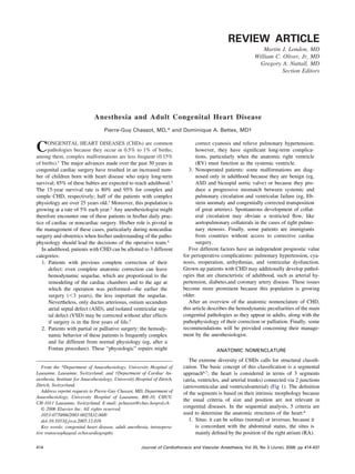
  • 16. ADULT CONGENITAL HEART DISEASE 429 lies in an oblique position because of the malalignment of the persist in an adult; it is responsible for an L-to-R shunt, which interventricular septum. may induce an LV dilatation and failure. It is diagnosed by a These patients become symptomatic after adolescence; they 4/6 continuous or systolic crescendo murmur in the upper left are not cyanotic, but their systemic ventricle is an anatomically sternal edge, radiating to the back. At echocardiography, there right ventricle; therefore, it dilates and fails in about 20 to 25 is a persistent diastolic turbulent flow in the distal main pul- years because of pressure overload.76 The diagnosis is fre- monary artery. Patent ductus arteriosus should be closed unless quently overlooked until the signs of progressive ventricular it is tiny. In case of a large patent ductus arteriosus with failure appear. AV blocks are frequent.77 gradient 60 mmHg between the aorta and pulmonary artery, The prevalence of coarctation of the aorta is 0.2% of births.78 the closure is recommended only if the associated PHT is A ridge of dense tissue narrows the lumen of the aorta at the reversible. level of the isthmus. The aortic arch above the lesion is dilated Coronary arteries may present anomalous origin from differ- and highly pulsatile, whereas the descending aorta distal to the ent aortic coronary sinuses or from a single common trunk or coarctation is much less expansive in systole. Aortic coarcta- from the pulmonary artery. They may also present anomalous tion is frequently associated with bicuspid aortic valve (50% of termination into the RV or the PA. This situation leads to a the cases) and ductus arteriosus (20% of the cases). The pres- fistula or a shunt between LV and RV. The effective shunt sure overload on the LV induces a concentric hypertrophy; the (Qp/Qs) is usually around 1.6.84 A biventricular dysfunction is systolic function is preserved, but diastolic dysfunction is frequently present. usual. The disease is characterized by arterial hypertension in the arms and hypotension in inferior limbs; the gradient of GENERAL ISSUES IN ANESTHESIA pressure is at least 20 mmHg. There is a 3/6 systolic murmur Adolescent or adult patients suffering from CHD share many audible on the left midthoracic area in the back. If the coarc- common features.85,86 They present 5 major independent risk tation is very narrowed, an important collateral network main- factors: PHT, cyanosis, reoperation, arrhythmias, and ventric- tains the distal perfusion through periscapular, internal mam- ular dysfunction. RV and/or LV failure is linked to the etiology mary, cervical, and intercostal arteries. Dilation and increased and to the degree of remodeling; it is directly associated with pulsatility of intercostal arteries cause a notching at the inferior the age and with the degree of cyanosis. Their average opera- edge of the ribs, which is typical on chest x-rays. The majority tive mortality of 7% is higher than the mortality of children of nonoperated patients die between 30 and 40 years from LV with similar pathologies. They show an elevated incidence of failure, endocarditis, aortic rupture, or cerebral hemorrhage.78 arrhythmias ( 50%) compared with children (15%). The pul- The operative risk is inversely proportional to the length of monary circulation is usually abnormal; there is PHT because the stenosis and to the degree of collateralization. When col- of volume and/or pressure overload, or the flow is limited and laterals are well developed, clamping of the aorta does not insufficient at exercise. They suffer from the systemic effects of increase upstream pressure more than 25 to 30 mmHg.79 Usu- hypoxia and increased hematocrit on peripheral organs: the ally, the stenosis is short, and the resected area is small; the kidneys, the lungs, the brain, and the hematologic system show surgeon can perform a direct end-to-end anastomosis. A patch signs of more or less severe dysfunction. Finally, they have with the left subclavian artery or with prosthetic material is coagulation disorders and an increased hemorrhagic risk sometimes necessary to prevent a residual stenosis. The clamp- (Table 1). ing time is 15 to 30 minutes. Meanwhile, the downstream The flow through a shunt is proportional to the diameter of pressure must be kept 60 mmHg with an arterial vasocon- the defect or the conduit and to the ratio of the impedances strictor (phenylephrine and norepinephrine). The operation is between the upstream and the downstream cavities. An R-to-L performed through a left thoracotomy and requires a double- shunt (TOF, TGA, VSD, and pulmonary atresia) increases lumen endotracheal tube for exclusion of the left lung. After the when systemic arterial resistances decrease or pulmonary re- procedure, the immediate decrease of the diastolic arterial sistances increase (Table 2). The SpO2 varies with the Qp/Qs pressure is a good marker of the absence of significant residual ratio and monitors very precisely the degree of mixing of gradient. Postoperative paraplegia because of medullary isch- arterial and venous blood. Increasing the inspired oxygen con- emia occurs in 0.1% to 0.4% of the patients.80 centration has minimal effect on cyanosis because of an R-to-L Postoperative hypertension is frequent and peaks at 12 to 24 shunt, whereas arterial vasoconstriction (phenylephrine and hours because of an acute firing of the baroreceptors adjusted to norepinephrine) increases arterial oxygen saturation. The pres- the previous high upstream pressure; there is a second peak ence of a shunt modifies the speed of induction of intravenous after 2 to 3 days linked to an excessive level of renin and and inhaled agents.87,88 angiotensin. Hypertension persists in 20% to 50% of the pa- An L-to-R shunt (ASD, VSD, and AV canal) decreases with tients.81 In the first postoperative days, a mesenteric arteritis is a drop in SVR (Table 3). The major problem is usually the frequent because of sympathetic hyperactivity; it produces se- presence of PHT. An elevated SpO2 is no guarantee that oxygen vere abdominal pain.82 Even if the lesion has been corrected in transport is satisfactory; with a Qp/Qs 3/1, the oxygen trans- childhood, it may recur in the young adult.83 Systemic hyper- port decreases even if the arterial blood is correctly oxygen- tension and left ventricular hypertrophy may persist after cor- ated.89 The purpose of a peripheral L-to-R shunt (Blalock- rection in up to one third of the cases. Taussig, Waterston, and aortopulmonary collateralization) is to Many arterial anomalies can happen at the level of the aortic make up for insufficient pulmonary flow; because its dimension arch including hypoplasia, ductus arteriosus, aortopulmonary is fixed, its output is proportional to the systemic arterial fistulae, and coronary anomalies. A small ductus arteriosus may pressure (the pulmonary flow falls in case of systemic hypo-
  • 17. 430 CHASSOT AND BETTEX Table 1. Risk Factors in Anesthesia in Case of Adult Congenital Table 3. Congenital Heart Diseases With L-to-R Shunt and Heart Disease Increased Pulmonary Flow (Qp *) Pulmonary hypertension (PVR 500 dynes/s/cm5) Pathologies: atrial septal defect (ASD), atrioventricular canal (AV Cyanosis (SaO2 85%, Hb 150 g/L, Ht 55%) canal), ventricular septal defect (VSD), anomalous venous Reoperation connections, ductus arteriosus. Arrhythmias ( 50% of the cases) Cyanosis absent, normal hematocrit Ventricular dysfunction Increased pulmonary flow ASD: RV volume overload VSD: LV volume overload Longstanding L-to-R shunt (VSD) leads to pulmonary hypertension tension). The SpO2 decreases with the SVR; arterial vasocon- and RV pressure overload. striction (phenylephrine and norepinephrine) increases arterial To decrease the shunt: + SVR, * PVR. oxygen saturation. These patients show a chronic low diastolic Anesthesia management aims at pressure that might endanger coronary perfusion. SVR decrease (isoflurane, intrathecal or epidural anesthesia) Cyanosis is caused by an insufficient pulmonary flow (pul- PVR increase (hypoventilation, slight hypercarbia, FIO2 0.3) monary atresia) or a contamination of arterial blood by venous Preload increase; hypovolemia poorly tolerated because of blood (R-to-L shunt). In an R-to-L shunt, the blood bypassing volume sequestration in pulmonary bed the lungs corresponds to an increase in nonoxygenated blood in Induction with intravenous substances is slowed; halogenated gas the lungs (deadspace effect). In this case, the measured end- uptake is increased tidal carbon dioxide (PETCO2) underestimates the actual Palliative shunts (Blalock-Taussig) or aortopulmonary collaterals Flow through the shunt is fixed and proportional to systemic PaCO2. These patients maintain a normal ventilatory response blood pressure to hypercapnia but show a blunted response to hypoxemia.90 SVR decrease leads to decrease in pulmonary flow They hyperventilate chronically to compensate for the poor Hypotension leads to low SpO2 CO2 disposal. Because of the low arterial oxygen saturation, the Abbreviations: SVR, systemic vascular resistances; PVR, pulmonary hematocrit increases up to 65% to 70% to match the oxygen vascular resistances; SpO2, pulse oximetry; Qp, pulmonary blood flow. transport. The hyperviscosity of a high hematocrit increases the cardiac ejection work and the risk of spontaneous thromboses, particularly when these patients are fasting or dehydrated. The cyanosis of these patients is less obvious when they are anemic Cyanotic diseases have the following effects on organs and because the degree of cyanosis depends on the concentration of systems: unsaturated hemoglobin. 1. Effects on myocardium: chronic systolic and diastolic ventricular dysfunction and increased ischemic risk.63 2. Hematologic effects: increase of the mass and stiffness Table 2. Congenital Heart Diseases With R-to-L Shunt and of erythrocytes, hyperviscosity, and gallstones because Decreased Pulmonary Flow (Qp +) of the excess of heme ring.91 Pathologies: single ventricle (univentricular AV connection, 3. Effects on coagulation: decrease in von Willebrand tricuspid atresia), tetralogy of Fallot, palliated Fallot, factor and platelet function and increased fibrinolysis;92 transposition of great arteries, pulmonary atresia the measured thrombocytopenia is usually artifactual Cyanosis present, increased hematocrit (Ht 55%). because of the increase in erythrocyte volume. When To reduce the shunt and increase SaO2: * SVR, + PVR. hematocrit is above 55%, the values of PT and PTT are Qp/Qs monitoring: SpO2 misleading because of the relative excess of citrate in PETCO2 underestimates PaCO2 Anesthesia management aims at: the samples compared with the reduced plasma vol- SVR increase* ( 1-stimulation); neuraxial blockade poorly ume93; false hypoglycemia might also occur because of tolerated the glycolysis from the excess number of erythrocytes. PVR decrease;* general anesthesia with normobaric 4. Effects on kidneys: the chronic hypoxemia leads to hyperventilation (hypocarbia, alcalosis) is prefered proliferative lesions in the glomeruli and to a thicken- Keep Ht 40% in patients with important R-to-L shunt or ing of the basal membrane, resulting in proteinuria and decreased pulmonary flow (SaO2 75%) uric acid increase; the level of uric acid in the plasma After Fontan procedures: spontaneous ventilation as possible is a good marker of the renal hemodynamics of cya- Decrease dynamic RV outflow tract obstruction by -blocker notic patients.94 To increase SaO2: * SVR ( 1-stimulation); * FIO2 without effect; 5. Neurologic effects: in adults with R-to-L shunts, the ideal SaO2: 80%-85% Induction with IV substances is accelerated; halogenated gas incidence of cerebral abscesses is increased but not the uptake is slowed incidence of cerebral vascular accidents.5,95,96 Fifty percent of the emergency hospitalizations are justified Abbreviations: SVR, systemic vascular resistances; PVR, pulmonary by threatening arrhythmias.97 Atrial tachyarrhythmias are fre- vascular resistances; SaO2, arterial oxygen saturation; SpO2, pulse oximetry; Qp, pulmonary blood flow; Qs, systemic blood flow; quent after correction of an ASD, Senning procedure, or when PETCO2, end-tidal carbon dioxide. atria are enlarged; AV blocks are usual after surgery at the level *In case of noncorrected transposition of great arteries or single of the AV junction (perimembranous VSD). Ventricular tachy- ventricle (rare in adults), Qp should be balanced with Qs (1:1) by an cardia may supervene in any congenital disease but is particu- adequate balance of SVR and PVR. larly frequent in corrected TOF with pulmonary valve insuffi-
  • 18. ADULT CONGENITAL HEART DISEASE 431 est PaCO2 with the lowest mean intrathoracic pressure, remem- PVR bering that the duration of inspiration raises the mean intratho- Extra-alveolar vessels racic pressure more than the peak value of the inspiratory pressure.104 Fearing the interferences of the IPPV, the anesthe- Alveolar vessels siologist frequently prefers an intrathecal or epidural blockade to general anesthesia. This might not be a good choice for 3 reasons. First, the incremental pressure of IPPV represents a Total PVR weak increase compared with the already elevated pulmonary pressure and the RV is well adapted to a high afterload; second, IPPV allows hyperventilation and therefore a decrease in PVR; and third, the arterial vasodilation secondary to neuraxial FRC blockade will worsen the R-to-L component of the shunt and aggravate the cyanosis of the patient. The tolerance to IPPV can be tested before induction by the observation of the arterial pressure tracing during a Valsalva maneuver; if the ventilatory pressure variations are stabilized and if the mean arterial pres- sure decreases 20% of its baseline value, IPPV can be started without fear. The inhaled and intravenous anesthetic agents have minimal effects on pulmonary circulation, except for ketamine, nitrous oxide, and desflurane, which increase Pulmonary volume PAP.103,105 Ketamine is useful in all sick children with CHD but Fig 16. PVR and ventilation. At low tidal volume, hypercarbia and is poorly adapted to the hemodynamic conditions of adults atelectasis increase vascular resistances in small alveolar vessels. AT because it increases PVR and myocardial oxygen consump- high tidal volume (hyperinflation), large extra-alveolar vessels are tion.104 mechanically compressed. Total PVR presents a “U”-shaped curve, The therapeutic measures to treat PHT are described in Table with its lowest value at the functional residual capacity (FRC). 4. Except for hyperventilation, inhaled prostanoids, and NO, all the substances used for reducing PHT have also some systemic vasodilating effect. The arterial vasodilators (phentolamine, ciency and RV dilatation.4,98 The incidence of sudden death is nitroprusside) are useless for the treatment of congenital PHT increased in CHD patients. because they aggravate the R-to-L shunt and because the pul- The criterion of PHT is a mean pulmonary arterial pressure monary vascular tree is almost devoid of 1 receptors.106 In the 25 mmHg at rest and 30 mmHg during exercise or PVR operating room or intensive care unit, the nonclosure or the 300 dynes/s/cm5. The most frequent origin is a nonrestrictive reopening of the pericardium and sternum after cardiac surgery L-to-R shunt with a pressure overload; it already appears dur- improves RV function and decreases ventricular interdepen- ing childhood in 50% of the patients with VSD or AV canal but dence. in only 10% of the adult patients with an ASD.99 The hyper- Poor ventricular performance is frequent among adult pa- tension is associated with a progressive muscular hypertrophy tients suffering from CHD. This is secondary to the following of the media layers of the arteries; it leads to a hypertension that phenomena67: is at first reactive for many years but becomes secondarily 1. Ventricular remodeling: concentric hypertrophy be- fixed. This last stage is the Eisenmenger syndrome, character- cause of pressure overload and spherical dilatation of a ized by PVR 800 dynes/s/cm5. The flow through an L-to-R single ventricle, for example; the LV ejection fraction shunt becomes first bidirectional and then reversed; it is the calculation is not valid when the shape of the ventricle main origin of an outbreak of cyanosis in adults.34 The survival is abnormal, and the dimensions in systole and diastole of patients with severe PHT is more favorable in adults suffer- are more adequate criteria. For the LV, the upper limit ing from CHD than from primary PHT.100,101 of short-axis diameter is 2.5 cm/m2 in end-systole and The limited compliance of the pulmonary vascular system 4.5 cm/m2 in end-diastole. precludes the possibility of adapting to blood volume varia- 2. Duration without correction: the longer the period be- tions. A decrease in arterial pressure because of hypovolemia or fore surgical correction, the more extensive the ven- vasodilation worsens the R-to-L component of the shunt. As tricular lesions. long as the pulmonary arterial pressure is not fixed, it overre- 3. Type of overload: a volume overload is better tolerated acts to sympathetic stimuli and to variations in pulmonary H than a pressure overload; the RV fails when the PAP is concentration; hypothermia, stress, pain, acidosis, hypercarbia, chronically above 50 mmHg. and hypoxia will aggravate the hypertension.102 4. Cyanosis: the myocardial oxygen consumption is be- Ventilating these patients is a compromise between active yond the possibility of the oxygen transport at exertion, hyperventilation and the preservation of a low intrathoracic and the myocardium is chronically ischemic. pressure. The ideal tidal volume corresponds to the functional 5. Ischemia: microvascular occlusions because of hyper- residual capacity103 (Fig 16). It is necessary to manipulate the viscosity and low perfusion pressure because of a shunt respiratory rate, the tidal volume, and the ventilatory mode induce chronic myocardial ischemia; coronary athero- (volume- or pressure-controlled ventilation) to obtain the low- matous disease may be superimposed in the adult.