

Recomendados
Mais conteúdo relacionado
Mais procurados
Mais procurados (20)
Semelhante a Cell Division Cancer
Semelhante a Cell Division Cancer (20)
Mais de shabeel pn
Mais de shabeel pn (20)
Último
Último (20)
Cell Division Cancer
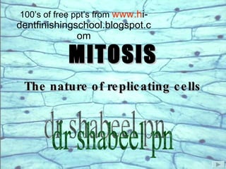
- 1. MITOSIS The nature of replicating cells 100’s of free ppt’s from www.h i-dentfinishingschool.blogspot.com dr shabeel pn
- 2. Reproduction: Not as simple as it looks.
- 3. Reproduction presents a major problem for cells and organisms: (how can information be transmitted faithfully to progeny) I II III IV = one bit of genetic information
- 4. = one bit of genetic information The information transfer problem becomes more challenging as more bits of information are incorporated into the organism
- 5. One of life’s solutions to this challenge: “Package” the bits of information into single units called chromosomes = one bit of genetic information
- 6. chromosomes Packaging of genetic material in prokaryotes and eukaryotes prokaryote cell eukaryote cell
- 7. Fig 2.4 The structure of a highly condensed, replicated chromosome. © 2003 John Wiley and Sons Publishers
- 8. A Chromosome
- 10. The cell cycle. © 2003 John Wiley and Sons Publishers
- 11. The S tages of the C ell C ycle Mitosis interphase G1 S G2 Cell division M
- 13. Every dividing tissue cell in the body is always at a stage of the cell cycle. Whether it is at :- STAGES OF MITOSIS Cytokinesis Diagram showing the Stages of Mitosis Prophase Metaphas e Anaphase Telophase Interphase Th us enabl ing the body to continuously make new body tissue for growth and repair .
- 14. Prophase Metaphase Anaphase Telophase The Stages of M itosis Interphase
- 16. Chromosomes attached to spindle during nuclear division
- 23. TELOPHASE Two new nuclei are formed when the chromosomes reach the opposite poles of the cell The nuclear membrane is formed- the nucleolus reappears The chromosomes disperse in the nucleus
- 29. Mitosis in animal cells. © 2003 John Wiley and Sons Publishers
- 32. Observed with place contrast microscopy. The work of Shinya Inoue and Rudolf Oldenbourge.The Mitosis World Website. The work of Mr Paul Maddox . The Mitosis World website . Kangaroo e pithelial kidney cell going through mitotic division. Mitosis and cell plate formation in a flattened endosperm cell of the African bloodlily Haementhus Katherininae.
Notas do Editor
- Introduction. Will automatically lead into the next slide.
- A bit of background knowledge on chromosomes in general. Genes are instructions for all activities of the cells and determine what they will be. Each chromosome contains approximately 4000 genes. The total of DNA in a human cell can consist of 60 000 genes. This is known as the genome.
- Click the “M Stage” button to find out more about what the M Stage is. If the graphics are distracting click once anywhere on the screen. The return button will take you to the last slide viewed. The house button will take you to the main menu.
- The return button will take you to the last slide viewed. The home button will take you to the contents page.
- Each button next to the text will take you to the indicated page. Press the “house button” (top right) at any time to return to the contents page. Mitosis is the life of a cell. This cycle is split into various seasons through its life to make it easier to understand
- A diagram showing the stages simplified. The “mitosis in action” button will show an animated version of this diagram. Although the two daughter cells shown straight after anaphase are far apart, this is simply to show that two individual cells have been produced. When in fact the daughter cells are sat very close together, which can be seen in the photographs shown on the ‘Telophase’ slide.
- Click the return button to return to the prophase slide. Or the house button to return to the main menu. The purpose of the spindle is to organise the chromosomes during mitosis. It is a cradle of microtubule fibres which cause constriction around the centre of the cell, causing the cytoplasm to split.
- A brief description of Interphase on the whole. The following pages go into greater depth about Interphase. The three stages follow on from each other and are in order-if these would like to be viewed then continue with the forward button. Alternatively an animated diagram can be viewed of the cell cycle, by clicking on the “cell cycle” button. Interphase is the time lapse stage between the end of telophase and the beginning of prophase. The cell is recovering its body mass and preparing for division once again, at the same time. A cell at interphase can be seen in the photograph. This could be mistaken for a cell at early prophase as there is only slight differences between the two, however the large nucleus is an indication of its stage.
- The photographs are of a cells at their resting states. The diagram is indicating the chromosomes condensing during prophase. Prophase is the first stage of mitosis, it prepares the cell for the next stage- Metaphase, by condensing the unravelled DNA into a helix which can be split into its two halves. The chromatin begins to condense to form chromosomes. Each chromosome has already replicated itself, fprming two sister chromatids which contain exactly the same genetic information. This could, however not be true if a mutation has occurred. Other events occuring in the cell are; the nucleolus begins to break down so the genetic information can be equally distributed between the two daughter cells. The nucleolus completely disappears because the genetic information is dispersed as condensing chromosomes within the cytoplasm. The contrioles (‘Points’ of the spindle) move to opposite ends of the nucleus as the spindle begins to form. The nuclear membrane begins to break down so there is no longer a nucleus as such in the parent cell.
- The information, ‘I’, will take you to a slide about the spindle and what it is made of. The dark area within the photographed cells are the developed spindles and is simplified with the diagram below it. Metaphase literally means ‘middle stage’, it is the moment before the chromosomes reach opposite ends of the cell at anaphase. The spindle can be seen in the green picture, and the chromosomes arranged upon it. The easy way to identify the start of metaphase is the disappearance of the nuclear membrane, this breaks up into individual vesicles, it then joins up onto the endoplasmic reticulum (so is not just lost within the cytoplasm). The spindle is now fully developed and takes the place where the nucleus was. The most obvious stage of metaphase;- the chromatid pairs align themselves along the centre of the spindle after being attached to the spindle fibres themselves.
- The photograph shows the cell at the proceeding stage of mitosis-anaphase and is highlighted to show the direction in which the spindle is moving. The aligned chromatid pairs are pulled apart from the centre of the spindle due to movement of the microtubule spindle fibres. They are pulled to opposite poles of the cell. This is the most recognisable event during mitosis, it can be seen clearly in the films featured in the ‘movies section’ which can be accessed through the contents page.
- The photographed cell now shows telophase. Two cells “stuck” next to each other. The forward button will now take you to a slide about Cytokinesis, or the home button will take you back to the contents page. The chromosomes have reached opposite ends of the spindle, they unravel and disperse into the cytoplasm so cannot be seen. The nuclear membrane reforms. The nucleolus reappears- much of the events that happen during telophase are the opposite of the events that happen during prophase.
- A mnemonic to help remember the stages of mitosis.
- Cytokinesis begins around anaphase and is the splitting of the cytoplasm, mitosis is mainly the replication of the DNA. Click the return button to return to the mitosis contents page, or the house button to return to the main menu.
- The desired button should be clicked to view the films. However a warning will come up which can be ignored, just press ‘ok’ and place the image where best suited. The films can be played, paused, rewound and fast-forwarded using the arrows at the base of the image.
- Extra information and pictures of tumours can be seen by clicking on the tumours button. The forward button will carry on discussing rapidly dividing cells, with pictures. The return button will take you back to the contents page. The rate at which cells die and are being made is known as cell turnover. Cells from different parts of the body replicate at different speeds. This is due to the need for new cells. Skin cells are permanently being damaged, so need new cells to replace the old ones which have died. Protected organs, such as the brain don’t need replicating cells for repair, as it rarely gets damaged and no longer needs to grow in size when it reaches adult size. The brain will have a very low cell turnover, especially compared to skin cells.
- Photographs of red blood cells and sperm cells. The forward button will take you to a slide about tumours. An animated sperm will appear without warning, simply click to finish the animation. Red blood cells have no nucleus so they themselves cannot divide mitotically. They are reproduced by the marrow in bones. Red blood cells are always needed for repairing the surface of skin etc, so will have a high turnover. Sperm cells do have a nucleus, but is a gamete or sex cell. This means it only has one set of chromosomes. The average healthy human male can produce up to 1000 new sperm every second.
- The continue button will take you to the questions page. Or return to the previous page or the contents page.