

Recomendados
Mais conteúdo relacionado
Mais procurados
Mais procurados (20)
Semelhante a 05 proteins %26 cell structure
Semelhante a 05 proteins %26 cell structure (20)
Último
Último (20)
05 proteins %26 cell structure
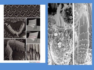
- 3. Neuronal structures and structural proteins Transmission Electron Microscopy Shows the different organelles in the cytosol
- 4. Golgi complex Is responsible for most posttranslational modification
- 8. Microtubules grow in a helical fashion. Their growth is stopped by “capping” their growing end with GDP-bound tubulin. In the absence of MAPs or other stabilizeing proteins they depolymerize, disrupting the structure of the corresponding process MAP2: dendrites MAP3 and tau prot: axons
- 9. Map-2 staining in dendrites not axons
- 16. Microtubule Domains in the axon
- 17. Microtubules and actin filaments are the TRACK along which proteins and organelles are moved by molecular motors
- 19. Axonal transport Fast transport is two ways: from the soma to the terminal and viceversa Slow transport is only from the soma to the terminal Fast transport: organelles and functional proteins Slow transport: structural proteins
- 20. Fast axonal transport 410mm/day
- 22. Tubulin Clathrin Neurofilament Actin Slow axonal transport = 0.2-2.5mm/day
- 23. Both techniques are used by neuroanatomists for understanding the connectivity of the brain: anterograde and retrograde dyes
- 24. Inside the muscle similar proteins organize muscle spindles where information is sent using an axon for conveying somatosensory information
- 25. Similar proteins permit to study synapses between sensory neurons in the dorsal horn (red) and motor neurons in the ventral horn (green) of the spinal cord
- 26. Central neurons have a different morphology from spinal cord neurons: Pyramidal cells of the hippocampus
- 27. Spines can be considered as specialized organelles for performing spatially localized dendritic (input) functions
- 28. Dendritic Spines