

Recomendados
Mais conteúdo relacionado
Mais procurados
Mais procurados (20)
Semelhante a Lecture8
Semelhante a Lecture8 (20)
Lecture8
- 1. DNA Replication and Protein Synthesis Patricia Caldani MS
- 3. Objective Genome structure Chromosome/Gene/DNA DNA Replication Protein Synthesis Transcription and Translation Mutations
- 4. The Human Genome The human genome is made up of 3 x 9 10 base pairs of DNA (haploid genome) This contains 30,000 genes arranged on 46 chromosomes Packaged within the nucleus of the cell
- 5. What is a Gene?? Genes are instruction manuals for our bodies They are the directions for building all the proteins that make our body function Genes are made up of DNA EXP. RBC use Hemoglobin
- 6. Chromosomes Long strands of DNA packaged and compressed very tightly Everyone has __23_________ copies of each chromosome 1 pair of each of the 22 ‘autosomes’ <22> • plus XX for a female (46XX) <1> • or XY for a male (46XY) <OR 1> DIPLOID GENOME
- 8. Chromosomes in Metaphase Telomere Short arm (p) Centromere Long arm (q) Telomere
- 9. DNA (DeoxyriboNucleic Acid) 2 major functions Direction of all protein synthesis Accurate transmission of this information from one generation to the next Fundamental to Cell metabolism Cell division
- 10. DNA (DeoxyriboNucleic Acid) String of deoxyribose sugars joined by _____phosphate______ groups. Each sugar is attached to one of 4 possible nucleotide bases ADENINE (A), CYTOSINE (C ), GUANINE (G) or THYMINE (T)
- 11. Each hydrogen bond hold two DNA strands together very tightly. To form Watson-Crick base pairs, DNA strands must be anti parallel & ____double stranded_______
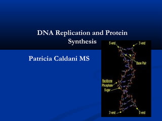
- 12. DNA Double helix structure 2 strands are held together by hydrogen bonds 4 bases pairing rule o Adenine = Thymine (A = T) o Guanine = Cytosine (G = C)
- 13. DNA Base Pairing A G C G A T C T G G T C G C T A G A C C Double helix consists of 2 complimentary strands of DNA.
- 14. Some Definitions Replicon: Unit in which replication occurs Origin: Position at which replication initiates Terminus: Position at which replication terminates Origin Replicon bo naA h sit pera s rep gene O ron D -ric r to Ite AT s xe e
- 16. DNA Replication is Semiconservative 3’ 5’ Semi- 5’ 3’ Conservative conservative Dispersive
- 17. Semiconservative Replication of Density-Labeled DNA DNA replication is semi-conservative: in the "next generation" molecule one strand is "old" and another is "new"
- 18. DNA Replication Semi – conservative replication C T ATC G A A TAG ATC T TAG TA DNA AT ATC C TAG unzips G Original double strand DNA separates and replicates 2 new double strands – each containing one parent and one daughter strand
- 19. Many enzymes are involved in DNA replication
- 20. The Enzymes of DNA Replication 1. Topoisomerase - initiates the unwinding of the super coiled DNA by clipping the DNA backbone. 2. Helicase - separates the double strand by "melting" the hydrogen bonds that hold the bases together. It requires energy (in the form of ATP ). 3. Primase - makes a short RNA segment (called a primer) that is complementary to the DNA strand at specific sites. It "primes" for DNA replication because it provides an - OH for DNA polymerase to attach the first DNA nucleotide. The RNA primer is later removed and the gap is filled in with DNA nucleotides. 4. DNA polymerase (III- major polymerase) - binds to one side of the DNA and nucleotides to bond with their complementary pair 5. Ligase – joins two large molecules via covalent bonds-repair discontinued SS-DNA strands
- 21. Common Features of Replication Origin Unique DNA sequence containing multiple short repeated sequence Origins contain DNA sequences recognized by replication initiator proteins AT-rich stretch
- 22. DNA replication starts within a special region of DNA called REPLICATION ORIGIN which is defined by a specific nucleotide sequence Replication of DNA is ____semiconservative_ ______ Two Y-shaped replication forks are moving in the opposite directions during DNA replication
- 23. Initiation of DNA Replication DnaB is a helicase. An enzyme moves along DNA duplex utilizing the energy of ATP hydrolysis to separate the strands. 5’-3’ SSB: single strand binding protein.
- 24. DNA synthesis always starts with a RNA primer
- 25. The Role of RNA Primer in DNA Replication E. coli primase catalyze the RNA Primer for DNA Synthesis dnaG Primer: <15 nts
- 26. DNA Polymerase Unable to separate the two strands of DNA Only elongate a pre-existing DNA or RNA (Primer) Only add nucleotides to the 3’-hydorxyl group, i.e., only 5’-3’ synthesis
- 27. DNA Synthesis Occurs in the 5’→3’ Direction P P P P OH 3’ 5’ 3’ 5’ P P P P P P P P P Incoming nuceolotide triphosphate 5’PPP OH3’ 5’ P P P P OH 3’ 3’ 5’ P P P P P P P P P Nucleotide monophosphate added to chain with release of diphosphate PP 5’ P P P P P OH 3’ 3’ 5’ P P P P P P P P
- 28. DNA Synthesis is Semidiscontinous Lagging strand synthesis 5’ 3’ 3’ 5’ 5’ 3’ Leading strand synthesis
- 29. DNA Polymerase Exonuclease Activity The enzyme that synthesizes DNA is ______POLYMERASE________ __________ self-correcting:: it has a proofreading activity
- 30. It is not trivial to replicate both DNA strands in the 5' to 3' direction!
- 31. Growing Fork Leading strand: continuous Lagging strand: discontinuous
- 32. Lagging strand synthesis Leading strand: continuous Lagging strand: discontinuous Okazaki fragment:
- 33. Gap Removal Pol I 5’-3’ exonuclease Fills gap DNA Pol I -exonuclease- reads the fragments and removes the RNA Primers. Ligase
- 34. Ligase DNA Ligase: adds phosphate in the remaining gaps of the phosphate - sugar backbone
- 35. Proofreading by 3’ to 5’ Exonuclease
- 36. Proofreading by 3’ to 5’ Exonuclease activity of DNA Pol
- 37. Final Step-Termination This process happens when the DNA Polymerase (enzymes) reaches to an end of both strands. The end of the parental strand where the last enzymes binds aren't replicated. These ends of chromosomal DNA consists of noncoding DNA.
- 39. From Genes to Proteins Patricia Caldani MS
- 40. DNA and RNA differ in 3 ways RNA DNA Single-stranded Double-stranded Ribose (sugar) Deoxiribose (sugar) Uracil (base) bonds to Thymine (base) Adenine bonds to Adenine
- 41. The Genetic Code Every three bases of DNA is called a ‘codon’ Each codon specifies an amino acid Codons specify amino acid sequence of protein
- 42. Amino Acid Code •64 possible triplet codons •Only 20 amino acids •Code is “degenerate or redundant” Alani ne Ala A Leuci ne Leu L Argi ni ne Arg R Lysi ne Lys K Asparagi ne Asn N Methi oni ne Met M Asparti c Aci d Asp D Phenylalani ne Phe F Cystei ne Cys C Proli ne Pro P Glutami ne Gln Q Seri ne Ser S Glutami c Aci d Glu E Threoni ne Thr T Glyci ne Gly G Tryptophan Trp W Hi sti di ne Hi s H Tyrosi ne Tyr Y Isoleuci ne Ile I Vali ne Val V
- 43. Genetic Code U URACIL C CYTOCINE A ADENINE G GUANINE U UUU Phe (F) CUU Leu (L) AUU Ile [I] GUU Val [V] U UUC Phe (F) CUC Leu (L) AUC Ile [I] GUC Val [V] C UUA Leu (L) CUA Leu (L) AUA Ile [I] GUA Val [V] A UUG Leu (L) CUG Leu (L) AUG Met [M] GUG Val [V] G C UCU Ser (S) CCU Pro [P] ACU Thr [T] GCU Ala [A] U UCC Ser (S) CCC Pro [P] ACC Thr [T] GCC Ala [A] C UCA Ser (S) CCA Pro [P] ACA Thr [T] GCA Ala [A] A UCG Ser (S) CCG Pro [P] ACG Thr [T] GCG Ala [A] G A UAU Tyr [Y] CAU His [H] AAU Asn [N] GAU Asp [D] U UAC Tyr [Y] CAC His [H] AAC Asn [N] GAC Asp [D] C UAA Ter [end] CAA Gln [Q] AAA Lys [K] GAA Glu [E] A UAG Ter [end] CAG Gln [Q] AAG Lys [K] GAG Glu [E] G G UGU Cys [C] CGU Arg [R] AGU Ser [S] GGU Gly [G] U UGC Cys [C] CGC Arg [R] AGC Ser [S] GGC Gly [G] C UGA Ter [end] CGA Arg [R] AGA Arg [R] GGA Gly [G] A UGG Trp [W] CGG Arg [R] AGG Arg [R] GGG Gly [G] G
- 44. Gene Structure Promoter Exons Introns AUG UAA start UAG ‘stop’ UGA Exon = coding sequence Intron= intervening sequence (non-coding)
- 45. Protein Synthesis Messenger - RNA DNA Code
- 46. The Flow of Information
- 47. Protein Synthesis transcription DNA RNA Protein translation
- 48. The Flow of Information
- 49. Transcription Double DNA strands separate DNA sense strand acts as template and is ‘transcribed’ into messenger RNA (mirror image of the DNA but Uracil instead of Thymine) Introns are sliced out of the sequence DNA ATCGG UAGCC mRNA
- 50. Transcription This is the first step in Protein Synthesis: 1. The instructions are ______________ (“transcribed”) to an RNA molecule.
- 51. To sum up Transcription… Info transferred from DNA to RNA What is the Enzyme involved in Transcription? Answer RNA Polymerase
- 52. Transcription has 3 steps… 1 – RNA Polymerase binds to the gene’s promoter (DNA) (like a starting line in a race).
- 53. 2 – RNA Polymerase UNWINDS the DNA molecule. The DNA nucleotides are exposed.
- 54. 3 – RNA Polymerase adds complimentary ______________ to separated DNA strand. ** Remember RNA has ______________ instead of Thymine for a base.
- 55. The RNA Polymerase will continue transcription until it reaches the “stop signal” on the DNA molecule (like a finish line). Then the RNA strand is released and goes on to the next step…Translation
- 56. Transcription
- 57. Transcription
- 58. Transcription RNA polymerase exon 1 2 3 intron 1 2 transcription factors 5’ 3’
- 59. Transcription RNA polymerase exon 1 2 3 intron 1 2 transcription factors 5’ 3’
- 60. Transcription 5’GGAUUCGUGCUGCUAA RNA polymerase exon 1 2 3 intron 1 2 transcription factors 5’GGATTCGTGCTGCTAA
- 61. Transcription primary transcript exon 1 2 3 RNA polymerase intron 1 2 transcription factors
- 62. Transcription mature mRNA AAAAAAAAAA RNA splicing primary transcript transcription factors RNA exon 1 2 3 polymerase intron 1 2 promotor
- 63. 3 types of RNA mRNA (messenger RNA) rRNA (ribosomal RNA) tRNA (transfer RNA)
- 64. Messenger RNA Delivers ______________ to the site of Translation.
- 65. mRNA instructions are written in “3-nucleotide” sequences. These sequences are called codons. Ex. UUU, CUG, ACU, etc. There are 64 possible codons.
- 66. Translation Remember what happens in Transcription? DNA to RNA In Translation…RNA is coded for Amino Acids.
- 67. Translation mRNA leaves the nucleus In the cytoplasm, ribosomes attach to the mRNA ensuring the correct amino acid, for each codon, is added to a growing chain of amino acids which forms the resulting protein.
- 68. Translation polyadenylation site (AATAAA) cap structure AAAAAAAAA 3’ mRNA molecule 5’ Ribosome cytoplasm
- 69. Translation Peptide chain AAAAAAAAA 3’ mRNA 5’ molecule Ribosome cytoplasm
- 70. Translation takes place in the ______________ tRNA (Transfer RNA) molecules carry single amino acids. They also have an OPPOSITE “3- nucleotide” sequence called anticodons.
- 71. rRNA (Ribosomal RNA) molecules are like assembly lines they carry: 1 mRNA 2 tRNA
- 73. 7 Steps in Translation 1 – mRNA start codon starts the process at the P site. 2 – the next tRNA bonds to the next codon at the A site. 3 – A & P are holding 2 tRNA’s…a ______________ bond is formed between 2 amino acids.
- 74. 4 – tRNA detaches from P-site, leaves behind amino acid, leaves Ribosome. 5 – tRNA at A-site moves to the P-site. Now a new codon is ready at the A-site for another tRNA.
- 75. 6 – tRNA detaches from P-site, leaves behind amino acid, ______________ ribosome. 7 – (Steps 2 – 6 repeat until a stop codon is reached). Ex. UAG, UAA, UGA. A new protein is then released into the cell.
- 76. Translation
- 77. Translation
- 79. The Human Genome Only ~5% of our DNA actually codes for proteins. Little variation exists from person to person.The remainder is ‘junk’ ‘Junk’ DNA includes repetitive sequences such as micro and minisatellites. Varies a lot between individuals allowing ‘DNA fingerprinting’
- 80. RNA polymerase responsible: transcription, and ribosome (which is responsible for translation) are very accurate enzymes and make very few mistakes. IF RNA polymerase or a ribosome make a mistake it is not highly detrimental to the cell
- 81. An alteration in the nucleotide sequence of the gene is called a ________________________ . Mutations in the gene affect the structure and the function of all the proteins expressed from the mutant gene.
- 82. Mutations A change in the DNA sequence of the gene o Germline mutation (inherited)– present in every cell in the body o Somatic mutation (acquired) – present only in the descendants of that cell All cells acquire mutations as they divide Mutations can alter protein product of DNA, stop gene working or activate gene
- 83. Types of Mutation (in coding sequence) AGC TTC GAC CCG Wild type AGC TGA CCC G Deletion AGC TTC CCG ACC CG Insertion AGC TTC TTC TTC GAC CCG Expansion ATC TGC GAC CCG Point mutation ATC TGA Nonsense ‘stop’
- 84. DNA Repair • Cells require mechanisms to ___________________ DNA damage • Mutation rate reflects the number of damaging events in DNA vs. the number of corrections • 2 broad types of changes: Single base mutations Structural distortions
- 85. Repair Mechanisms • Direct repair, e.g., photoreactivation of thymine dimers by a light-dependent enzyme (photolyase) • Nucleotide excision repair • Mechanism • Multiple systems, some of which act generally, others more specifically e.g. glycosylases and AP endonucleases • Found in bacteria, archaea and eucaryotes • UV damage and repair of bulky lesions • Tolerance systems allow replication of damaged DNA but with higher error frequencies • Recombination repair uses homologous recombination to obtain the correct sequence from an undamaged source • Mismatch repair removes mismatches which arise during DNA replication
- 86. Nucleotide Excision Repair Base mutation/distortion 5’ and 3’ incisions Exonuclease excision Replacement strand synthesis Ligase seals backbone nick
- 87. Lectures 1-4 Overview • DNA replication is semi-conservative: both strands are used as templates for new strands • DNA synthesis occurs exclusively 5’→3’ • DNA synthesis is semi-discontinuous (except in rolling circle replication where the two strands are replicated from independent origins): Lagging strand synthesis 5’ 3’ 3’ 5’ 5’ 3’ Leading strand synthesis • Leading strand synthesis is primed once with a primase-synthesized RNA primer • Lagging strand synthesis is primed repeatedly with Okazaki fragments
- 88. Summary • DNA damage can be of a variety of types • Cells require mechanisms to repair DNA damage • Different mechanisms exist • Nucleotide excision repair is a sequential and coordinated series of enzymatic events by which DNA damage cane be repaired
- 89. Lectures 1-4 Overview • DNA polymerases have proof-reading activity which corrects incorporation of incorrect nucleotides • Proteins at the replication fork: DNA PolIII Primase Lagging strand Helicase (Okazaki fragment) Parental DNA SSB 5’ 3’ 3’ 5’ 5’ 3’ Leading strand + DNA PolI + Ligase In eukaryotes replication is initiated at multiple ori on different chromosomes; termination is poorly understood
- 90. Exercise 1. Each column in the table below represents three nucleotides. Within each column, fill in the cells that are blank by using information from the cell that is not blank. DNA strand TAC GGG mRNA UCG CCU Amino Acid Leu
- 91. Transcription and Translation problems 1. polypeptide: proline-tyrosine-histidine-valine-glutamic acid What is the base sequence of the mRNA that codes for the polypeptide? A. CCG-UAU-CAU-GUA-GAA B. GAA-GUA-CAU-UAA-CCG C. CCG-GUA-GAA-UAA-AUG D. CCG-GUA-CAU-CUA-CCG 2. Use a genetic code table to determine the polypeptide sequence synthesized from the mRNA below. Assume that translation begins at the first nucleotide at the 5' end. 5' AUGAAGUGUUAACCC 3‘ 3. What RNA sequence is transcribed by the gene with the DNA template strand 5’ TTGAGCGCGTA 3’
- 92. 1. Major features of the Watson and Crick model of the double helix 2. How DNA is replicated in Eukaryotes and Prokaryotes 3. The major steps involved in DNA replication 4. How the process of DNA replication ensures accuracy 5. Mutations and how they can be harmful or beneficial 6. Scientists Watson Crick, Meselson and Stahl, and Chargaff discoveries 7. Chromosome structure and function 8. The purpose of Transcription and the differences between RNA and DNA 9. How amino acids are designated in a stretch of RNA 10. Steps involved in protein synthesis, their locations, and the important players involved 11. Be able to transcribe and translate DNA sequence
Notas do Editor
- The human genome is made up of 3 x 10 to the 9 base pairs of DNA. This contains 30 000 genes arranged on 46 chromosomes that are packaged within the nucleus of the cell.
- The human genome is made up of 3 x 10 to the 9 base pairs of DNA. This contains 30 000 genes arranged on 46 chromosomes that are packaged within the nucleus of the cell.
- Correct human chromosome compliment of 46 chromosomes was established in 1956. Chromosomes are stably inherited packages of double stranded DNA which store the genetic blueprint for all the inherited characteristics of an individual. They are long strands of DNA packaged and compressed very tightly. Everyone has 2 copies of each chromosome, 23 pairs in total. 22 of the pairs are the same in both males and females and are called the autosomes. The last pair, the sex chromosomes, are different in males and females – a female has 2 X chromosomes and a male has an X and a Y. The presence of chromosome pairs implies that human cells requires 2 copies of the DNA blueprint (also called the genome) for normal gene function, this is called a DIPLIOD GENOME.
- In metaphase the chromosomes have a constriction at one point along their length called a centromere. The centromere divides the chromosome into a long arm and a short arm. The short arm is named p for petite and the long arm q – the opposite of p. The ends of the chromosomes are called telomeres.
- Within chromosomes the information carrying component is DNA. The DNA has 2 major functions. It is the direction of all protein synthesis and it also accurately transmits the information from one generation to the next. Accurate replication of DNA is essential in cell metabolism and cell division.
- DNA is a string of deoxyribose sugars, joined by phosphate groups between 2 of their carbon atoms at the positions called the 5’ and 3’ . This sugar phosphate chain has direction - at one end it has an unconnected 5’ sugar group and at the other it has a 3’ end. To the side of each sugar is attached one of 4 possible nucleotide bases: adenine, cytosine, guanine or thymine. (A,C, T or G)
- Human DNA is double stranded and exists in a double helix structure, as described by Crick and Watson in the 1950’s. The 2 strands are held together by hydrogen bonds between the nucleotide bases. The 4 bases , ACTG have a pairing rule in that A always bonds to T and G always bonds to C. This specific pairing is fundamental to DNA replication during which 2 DNA strands separate and each act as a template for the synthesis of a new strand, maintaining the genetic code during cell division.
- The 2 strands are therefore complimentary to each other - One strand can be predicted from the other. Here is a sequence of code. Can you tell what the complementary sequence is? Using this process of replication the genetic code is maintained during cell division. The process is called semi-conservative replication.
- As I have said the genetic code - this sequence of bases is the instructions for the production of a protein. The code is read in groups of three bases and each of these 3 bases is called a codon. Each codon specifies an amino acid which are then formed together in sequence to make up the protein.
- As the 4 bases are grouped into 3 codons there are 64 possible combinations, but there are only 20 amino acids. Therefore each amino acids may be encoded for by more than one codon. The code is therefore said to be redundant or degenerate. This table shows the recognised abbreviations for each of the 20 amino acids.
- This is the table for the Genetic Code with the bases, shown along the top and sides of the table. You will notice that the base thymine has been replaced by a different base called uracil, the reason for this will become clear later but bear with me just now. The code is said to be degenerate as each amino acid can be coded for by more than one triplet codon. As you see the amino acid SERINE is coded for by the codons UCU, UCC, UCA AND UCG. However some of the amino acids only have one codon, such as AUG for methionine, which is a start signal for the production of a protein. The end codons to signal the end of a coding region are UGA, UAA or UAG.
- This is a diagram of a gene, a coding sequence for a protein. At the start of the gene there is a AUG start codon and at the end will be one of the 3 end codons. The parts of the genes that carry the code for the amino acids are called the exons of the gene but these are interspersed by non-coding areas called introns. Not all genes are expressed by every cell.
- The function of DNA is to produce proteins. The DNA is contained in the nucleus of the cell. The proteins however are manufactured at cellular assembly plants (ribosomes) outside the nucleus in the cytoplasm of the cell. To deal with this lack of direct contact the gene uses a messenger molecule to carry the genetic information from the gene to the ribosomes. This molecule is called messenger ribonucleic acid and is made up of a string of nucleotides much the same as the ones used in DNA, except that the base thymine is replaced by uracil. The mRNA which is single stranded is made as the genes DNA sequence is being read, the transcription process adding the correct nucleotides to the growing RNA molecule by using the same base pairing rule as used for DNA replication. In this way there is faithful transfer from gene to mRNA of the series of codons for amino acids that represent the information necessary to build the protein encoded by the gene. The ribosomes in the cytoplasm lock on to the long ribbon like mRNA molecules as they pass out of the nucleus and move along them translating the nucleotide code into the appropriate chain of amino acids. When translation is complete the newly formed chain of amino acids breaks away form the ribosome and folds up into the mature protein.
- In summary the DNA is TRANSCRIBED into mRNA which is then translated into the protein.
- The protein synthesis process is initiated by an RNA polymerase which recognises a specific DNA sequence at the 5’ end of the gene. The gene is opened up, the 2 DNA strands separating for a short distance and then the DNA code is transcribed into a single stranded mRNA molecule complementary to the DNA strand. As the RNA is transcribed the DNA strands reassociate behind it. DNA sense strand acts as template and is ‘transcribed’ into messenger RNA (mirror image of the DNA but Uracil instead of Thymine) Transcription is terminated when a stop codon is reached. The mRNA is then modified so that the sequences that are transcribed from introns are spliced out and the sequences that are transcribed from exons are joined together to produce mature RNA. The RNA then diffuses into the cytoplasm to a ribosome. The mRNA is translated into a protein by transfer RNA molecules. Introns are sliced out of the sequence
- mRNA leaves the nucleus and enters the cytoplasm. It undergoes a number of structural changes including the attachment of a special nucleotide to the 5’ end, capping the molecule and a poly (A) tail to the 3’ end (polyadenylation). The ribosomes attach to the mRNA and move along its length ensuring the correct amino acid, for each codon, is added to a growing chain of amino acids which forms the resulting protein.
- Very little of our total DNA actually encodes for protein. Only about 5% and there is very little variation from person to person. About 95% of our DNA is called JUNK DNA. This includes the introns of genes, the non-coding sequences and also the non-coding sequences between genes. The junk DNA includes repetitive sequences such as micro and minisatellites which vary greatly between individuals - allowing DNA fingerprinting.