J ENDOVASC THER 2010
•
0 gostou•417 visualizações

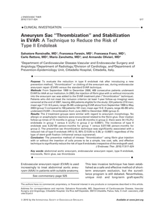
J ENDOVASC THER 2010;17:517–524-Clinical Investigation- Aneurysm Sac ‘‘Thrombization’’ and Stabilization in EVAR: A Technique to Reduce the Risk of Type II Endoleak (Chirurgia Vascolare-ULSS 15 Alta Padovana) (Vascular Surgery -ULSS 15 Alta Padovana)

Recomendados
Recomendados
Mais conteúdo relacionado
Mais procurados
Mais procurados (20)
Destaque
Destaque (20)
Semelhante a J ENDOVASC THER 2010
Semelhante a J ENDOVASC THER 2010 (20)
Mais de Salvatore Ronsivalle
Mais de Salvatore Ronsivalle (7)
Último
Último (20)
J ENDOVASC THER 2010
- 1. ¤CLINICAL INVESTIGATION ¤ Aneurysm Sac ‘‘Thrombization’’ and Stabilization in EVAR: A Technique to Reduce the Risk of Type II Endoleak Salvatore Ronsivalle, MD1; Francesca Faresin, MD1; Francesca Franz, MD1; Carlo Rettore, MD2; Mario Zanchetta, MD3; and Armando Olivieri, MD4 1Department of Cardiovascular Disease–Vascular and Endovascular Surgery and Angiology,2Department of Radiology,3Division of Cardiology, and4Department of Prevention–Epidemiology Unit, Cittadella Hospital, Cittadella, Italy. ¤ ¤ Purpose: To evaluate the reduction in type II endoleak risk after introducing a new prevention method, ‘‘thrombization’’ or clotting of the aneurysm sac, during endovascular aneurysm repair (EVAR) versus the standard EVAR technique. Methods: From September 1999 to December 2008, 469 consecutive patients underwent EVAR for AAA at our institution. In 2003, the injection of fibrin glue with or without microcoils into the aneurysm sac was added to the EVAR treatment plan (‘‘thrombization’’ technique). Patients who did not meet the inclusion criterion (at least 1-year follow-up imaging) were censored at the end of 2007, leaving 404 patients eligible for the study: 224 patients (210 men; mean age 71.968.5 years, range 25–88) undergoing EVAR alone from September 1999 to May 2003 (group 1) compared to 180 patients (161 men; mean age 72.668 years, range 46–89) who underwent EVAR + thrombization from June 2003 to December 2006 (group 2). Results: The 2 treatment groups were similar with regard to aneurysm morphology. No allergic or anaphylactic reactions were encountered related to the fibrin glue. Over median follow-up times of 72 months in group 1 and 26 months in group 2, there were 34 (15.2%) endoleaks in group 1 versus 4 (2.2%) in group 2 (p,0.0001). The incidence of type II endoleak was 0.25/100 person-months for group 1 versus 0.07/100 person-months for group 2. The preventive sac thrombization technique was significantly associated with a reduced risk of type II endoleak (HR 0.13, 95% CI 0.05 to 0.36; p,0.0001) regardless of the type of stent-graft fixation (infrarenal versus suprarenal). Conclusion: The preventive method of intrasac ‘‘thrombization‘‘ using fibrin glue injection with or without the insertion of coils proves to be a simple, low cost, safe, and effective technique to significantly reduce the risk of type II endoleaks irrespective of the endograft used. J Endovasc Ther. 2010;17:517–524 Key words: abdominal aortic aneurysm, endovascular aneurysm repair, type II endoleak, microcoils, fibrin glue, sac thrombosis ¤ ¤ Endovascular aneurysm repair (EVAR) is used increasingly to treat abdominal aortic aneu- rysm (AAA) in patients with suitable anatomy. See commentary page 525 This less invasive technique has been estab- lished as a safe and effective method of short- term aneurysm exclusion, but the surveil- lance program is still debated. Nevertheless, unique mid- and long-term graft-specific The authors have no commercial, proprietary, or financial interest in any products or companies described in this article. Address for correspondence and reprints: Salvatore Ronsivalle, MD, Department of Cardiovascular Disease, Vascular Surgery and Angiology, Cittadella Hospital, Via Riva dell’Ospedale, 35013 Cittadella, Padua, Italy. E-mail: vascolare_cit@ ulss15.pd.it J ENDOVASC THER 2010;17:517–524 517 ß 2010 by the INTERNATIONAL SOCIETY OF ENDOVASCULAR SPECIALISTS Available at www.jevt.org
- 2. complications related to EVAR continue to present management dilemmas for clinicians. Principal among these adverse events is the presence of a type II endoleak, which occurs at some interval after EVAR in 20% to 30% of patients.1,2 Type II endoleak, the most common se- quelae of the EVAR procedure, is due to partial or incomplete thrombosis of the aneurysm sac after successful aneurysm exclusion in conjunction with retrograde perfusion from aortic collateral branches (lumbar, inferior mesenteric, sacral, and renal accessory arteries). The treatment approach- es to types I and III endoleak are well established, while the management of type II is still a matter of debate. A conservative approach is usually used when there is gradual sac shrinkage, although its impact on long-term outcome after EVAR is still un- known.3–5 On the other hand, aneurysm sac enlargement within 6 to 12 months calls for more aggressive techniques, such as percuta- neous transarterial or direct translumbar em- bolization with microcoils or liquid embolic agents, laparoscopic retroperitoneal branch ligation, or endoscopic aneurysm sac fenes- tration.6–16 These procedures seldom resolve the problem, and the best results are achieved with open surgery, which is therefore the most appropriate choice in most cases. The natural history of a type II endoleak led us to believe that prevention is the best strategy in managing this complication. In 2003, we began stimulating and accelerating complete aneurysm sac thrombosis by intro- ducing biocompatible materials during EVAR, primarily fibrin glue.14,15 Several years later, we introduced microcoils along with the fibrin glue to stabilize the sac in a process we now call ‘‘thrombization.’’ The aim of this study was to evaluate the effect that thrombi- zation of the aneurysm sac has on the risk of type II endoleak compared to the standard EVAR technique alone. METHODS Study Design This observational retrospective study was designed to encompass all consecutive pa- tients who underwent EVAR for AAA at our institution from September 1999 to December 2008 and had at least 1-year follow-up imaging. A contrast-enhanced helical com- puted tomography scan with 2.5-mm cuts was used preoperatively to determine if a patient met the radiological criteria for stent- graft repair based on the manufacturer’s Instructions for Use. Five different stent-graft designs were used in the study period: Talent, AneuRx, and Endurant (Medtronic CardioVas- cular, Santa Rosa, CA, USA); Excluder (W.L. Gore & Associates, Inc. Flagstaff, AZ, USA); and Anaconda (Vascutek, a Terumo Compa- ny, Inchinnan, Scotland, UK). The follow-up protocol included a color duplex ultrasound (CDU) scan at discharge; at 3, 6, and 12 months; and at 6-month intervals thereaf- ter. Abdominal radiography was also per- formed at discharge and a year later. A CT scan was scheduled at 6 months. At the end of the observation period, all imaging tests were collected and separately viewed in a double-blinded manner by 2 EVAR experts (a vascular surgeon and an interventional cardiologist). The size of the aneurysm sac and the presence and type of endoleak were evaluated. In accordance with current standard reports for EVAR,17 primary clinical success was defined as absence of aneurysm-related death, aneurysm rupture, conversion to open surgery, and secondary endovascular or surgical procedures. Assist- ed primary and secondary clinical success were applicable when success was achieved with additional endovascular or surgical pro- cedures, respectively. Fibrin Sealant Fibrin glue (Tisseel/Tissucol; Baxter-Hyland Immuno AG, Vienna, Austria) is a fully absorbable biological adhesive matrix with- out cytotoxic effects made from a fibrinogen solution containing plasma proteins and factor XIII and a thrombin solution containing calcium chloride and aprotinin. These com- ponents are commercially prepared from human plasma, except for aprotinin, which is extracted from bovine lung. When mixed together, the 2 solutions recreate the final phase of the natural coagulation cascade 518 ANEURYSM SAC STABILIZATION IN EVAR Ronsivalle et al. J ENDOVASC THER 2010;17:517–524
- 3. forming a structured fibrin clot similar to the physiological clot, susceptible to fibrinolytic degradation by proteolytic enzymes such as plasmin.18 Over the last 3 decades, fibrin glue has been used extensively by surgeons.19,20 Fibrin glue does not interfere with magnetic resonance imaging (MRI), CT, or CDU. The required dose of sealant to cover 40 cm2 or 3.5 cm3 is about 5 mL; however, its use in aneurysm sac embolization is an off-label indication. In Italy, 5 mL of Tissucol costs 435 euros (,US$565). Coils MReye embolization coils (IMWCE 35-20- 20; Cook Medical, Bloomington, IN, USA) are made of a radiopaque nickel and cobalt alloy (inconel) that allows the use of MRI without altering CT or CDU imaging. Most commonly utilized in arterial and venous embolization, these coils exert a greater radial strength than platinum coils; they are introduced through 0.035- or 0.038-inch catheters. In Italy, one coil costs 48 euros (,US$62). Thrombization Technique After deployment of the main stent-graft component with its contralateral iliac exten- sion, the angiographic pigtail catheter is withdrawn, leaving a 180-cm-long 0.035-inch standard J guidewire between the endograft and the native aorta. A 23- to 35-cm-long 5-F Brite Tip introducer (Cordis, a Johnson & Johnson company, Miami Lakes, FL, USA) is then fed through the standard guidewire under fluoroscopic monitoring and released into the desired position inside the aneurysm sac. After the guidewire and 5-F cannula have been removed, an aneurysmogram is per- formed by manually injecting 10 mL of contrast into the sac to identify the number and site of lumbar and inferior mesenteric arteries. One or more MReye embolization coils are then advanced into the sac through the 5-F dilator and released by advancing the 0.035-inch J guidewire inside the dilator toward the end of the introducer sheath. A 25- to 35-cm-long Duplocath catheter (Baxter International) inserted on a Duploject syringe clip (Baxter International) is fed into the introducer until it reaches the aneurysm sac. Then a latex balloon is inflated into the distal end of the iliac graft extension so as to prevent distal embolization of the fibrin glue. With the balloon inflated, 2.5 mL of each fibrin glue solution are simultaneously inject- ed into the sac through a 2-way catheter; a second injection with the same amount can be done if necessary. After the Duplocath catheter is removed, an aneurysmogram is performed to verify sac thrombization with root occlusion of the lumbar and inferior mesenteric arteries. The Brite Tip introducer is then removed, and the completion angio- gram is performed (Fig. 1). Patient Sample During the study period, 469 patients underwent temporally sequential EVAR techniques. From September 1999 to May 2003, 224 patients (210 men; mean age 71.968.5 years, range 25–88) had standard EVAR. From June 2003 to December 2006, 124 patients (114 men; mean age 73.268 years, range 51–89) underwent EVAR with preven- tive fibrin glue intrasac thrombization. Finally, from January 2007 to December 2008, 121 patients (110 men; mean age 71.767 years, range 46–88) underwent EVAR with enhanced preventive intrasac thrombization featuring insertion of $1 microcoils followed by a fibrin Figure 1 ¤(A) Control CT with visible inconel (radiopaque nickel and cobalt alloy) coils and (B) final aneurysmogram performed to verify sac thrombization, with occlusion of lumbar and inferior mesenteric arteries. J ENDOVASC THER 2010;17:517–524 ANEURYSM SAC STABILIZATION IN EVAR 519 Ronsivalle et al.
- 4. glue injection to achieve better aneurysm sac stabilization. Patients who did not meet the criteria for study inclusion were censored at the end of 2007, along with those who died or were lost to follow-up. Thus, 65 patients were excluded owing to lack of 1-year follow-up due to death or loss to follow-up, leaving 404 patients eligible for the study. For purposes of analy- sis, the 224 patients undergoing EVAR alone (Table 1) were compared to the 180-patient cohort (161 men; mean age 72.668 years, range 46–89) who had either form of throm- bization. Statistical Analysis Continuous data are presented as the mean 6 standard deviation or the median for nonparametric data; categorical data are given as counts (percentages). Time to iden- tification of a type II endoleak was the primary outcome in the study: freedom from endoleak was estimated with Kaplan-Meier analysis from the date of surgery/intervention to the date at which type II endoleak was diagnosed. The log-rank test was used to compare the outcomes in patients with and without sac thrombization. Cox proportional hazard mod- eling was used to examine the risk of type II endoleak between the 2 groups after adjust- ing for potential confounders [age, gender, smoking habit, family history of AAA, chronic renal failure, carotid artery disease, peripheral artery disease, obesity (body mass index .30 kg/m2 ), hypertension, cardiac disease, hyperlipidemia, stent-graft type, aneurysm diameter, aneurysm length, neck diameter, neck length, and number/size of patent lum- bar and inferior mesenteric arteries]. Out- comes of the model are given as the hazard ratio (HR) and the 95% confidence intervals (CI). Statistical analysis was performed with Stata software (version 8.2; Stata Corpora- tion, College Station, TX, USA). RESULTS Both groups were homogeneous for all anatomical parameters assessed (Table 2; sac and neck size, diameter of the iliac arteries, and number of sacral and/or renal accessory arteries). Mean AAA diameter was 58.1613.1 mm in the EVAR-only patients (group 1) and 58.2614.1 mm in the EVAR + thrombization patients (group 2). The patients with suprarenal fixation of the main stent- graft (Talent, Endurant) were homogeneous for all anatomical parameters with patients receiving infrarenally fixed stent-grafts (An- euRx, Excluder, Anaconda; Table 3). Patients had an average of 3 to 4 patent lumbar arteries. Endoleak Analysis Over median follow-up times of 72 months in group 1 and 26 months in group 2 (composite 19,065 months of follow-up after surgery), there were 38 episodes of type II endoleak: 34 (15.2%) in group 1 and 4 (2.2%) in group 2. Half of the type II endoleaks in group 1 arose within the first month of follow- up. Among the 34 type II endoleaks detected, 16 (47%) resolved spontaneously, 3 (9%) were treated with open surgery (complete conver- sion) after failed transarterial embolization, 1 (3%) underwent surgical ligation of a lumbar ¤ ¤ TABLE 1 Baseline Characteristics of the Study Cohort EVAR Alone (n5224) EVAR + Thrombi- zation (n5180) p Men 210 (93.7%) 161 (89.4%) 0.116 Age, y 71.968.5 72.668 0.385 Smoking 51 (22.7%) 19 (10.5%) 0.001 Family history of AAA 2 (0.8%) 1 (0.5%) 0.695 Chronic renal failure 54 (24.1%) 38 (21.1%) 0.475 Carotid artery disease 88 (39.2%) 103 (57.2%) ,0.001 Peripheral artery disease 80 (35.7%) 24 (13.3%) ,0.001 BMI .30 kg/m2 47 (20.9%) 41 (22.7%) 0.664 Hypertension 190 (84.8%) 172 (95.5%) ,0.001 Cardiac disease 125 (55.8%) 130 (72.2%) 0.001 Diabetes mellitus 40 (17.8%) 26 (14.4%) 0.356 Hyperlipidemia 150 (66.9%) 158 (87.7%) ,0.001 ¤ ¤ Continuous data are presented as means 6 standard deviation; categorical data are given as counts (percentages). EVAR: endovascular aneurysm repair, AAA: ab- dominal aortic aneurysm, BMI: body mass index. 520 ANEURYSM SAC STABILIZATION IN EVAR Ronsivalle et al. J ENDOVASC THER 2010;17:517–524
- 5. artery (semi-conversion), 4 (12%) were un- available for follow-up, and 10 (29%) were stable at follow-up. All patients having been treated with transarterial embolization of the collateral aortic arteries with a negative outcome underwent open surgery. Of the 4 type II endoleaks in the EVAR + thrombization group (all occurring after 1 month), 1 resolved spontaneously, 1 was unavailable for follow- up, and 2 were stable. The reduction in sac diameters averaged 5 mm in both groups (p5NS). The incidence of type II endoleak was 0.25/ 100 person-months for the EVAR-alone group and 0.07/100 person-months for the EVAR + thrombization group. The Kaplan-Meier anal- ysis (Fig. 2) showed significantly better (p,0.0001) freedom from type II endoleak in the EVAR + thrombization group. In the hazard model, preventive sac thrombization was shown to significantly reduce the risk of type II endoleak (HR 0.13, 95% CI 0.05 to 0.36; p,0.0001). Among the other factors analyzed, only female gender (HR 0.32, 95% CI 0.14 to 0.74; p50.007) and obesity (HR 0.10, 95% CI ¤ ¤ TABLE 2 Anatomical Parameters for the EVAR Alone Versus EVAR + Thrombization Groups EVAR Alone EVAR + Thrombization p AAA Diameter, mm 58.1613.1 58.2614.1 0.949 Length, mm 70.9625.1 69.0622.3 0.432 Neck Diameter, mm 23.162.7 23.462.9 0.299 Length, mm 27.3610.9 27.7612.8 0.734 Right CIA 15.566.8 17.1610.6 0.089 Left CIA 17.1610.2 15.766.1 0.079 ¤ ¤ Data are presented as means 6 standard devia- tion. EVAR: endovascular aneurysm repair, AAA: ab- dominal aortic aneurysm, CIA: common iliac artery. ¤ ¤ TABLE 3 Anatomical Parameters for the EVAR Alone Versus EVAR + Thrombization Groups According to the Level of Stent-Graft Fixation Suprarenal Fixation Infrarenal Fixation EVAR Alone EVAR + Thrombization EVAR Alone EVAR + Thrombization AAA Diameter, mm 60.7612.6 59.1614.0 52.9612.5 55.4614.4 Length, mm 71.1626.4 69.6622.7 70.4622.5 67.2621.1 Neck Diameter, mm 23.562.7 23.762.9 22.462.6 22.462.6 Length, mm 27.069.8 26.5612.4 28.0612.9 31.7613.3 Right CIA 15.466.4 17.3611.7 15.867.5 16.466.2 Left CIA 17.6610.9 15.665.8 16.268.6 15.867.0 ¤ ¤ Data are presented as means 6 standard deviation. EVAR: endovascular aneurysm repair, AAA: abdominal aortic aneurysm, CIA: common iliac artery. Figure 2 ¤The Kaplan-Meier analysis comparing the freedom from type II endoleak for the 2 treatment groups. Numbers under the curves represent the patients at risk at each time interval. Standard error #10% up to 113 months. J ENDOVASC THER 2010;17:517–524 ANEURYSM SAC STABILIZATION IN EVAR 521 Ronsivalle et al.
- 6. 0.01 to 0.73; p50.023) were independently associated with type II endoleak. Late Complications In group 1, 6 (2.6%) patients presented with late type Ia endoleak. Five were treated with surgical conversion and 1 had additional cuff implantation. Six (2.6%) patients in this subgroup developed type Ib endoleak and received an iliac extension. One (0.4%) patient had a type III endoleak that was treated with additional cuff implantation. Three (1.3%) patients developed a partial graft limb throm- bosis that was treated with angioplasty accompanied by stenting in 2 and by addi- tional iliac cuff implantation in the third. Nine (4%) patients had graft limb occlusion; 4 underwent femorofemoral bypass, 4 were stable at regular follow-up, and the last patient was not available for follow-up. One (0.4%) patient developed acute renal failure due to partial renal artery occlusion, which was treated by stenting. In group 2 patients, 2 (1.1%) type Ia endoleaks were detected; 1 was treated with open surgery and the other with an additional cuff implantation. Four (2%) type Ib endoleaks were resolved by iliac extension. There were 3 (1.6%) partial graft limb thromboses treated with stent insertion in 2 and iliac cuff implantation in the other. Three (1.6%) graft limb occlusions required femorofemoral by- pass crossover in 2 (the other is stable). Two (1.1%) cases of acute renal failure due to partial renal artery occlusion were identified; 1 was treated with stenting and the other underwent surgical conversion. There was also 1 (0.5%) case of colon ischemia that was treated with partial colon resection. There were no signs of allergic, anaphylac- tic, or tissue reaction to the microcoils or fibrin glue in any patient during the follow-up period. Survival Analysis In group 1, 21 (9%) patients died of multiple causes at a median follow-up of 24 months, whereas 20 (9%) patients were lost to follow- up at a median 12 months. In group 2, 20 (11%) patients died of multiple causes at a median follow-up of 12 months and 14 (8%) patients were lost at a mean follow-up of 11 months. There were no statistically signif- icant differences (p.0.05) in the all-cause mortality rates between group 1 (10.7%) and group 2 (11.1%). DISCUSSION Type II endoleak represents the most frequent form of endoleak after EVAR. Several treat- ment options are available for the manage- ment of type II endoleak. A few of these, for example, transarterial chemical or coil embo- lization, as well as translumbar sac emboliza- tion, have been well described in the litera- ture.6–11 Laparoscopic or open ligation of feeding vessels has also been advocated as a potential option.12 However, the success of these techniques varies widely. Baum et al.10 compared transarterial coil embolization with translumbar embolization: transarterial treat- ment had an 80% failure rate, while 92% of cases were treated successfully with translum- bar embolization. Timaran et al.21 had similar results in catheter-based treatment of persis- tent endoleaks: a transfemoral approach achieved only a 38% success rate, although a 71% success rate was noted with a translumbar approach. Muthu et al.22 tried a branch vessel management strategy consisting of routine intraoperative embolization of all patent mes- enteric arteries, but there were no significant differences in the incidence of type II endoleak rate between the pre-protocol group compared with the post-protocol group. Evidently, inferi- or mesenteric artery (IMA) embolization does not avoid type II endoleak, which can develop in the setting of chronic IMA occlusion. Most of the treatment techniques that are available for type II endoleak seldom solve this problem once present; hence, we believe that the best strategy is prevention. Our experience indicates a 13% lower risk of type II endoleak in patients who received intrasac thrombization during EVAR either with fibrin glue alone or with combined inconel coils and fibrin glue compared to patients having undergone standard EVAR alone. In the thrombization technique, biomateri- als used for intrasac embolization are inserted between the main stent-graft and aneurysm 522 ANEURYSM SAC STABILIZATION IN EVAR Ronsivalle et al. J ENDOVASC THER 2010;17:517–524
- 7. wall as a means of forming a scaffold. The addition of a fibrin sealant accelerates and consolidates clot formation into a concretion, resulting in a durable, long-lasting, sturdy stabilization of the entire complex en bloc. Centripetal backflow inside aortic collateral branches reduces the risk of peripheral micro- embolization during the procedure. In our cohort, fibrin glue injections did not cause any allergic or anaphylactic reactions nor were there any intra- or perisac tissue reac- tions when using fibrin glue. The addition of preventive sac thrombization adds ,US$630 to the total cost of the EVAR procedure, which is far less that the time and resources needed to follow type II endoleaks and treat those that persist. In addition to documenting the type II endoleak risk reduction potential of our preventive strategy, Cox proportional hazard modeling identified only gender and obesity as factors associated with type II endoleak. Indeed, more than a third (36%) of the women in group 1 developed a type II endoleak and 11% in group 2. However, identification of positive predictive factors for type II endoleak was not an endpoint of this study. The prognostic role of gender and obesity (as well as other variables) might be considered in future studies. Limitations This study was limited because it was observational and nonrandomized; nonethe- less, the results were positive. The EVAR procedures without thrombization were all performed before 2004, and although some studies23,24 have indicated that the more experienced the operator or center the better the patient outcome, Shackley et al.23 docu- mented an unchanged endoleak rate based on patient volume in their review. Further, all interventions in the study period (1999–2008) were performed by the same primary surgeon together with the same interventional cardi- ologist; both had passed the EVAR learning curve before the 1999 start date. The EVAR procedures with thrombization actually consisted of 2 groups of patients: those who received only fibrin glue and those who received fibrin glue and coils. Therefore, it is unclear which technique resulted in endoleak reduction. Conclusion The natural history of a type II endoleak leads us to consider that prevention is the best strategy to manage this complication. Intrasac thrombization performed during EVAR, with coil insertion followed by fibrin glue injection, seems to be a quick, money- saving, and safe technique, regardless of the stent-graft used. It is effective in demonstrat- ing a significant reduction of type II endoleak incidence without complications, thereby in- creasing EVAR success and reducing the need of a close follow-up. REFERENCES 1. Jones JE, Atkins MD, Brewster DC, et al. Persistent type 2 endoleak after endovascular repair of abdominal aortic aneurysm is associ- ated with adverse late outcomes. J Vasc Surg. 2007;46:1–8. 2. Gelfand DV, White GD, Wilson SE. Clinical significance of type II endoleak after endovas- cular repair of abdominal aortic aneurysm. Ann Vasc Surg. 2006;20:69–74. 3. Veith FJ, Baum RA, Ohki T, et al. Nature and significance of endoleaks and endotension: sum- mary of opinions expressed at an international conference. J Vasc Surg. 2002;35:1029–1038. 4. van Marrewijk C, Buth J, Harris PL, et al. Significance of endoleaks after endovascular repair of abdominal aortic aneurysm: the EUROSTAR experience. J Vasc Surg. 2002;35: 461–473. 5. Faries PL, Briggs VL, Bernheim J, et al. In- creased recognition of type II endoleaks using a modified intraoperative angiographic proto- col: implications for intermittent endoleak and aneurysm expansion. Ann Vasc Surg. 2003;17: 608–614. 6. Laheij RJ, Buth J, Harris PL, et al. Need for secondary interventions after endovascular repair of abdominal aortic aneurysms. Inter- mediate-term follow-up results of a European collaborative registry (EUROSTAR). Br J Surg. 2000;87:1666–1673. 7. Gould DA, McWilliams R, Edwards RD, et al. Aortic side branch embolization before endo- vascular aneurysm repair: incidence of type II endoleak. J Vasc Interv Radiol. 2001;12:337– 341. J ENDOVASC THER 2010;17:517–524 ANEURYSM SAC STABILIZATION IN EVAR 523 Ronsivalle et al.
- 8. 8. Kasirajan K, Matteson B, Marek JM, et al. Technique and results of transfemoral super- selective coil embolization of type II lumbar endoleak. J Vasc Surg. 2003;38:61–66. 9. Baum RA, Cope C, Fairman RM, et al. Trans- lumbar embolization of type 2 endoleaks after endovascular repair of abdominal aortic aneu- rysms. J Vasc Interv Radiol. 2001;12:111–116. 10. Baum RA, Carpenter JP, Golden MA, et al. Treatment of type 2 endoleaks after endovas- cular repair of abdominal aortic aneurysms: comparison of transarterial and translumbar techniques. J Vasc Surg. 2002;35:23–29. 11. Mansueto G, Cenzi D, Scuro A, et al. Treatment of type II endoleak with a transcatheter trans- caval approach: results at 1-year follow-up. J Vasc Surg. 2007;45:1120–1127. 12. Ho P, Law WL, Tung PH, et al. Laparoscopic transperitoneal clipping of the inferior mesen- teric artery for the management of type II endoleak after endovascular repair of an aneurysm. Surg Endosc. 2004;18:870. 13. van Nes JG, Hendriks JM, Tseng LN, et al. Endoscopic aneurysm sac fenestration as a treatment option for growing aneurysms due to type II endoleak or endotension. J Endovasc Ther. 2005;12:430–434. 14. Zanchetta M, Faresin F, Pedon L, et al. Fibrin glue aneurysm sac embolization at the time of endografting. J Endovasc Ther. 2005;12:579–582. 15. Zanchetta M, Faresin F, Pedon L, et al. Intra- operative intrasac thrombin injection to pre- vent type II endoleak after endovascular ab- dominal aortic aneurysm repair. J Endovasc Ther. 2007;14:176–83. 16. Bush RL, Lin PH, Ronson RS. Colonic necrosis subsequent to catheter directed thrombin embolization of the inferior mesenteric artery via the superior mesenteric artery: a complica- tion in the management of a type II endoleak. J Vasc Surg. 2001;34:1119–1122. 17. Chaikof EL, Blankensteijn JD, Harris PL, et al. Reporting standards for endovascular aortic aneurysm repair. J Vasc Surg. 2002;35:1048– 1060. 18. Sierra DH. Fibrin sealant adhesive systems: a review of their chemistry, material properties and clinical application. J Biomaterial Appl. 1993;7:309–352. 19. Clark RA. Fibrin glue for wound repair: facts and fancy. Thromb Haemost. 2003;90:1003– 1006. 20. Radosevich M, Goubran HI, Burnouf T. Fibrin sealant: scientific rationale, production meth- ods, properties, and current clinical use. Vox Sang. 1997;72:133–143. 21. Timaran CH, Ohki T, Rhee SJ, et al. predicting aneurysm enlargement in patients with persis- tent type II endoleaks. J Vasc Surg. 2004;39: 1157–1162. 22. Muthu C, Maani J, Plank LD, et al. Strategies to reduce the rate of type II endoleaks: routine intraoperative embolization of the inferior mesenteric artery and thrombin injection into the aneurysm sac. J Endovasc Ther. 2007;14: 661–668. 23. Shackley P, Slack R, Booth A, et al. Is there a positive volume-outcome relationship in pe- ripheral vascular surgery? Results of a system- atic review. Eur J Vasc Endovasc Surg. 2000; 20:326–335. 24. Killeen SD, Andrews EJ, Redmond HP, et al. Provider volume and outcomes for abdominal aortic aneurysm repair, carotid endarterecto- my, and lower extremity revascularization procedures. J Vasc Surg. 2007;45:615–626. 524 ANEURYSM SAC STABILIZATION IN EVAR Ronsivalle et al. J ENDOVASC THER 2010;17:517–524