Synechia
•Download as DOCX, PDF•
24 likes•15,909 views

Ocular synechiae are abnormal adhesions of the iris to other ocular structures that can be caused by inflammation or trauma. Anterior synechiae involve the iris adhering to the cornea, while posterior synechiae involve the iris adhering to the lens or vitreous. This can block the normal flow of aqueous humor and cause glaucoma. Synechiae are generally treated by breaking up adhesions with mydriatic drugs or surgery like laser iridotomy. Managing any underlying conditions like uveitis is also important to prevent future synechiae formation.

Recommended
More Related Content
What's hot
What's hot (20)
Viewers also liked
Viewers also liked (20)
Similar to Synechia
Similar to Synechia (20)
More from Raju Kaiti
More from Raju Kaiti (20)
Recently uploaded
Recently uploaded (20)
Synechia
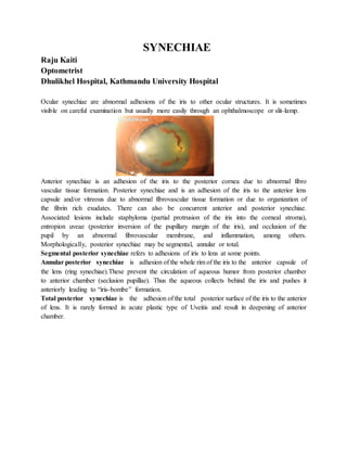
- 1. SYNECHIAE Raju Kaiti Optometrist Dhulikhel Hospital, Kathmandu University Hospital Ocular synechiae are abnormal adhesions of the iris to other ocular structures. It is sometimes visible on careful examination but usually more easily through an ophthalmoscope or slit-lamp. Anterior synechiae is an adhesion of the iris to the posterior cornea due to abnormal fibro vascular tissue formation. Posterior synechiae and is an adhesion of the iris to the anterior lens capsule and/or vitreous due to abnormal fibrovascular tissue formation or due to organization of the fibrin rich exudates. There can also be concurrent anterior and posterior synechiae. Associated lesions include staphyloma (partial protrusion of the iris into the corneal stroma), entropion uveae (posterior inversion of the pupillary margin of the iris), and occlusion of the pupil by an abnormal fibrovascular membrane, and inflammation, among others. Morphologically, posterior synechiae may be segmental, annular or total. Segmental posterior synechiae refers to adhesions of iris to lens at some points. Annular posterior synechiae is adhesion of the whole rim of the iris to the anterior capsule of the lens (ring synechiae).These prevent the circulation of aqueous humor from posterior chamber to anterior chamber (seclusion pupillae). Thus the aqueous collects behind the iris and pushes it anteriorly leading to “iris-bombe” formation. Total posterior synechiae is the adhesion of the total posterior surface of the iris to the anterior of lens. It is rarely formed in acute plastic type of Uveitis and result in deepening of anterior chamber.
- 2. Anterior synechiae causes closed angle glaucoma, which means that the iris closes the drainage way of aqueous humor which in turn raises the intraocular pressure. Posterior synechiae also cause glaucoma, but with a different mechanism. In posterior synechiae, the iris adheres to the lens, blocking the flow of aqueous humor from the posterior chamber to the anterior chamber. This blocked drainage raises the intraocular pressure. Etiology: Infective uveitis : such as herpes simplex, herpes zoster, tuberculosis and syphilis Allergic (hypersensitivity) uveitis Toxic uveitis Traumatic uveitis Uveitis associated with non-infective systemic diseases Posterior synechiae are the most common ocular complications in chronic or recurrent anterior uveitis, such as HLA B27-associated uveitis, idiopathic anterior uveitis, and iridocyclitis in juvenile idiopathic arthritis, sarcoidosis, intermediate uveitis, lens-induced uveitis and uveitis-glaucoma-hyphema (UGH) syndrome. Intraocular inflammation, especially of the iris and ciliary body. Synechiae can also be squeal of many ocular diseases, such as cataract, increased intraocular pressure, compressive or invasive intraocular neoplasms, and inflammation resulting from various causes. Idiopathic uveitis Signs: Central iridocorneal synechiae are frequently associated with rubeotic iris vessels Annular Posterior synechiae Total posterior synechiae FestoonedPupil
- 3. Pupil is irregular/ festooned pupil Synechiae associated with uveitis have signs like Keratic precipitates, anterior chamber cells and flares, irregular pupils, ciliary injections, vitreous cells, iris abnormalities, fundal changes as well. These signs depend on type of uveitis anterior, intermediate uveitis, posterior uveitis and pan uveitis. Peripheral anterior synechiae are a well-recognized consequence of altered anterior chamber (AC) anatomy and anterior chamber inflammation. Peripheral anterior synechiae can subsequently result in significant morbidity as a precipitant to secondary angle-closure glaucoma. Symptoms Peripheral anterior synechiae are usually asymptomatic unless large areas of at least 270° are involved. Peripheral anterior synechiae can present in the following manners: Acute angle closure with the classic constellation of symptoms, including ocular pain, headaches, blurred vision, photophobia, watering and halos.. Reduced vision due to corneal edema or end-stage glaucomatous optic neuropathy If associated with systemic diseases may have recurrent attacks Differential Diagnosis: Cataract, Traumatic Filtering Bleb Complications Uveitis, Anterior, Granulomatous/Nongranulomatous Uveitis, Intermediate, Juvenile Idiopathic Arthritis Sarcoidosis Glaucoma, Angle Closure, Acute/ Chronic Glaucoma, Aphakic and Pseudophakic Glaucoma, Phacolytic/ Phacomorphic Herpes Simplex/Herpes Zoster HLA-B27 Syndromes Melanoma: Choroidal/ Ciliary Body/ Iris Neurofibromatosis-1 Retinopathy of Prematurity Management: Mydriatic/cycloplegic agents, such as topical homatropine, which is similar in action to atropine, are useful in breaking and preventing the formation of posterior synechiae by keeping the iris dilated and away from the crystalline lens. Dilation of the pupil in an eye with synechiae can cause the pupil to take an irregular (non-circular) shape. If the pupil can be fully dilated during
- 4. the treatment of iritis, the prognosis for recovery from synechiae is good. Inflammation from synechiae or synechia may be treated with topical corticosteroids. In some cases, surgical interventions might be required. In annular posterior synechiae, a complete iridectomy or laser irodotomy might be required. In cases with total posterior synechiae with complicated cataract, removal of the lens is after rupturing the posterior synechiae with iris repository. No specific medical management exists pertaining to the treatment of peripheral anterior synechiae (PAS). In general, the treatment of the underlying etiology prevents the formation of peripheral anterior synechiae. The appropriate management of peripheral anterior synechiae depends on the disease process that leads to peripheral anterior synechiae formation. The following drug categories may be considered depending on the primary diagnosis: topical beta-blockers, topical alpha-agonists, topical carbonic anhydrase inhibitors, oral carbonic anhydrase inhibitors, topical prostaglandin analogs, miotics, cycloplegic, and topical corticosteroids. Treat intraocular pressure (IOP) as necessary. o Topical alpha-agonists, beta-blockers, CAIs, and prostaglandin analogs may be useful in lowering intraocular pressure in eyes with peripheral anterior synechiae. o Miotics are useful in pupil block due to primary angle closure but may accentuate angle closure in posterior pushing mechanisms. o Miotics or prostaglandin analogs likely will not be useful in cases where 360° peripheral anterior synechiae exist. Inflammatory states o Topical steroids minimize inflammation and therefore, PAS formation. o Cycloplegics should be used to prevent posterior synechiae. o Mitotic and epinephrine should be avoided because they can increase inflammation. Surgical care: Nd:YAG/argon laser irodotomy Surgical iridectomy Argon laser peripheral iridoplasty Argon laser pupilloplasty is used to expand/enlarge pupil, which may break acute angle- closure attack and/or posterior synechiae. Nd: YAG peripheral synechialysis can be attempted in early synechial closure but may not be effective if the synechiae are firm. Surgical goniosynechialysis Glaucoma filtering procedures Optometric management:
- 5. Mydriatic/cycloplegic agents can be prescribed and are useful in breaking and preventing the formation of posterior synechiae. Prescribing protective sunglasses will help the patients with photophobia. Inflammatory conditions can be treated with topical steroids. Measuring intraocular pressure is important and if raised should be treated with anti- glaucoma medications. Apart from these the causative conditions should be ruled out and treated. Proper counseling should be provided and in cases of recurrent attacks systemic evaluations should be advised.