

Recommended
More Related Content
What's hot
What's hot (20)
Viewers also liked
Similar to Difficult vaginal hysterectomy
Similar to Difficult vaginal hysterectomy (20)
More from MOHAMMAD QUAYYUM
Recently uploaded
Recently uploaded (20)
Difficult vaginal hysterectomy
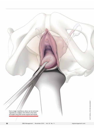
- 1. scott bodell for obg Management Even a large, myomatous uterus can be removed vaginally by a surgeon who follows a few basic principles using well-honed surgical techniques. 32 OBG Management | November 2010 | Vol. 22 No. 11 obgmanagement.com
- 2. Surgical Techniques The difficult vaginal hysterectomy: 5 keys to success Challenges can be overcome by ensuring overall surgical expertise, adequate exposure, proper entry into the anterior cul-de-sac, good mobility, and competent morcellation John A. Occhino, MD, and John B. Gebhart, MD, MS CASE Is the vaginal route appropriate? A 46-year-old woman (para 2 with 1 cesarean delivery) who has a history of benign menorrhagia comes to your office seeking definitive treatment after medical therapy fails to alleviate her bleeding. Pelvic examination reveals a uterus of 14-weeks’ size that descends to the distal vagina, with good vaginal access. What options for hysterectomy do you offer to the patient? T here are few absolute contraindications to a vaginal approach to hysterectomy. Among them are advanced pelvic malignancy, severe endometriosis, and a suspicious adnexal mass. Contraindications do not include a history of pelvic surgery, cesarean delivery, or an enlarged uterus. Such circumstances may increase the challenges involved in performing vaginal hysterectomy, but data suggest that it is achievable in these settings.1–7 Dr. Occhino is Assistant Professor in the Department of Obstetrics and Gynecology at the University of Missouri in Columbia, Mo. Dr. Gebhart is Associate Professor in the Department of Obstetrics and Gynecology, Division of Gynecologic Surgery, and Fellowship Program Director at the Mayo Clinic in Rochester, Minn. The authors report no financial relationships relevant to this article. o b g m a n a g e m e n t . c om Vaginal hysterectomy offers substantial benefits, making the challenges worthwhile in most cases. It is the original minimally invasive approach to hysterectomy. It yields outcomes, postoperative discomfort levels, and recovery times similar to those of laparoscopic-assisted vaginal hysterectomy, total laparoscopic hysterectomy, and roboticassisted hysterectomy—but the vaginal approach is more cost-effective.3,8–11 This article focuses on strategies and techniques for accomplishing the difficult vaginal hysterectomy, describing five keys to success: • surgical experience • adequate exposure • entry into the anterior cul-de-sac • terine mobility (or the ability to create it) u • good morcellation technique. For clarity throughout this article, we assume that hysterectomy is being performed for benign indications. 1 # Surgical experience Vaginal hysterectomy can be performed successfully in the setting of nulliparity, uterine enlargement, and a history of cesarean delivery, provided the surgeon has the appropriate skill set, assistance, and patience. Little is lost if the operation is attempted vaginally but needs to be converted Vol. 22 No. 11 | November 2010 | OBG Management In this Article Entry into the anterior cul-de-sac page 40 Video at obgmanagement.com When cystotomy happens page 44 Video at obgmanagement.com Good morcellation technique page 46 Video at obgmanagement.com continued on page 39 33
- 3. Surgical techniques / difficult Vaginal hysterectomy FIGURE 1 Fixed retraction A Magrina-Bookwalter fixed vaginal retractor in place at the time of surgery. to a laparoscopic or open approach. If the surgeon persists in attempting to complete each hysterectomy vaginally, he or she will gradually improve in skill and eventually gain the ability to complete tougher cases without the need to convert. Chen and colleagues developed and validated the Vaginal Surgical Skills Index (VSSI), identifying 13 aspects of successful vaginal surgery: • inspection • incision • maintenance of visibility • use of assistance • knowledge of instruments FIGURE 2 • tissue and instrument handling • electrosurgery • knot-tying and ligation • hemostasis • procedure completion • time and motion • flow and forward planning • knowledge of the procedure.12 Thirty-seven trainees from two institutions were evaluated during 76 surgical procedures. The trainees were supervised by five surgeons, who completed the evaluations immediately after each procedure. A sixth surgeon from a different institution watched videos of each procedure and acted as a blinded external reviewer. Chen and colleagues found good interrater and intra-rater reliability and high internal consistency for the VSSI, one of the first tools to objectively assess vaginal surgery skills. 2 # Obtaining adequate exposure Good anesthesia, proper lighting, and fixed retraction are invaluable when operating vaginally. A weighted speculum with Deaver retractors at 3, 9, and 12 o’clock provide good visualization if assistants are available. Selfretaining retractors are also useful (Figure 1). We prefer to empty the bladder before Palpate the bladder reflection A B A. Use the index finger to palpate the bladder reflection. B. Note it with a marking. o b g m a n a g e m e n t . c om Vol. 22 No. 11 | November 2010 | OBG Management 39
- 4. Surgical techniques / difficult Vaginal hysterectomy FIGURE 3 Incise the vaginal epithelium A sharp and deep incision aids in identification of the appropriate plane. Dissect the bladder free of the uterus FIGURE 4 We recommend sharp dissection to free the bladder from the uterus. making the vaginal incision, although no data suggest that doing so helps to avoid inadvertent bladder injury. 3 # Entry into the anterior cul-de-sac We prefer to enter the anterior cul-de-sac first. The pertinent risk in vaginal hysterectomy is injury to the bladder. Because anatomic planes are undisturbed at this point, we feel entry into the anterior cul-de-sac gives the surgeon the best opportunity to avoid injury. If it is a struggle or lack of uterine descent Use traction and counter-traction FIGURE 5 Sharp entry into the peritoneal cavity is enhanced through the use of traction and counter-traction. 40 FIGURE 6 makes it difficult, then start with entry into the posterior cul-de-sac (see “gaining mobility” below). In a patient who does not have a history of surgery, palpation of the bladder reflection on the anterior uterus can help determine the appropriate site for the initial incision (Figure 2, page 39). Place a Deaver retractor anteriorly to assist with retraction. It is important to make the first incision deep enough to set up entry into the anterior cul-de-sac (Figure 3). With traction on the uterus, grasp the anterior vaginal epithelium and elevate it to Open the peritoneal defect Place scissors into the peritoneal defect and spread the blades wide. OBG Management | November 2010 | Vol. 22 No. 11 obgmanagement.com
- 5. Surgical techniques / difficult Vaginal hysterectomy allow sharp dissection and mobilization of the bladder (Figure 4, page 40). We prefer sharp dissection rather than blunt dissection because it maintains surgical planes and is more precise. Once the peritoneum is identified, grasp, elevate, and incise it (Figure 5 , page 40). Insert scissors through the peritoneal defect, spread the tips widely, and place the anterior Deaver retractor intraperitoneally (Figure 6 , page 40) so that bowel can be visualized (Figure 7). (This step can be viewed in Video 1, which accompanies this article at obgmanagement.com.) If the patient has a history of cesarean delivery, entry into the anterior cul-de-sac can be more challenging. Several maneuvers can help avert bladder injury: • tay on the uterus during dissection into S the vesicovaginal space. It is better to stay deep and cut into the uterus than to dissect superficially and end up with a cystotomy. • etrograde fill the bladder to identify R the plane between the bladder and the uterus. • ostpone entry into the anterior cul-deP sac until after posterior entry, ligation of the uterosacral ligaments, and the first “bite” of the cardinal ligaments. • se a uterine sound, bent into a “U” U shape, passing it through the urethra into the bladder and allowing the point to come back toward the surgeon (while it is in the bladder). Manipulation of this sound through external palpation should make it possible to identify the bladder reflection. • n the setting of a small uterus, after I entering the posterior cul-de-sac, pass a finger along the back of the uterus, around the fundus, and back toward the surgeon. This maneuver identifies the optimal spot for dissection between the bladder and the uterus. When cervical elongation is encountered during entry into the cul-de-sac, the peritoneal reflection will be higher (both anteriorly and posteriorly), and additional bites on the pedicles, as well as additional o b g m a n a g e m e n t . c om FIGURE 7 Visualize the bowel Visualization of the bowel confirms an intraperitoneal location. When the cervix is elongated FIGURE 8 To help avert bladder injury during entry into the anterior culde-sac in a patient who has a history of cesarean delivery, stay on the uterus during dissection into the vesico vaginal space When the cervix is elongated, the peritoneal reflection, both anteriorly and posteriorly, is much higher on the uterus (near the small myoma). Vol. 22 No. 11 | November 2010 | OBG Management 43
- 6. Surgical techniques / difficult Vaginal hysterectomy Begin morcellation FIGURE 10 Bi-valve the cervix JilL Rhead for obg Management FIGURE 9 Once the uterine vessels have been controlled, morcellation may begin. Bi-valve the cervix in the midline, following the endocervical canal. d issection, may be required before entry is accomplished (Figure 8 , page 43). When cystotomy happens If cystotomy occurs during an attempt to enter the anterior cul-de-sac, a number of steps can lead to successful repair. Rather than repair the defect immediately, mark it with a suture for later identification. Once the uterus is removed, inspect the bladder carefully to identify any additional injuries, FIGURE 11 Excise the fibroid then repair the cystotomy using absorbable 2-0 suture on a tapered needle (we prefer chromic suture). Begin by taking a full-thickness bite of tissue, just lateral to the edge of the cystotomy. Then run the suture, incorporating the bladder epithelium into the closure. Place a second, imbricating layer of the same suture. Last, if possible, sew the peritoneum beneath the bladder over the repair for an additional layer of reinforcement. (View this step in Video 2 at obgmanagement.com.) Cystoscopy helps to visualize the repair and test for water-tightness, and assess ureteral patency. Keep the bladder on catheter drainage for 10 to 14 days. 4 # Fibroids may be excised sharply with the aid of a scalpel and traction supplied by a tenaculum. 44 OBG Management | November 2010 | Vol. 22 No. 11 Gaining mobility In the setting of nulliparity or a small, well-supported uterus, it may be necessary to create mobility to accomplish the hysterectomy vaginally. Begin by entering the posterior cul-de-sac and cutting and suture ligating the uterosacral ligaments. Then take the first bite of the cardinal pedicles bilaterally. This typically facilitates uterine descent, making it possible to enter the anterior culde-sac and accomplish the hysterectomy. On occasion, once the uterine arteries have been secured, you can split (bi-valve) obgmanagement.com
- 7. Surgical techniques / difficult Vaginal hysterectomy Don’t settle on a route prematurely It is important to understand the individual patient’s anatomy and underlying disease process before deciding on an appropriate surgical route. For this reason, a general medical and surgical history and a focused physical exam should precede any decision to operate. During the pelvic examination, note the size and mobility of the uterus, any associated uterovaginal prolapse, the presence of any adnexal mass or tenderness, vaginal capacity, and the adequacy of the pubic arch. If you are unable to determine the size of the uterus on examination, owing to the patients’ body habitus or discomfort, pelvic ultrasonography may be helpful. When office examination is difficult, or when it is impossible to gather substantial information about uterine characteristics, an examination under anesthesia is an excellent way to help determine the optimal route of hysterectomy. Provided the patient is properly apprised about this examination beforehand, the surgeon can then proceed to the appropriate surgical route once the exam is completed. Ensure consent for all aspects of the procedure As for any surgery, the informed consent discussion is important. Regardless of the hysterectomy approach, this discussion should include a mention of risks, benefits, and alternatives to surgery; the possible need for additional procedures (in the setting of unexpected pathology); and consent or decline of blood products, if needed. If photography or videotaping of the procedure is desired, this option needs to be discussed as well. When a vaginal approach is planned, there is always a small chance that it will have to be converted to a laparoscopic or open approach. This possibility should be relayed to the patient during the preoperative discussion. Inevitably, some cases fall on the border between the vaginal approach and another route. When this happens, we prefer to ask the patient to consent to the aforementioned examination under anesthesia, with the understanding that we may proceed as indicated to hysterectomy, based on the findings of that exam. For example, in the opening case, the informed consent discussion would likely go something like this: Mrs. Smith, because of fibroids, your uterus is enlarged to about the size of a small cantaloupe. Because you have had a vaginal delivery and your uterus is mobile, I think I will be able to remove it through the vagina. If vaginal removal is possible, you are likely to have a shorter recovery and a lower risk of complications than if a different approach is required. However, if I am unable to do a vaginal hysterectomy, an abdominal operation may need to be performed and would involve either a laparoscopy or an incision in the lower abdomen. I would like to evaluate things after you are asleep in the operating room. At that time, I will make the final decision about the best route for your hysterectomy. For the exam, the anesthetized patient should be placed in the dorsal lithotomy position with her legs in stirrups. Often, there is greater vaginal access and uterine mobility at this time. the uterus to gain access to the utero-ovarian pedicles and complete the hysterectomy. 5 # Good morcellation technique Morcellation facilitates removal of the large uterus. As experience with morcellation increases, the surgeon will be able to remove larger and larger uteri vaginally. However, it is critical to secure the uterine vessels before morcellation begins, and it is preferable to have entered both cul-de-sacs as well. 46 OBG Management | November 2010 | Vol. 22 No. 11 Once those steps have been accomplished, bi-valve the cervix in the midline, following the endocervical canal to stay in the midline (Figures 9,10 , page 44). Use a tenaculum to grasp bites of the uterus in an anterior and posterior fashion (Figure 11 , page 44). This step reduces uterine size until the fundus can be inverted and the utero-ovarian pedicles secured. Be sure to excise uterine tissue under direct visualization to avoid inadvertent injury to the bowel and bladder. (See Video 3 at obgmanagement.com for a demonstration of morcellation technique.) obgmanagement.com
- 8. We need to do more vaginal procedures Of the roughly 600,000 hysterectomies performed each year in the United States, roughly 60% are performed abdominally.13,14 More and more hysterectomies are being done laparoscopically or with robotic assistance, and fewer straight vaginal hysterectomies are performed.15 Recent graduates are less likely than their predecessors to be up-to-date on this important skill set—a fact that may lead to further decreases in the number of hysterectomies performed vaginally each year.16 We need to make every effort to increase the rate of vaginal hysterectomy. Not only is it better for the patient; it saves precious health-care dollars. CASE Resolved The vaginal approach was chosen for this patient. After ligation of the uterine vessels, morcellation allowed for a successful hysterectomy without complication. References 1. Figueiredo O, Figueiredo EG, Figueiredo PG, Pelosi MA 3rd, Pelosi MA. Vaginal removal of the benign nonprolapsed uterus: experience with 300 consecutive operations. Obstet Gynecol. 1999;94(3):348–351. 2. Rooney CM, Crawford AT, Vassallo BJ, Kleeman SD, Karram MM. Is previous cesarean section a risk for incidental cystotomy at the time of hysterectomy? A case-controlled study. Am J Obstet Gynecol. 2005;193(6):2041–2044. 3. Sesti F, Calonzi F, Ruggeri V, Pietropolli A, Piccione E. A comparison of vaginal, laparoscopic-assisted vaginal, and minilaparotomy hysterectomies for enlarged myomatous uteri. Int J Gynaecol Obstet. 2008;103(3):227–231. 4. Sheth SS, Malpani AN. Vaginal hysterectomy following previous cesarean section. Int J Gynaecol Obstet. 1995;50(2):165–169. 5. Kovac SR. Guidelines to determine the route of hysterectomy. Obstet Gynecol. 1995;85(1):18–23. 6. Unger JB, Meeks GR. Vaginal hysterectomy in women with history of previous cesarean delivery. Am J Obstet Gynecol. 1998;179(6 Pt 1):1473–1478. 7. Paparella P, Sizzi O, Rossetti A, De Benedittis F, Paparella R. Vaginal hysterectomy in generally considered contraindications to vaginal surgery. Arch Gynecol Obstet. 2004;270(2):104–109. 8. Schindlbeck C, Klauser K, Dian D, Janni W, Friese K. Comparison of total laparoscopic, vaginal and abdominal hysterectomy. Arch Gynecol Obstet. 2008;277(4):331–337. 9. Nazah I, Robin F, Jais JP, et al. Comparison between bisection/ morcellation and myometrial coring for reducing large uteri during vaginal hysterectomy or laparoscopically assisted vaginal hysterectomy: results of a randomized prospective study. Acta Obstet Gynecol Scand. 2003;82(11):1037–1042. 10. Wexner SD, Bergamaschi R, Lacy A, et al. The current status of robotic pelvic surgery: results of a multinational interdisciplinary consensus conference. Surg Endosc. 2009;23(2):438–442. 11. Johnson N, Barlow D, Lethaby A, Tavender E, Curr E, Garry R. Surgical approach to hysterectomy for benign gynaecological disease. Cochrane Database Syst Rev. 2006;(2):CD003677. 12. Chen CC, Korn A, Klingele C, et al. Objective assessment of vaginal surgical skills. Am J Obstet Gynecol. 2010;203(1):79.e1–8. 13. Keshavarz H, Hillis SD, Kieke BA, Marchbanks PA. Hysterectomy surveillance—United States, 1994-1999. MMWR CDC Surveill Summ. 2002;51(SS05):1–8. http:// www.cdc.gov/mmwr/preview/mmwrhtml/ss5105a1.htm. Published July 12, 2002. Accessed October 3, 2010. 14. Whiteman MK, Hillis SD, Jamieson DJ, et al. Inpatient hysterectomy surveillance in the United States, 2000-2004. Am J Obstet Gynecol. 2008;198(1):34.e1–7. 15. Merrill, RM. Hysterectomy surveillance in the United States, 1997 through 2005. Medical Science Monitor, 2008;14(1):CR24–31. 16. Julian TM. Vaginal hysterectomy: an apparent exception to evidence-based decision making. Obstet Gynecol. 2008;111(4):812–813. 2010 Pelvic Anatomy and GYNECOLOGIC surgery symposium Join Dr. Gebhart, along with Drs. Tommaso Falcone, Mark Walters, Javier Magrina, Michael Baggish, and Mickey Karram December 9–11, 2010 at the WYNN Las Vegas See www.PAGS-cme.org for details. o b g m a n a g e m e n t . c om Vol. 22 No. 11 | November 2010 | OBG Management 47