Call Girls Aurangabad Just Call 8250077686 Top Class Call Girl Service Available
Historia de la Radiología 2
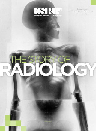
1. THE STORY OF RADIOLOGY
AN INTRODUCTION
THE STORY OF
RADIOLOGY
1
Volume 2
2. THE STORY OF RADIOLOGY
AN INTRODUCTION
THE STORY OF
RADIOLOGY
CONTENTS
Published by the
ESR – European Society of Radiology
In cooperation with
ISHRAD – The International Society for the History of Radiology
Deutsches Röntgen Museum
October 2013
Coordination: ESR Office
Neutorgasse 9, 1010 Vienna, Austria
Phone: (+ 43 1) 533 40 64-0
E-mail: communications@myESR.org
www.myESR.org
Associate Editors: Adrian Thomas & Uwe Busch
Managing Editor: Julia Patuzzi
Editors: Simon Lee, Michael Crean
Art Direction & Layout: Robert Punz at zerozero.graphicdesign
Photo Credits:
see page 102
The content of this book is the work of the authors and does not necessarily
represent the views of the European Society of Radiology.
05 07 25
49 67
INTRODUCTION
THE PROGRESS
IN RADIOLOGY
IN 1896
A SHORT HISTORY
OF EARL RADIATION
Y
PROTECTION
THE HISTORY OF
CONVENTIONAL
TOMOGRAPHY
TWO CENTENARIES:
WILLIAM COOLIDGE &
GUSTAV BUCKY
87
FLORENCE STONEY:
X-RAY NOTES
FROM THE U.S.
3
3. THE STORY OF RADIOLOGY
AN INTRODUCTION
THE STORY OF RADIOLOGY
AN INTRODUCTION
THE STORY OF
RADIOLOGY
AN INTRODUCTION
The world of radiology is celebrating again. This time it’s the second International Day of Radiology, and this
book marks the second instalment in the Story of Radiology; covering more than a century of scientific discovery and innovation which has revolutionised medicine.
The first International Day of Radiology was organised by the European Society of Radiology (ESR), the Radiological Society of North America (RSNA) and the American College of Radiology (ACR) as a joint initiative.
Though the main aim of the campaign is to promote the benefits of radiology to the wider public, we haven’t
forgotten about the radiological community and that is why we have put together this second volume on the
history of radiology.
WILHELM
CONRAD
RÖNTGEN
(1845–1923),
FATHER OF
RADIOLOGY
However, as was the case last year, we could not have produced this book without help from the experts at the
International Society for the History of Radiology (ISHRAD) and the German Röntgen Museum, who were kind
enough to provide the content of this book. Given the rapid technological progress and development taking
place in radiology, it is more important than ever that we understand the history of the discipline in order to
foresee what future developments might bring.
This book is only a snippet of the vast history of radiology, and so it is best to view it as merely another chapter
in the Story of Radiology. We are a confident that this second volume will prove as popular as the last, and we
look forward to adding many more chapters to the story.
4
5
4. THE PROGRESS
IN RADIOLOGY
IN 1896
BY UWE BUSCH
:
FIRST ANGIOGRAM OF A HAND, MADE BY HASCHEL AND
IN JANUARY 1896 WITH TEICHMANN SOLUTION
6
LINDENTHAL
7
5. THE STORY OF RADIOLOGY
THE STORY OF RADIOLOGY
THE PROGRESS
IN RADIOLOGY
IN 1896
THE PROGRESS
IN RADIOLOGY
IN 1896
The year 1896 was a momentous one, with major discoveries and
The news spread like wildfire. Shortly afterwards, articles were pub-
inventions in the field of radiodiagnostics and the emergence of radio-
lished in the Frankfurter Zeitung (7.1.), the Electrical Engineer, New
therapy. In the first year after the discovery of x-rays, a total of 49 books
York (8.1), the Würzburger Anzeiger (9.1), The Electrician, London
and brochures, and 1,044 scientific essays were written on the scien-
(10.1), The Lancet (11.1), the British Medical Journal (11.1), Le Matin,
tific aspects and possible applications of the newly discovered rays1. A
Paris (13.1), Nature, London, The New York Times, (16.1), Science,
multitude of these publications dealt specifically with possible appli-
New York (24.1.), and La Settimana, Florence (25.1.). Due to the global
cations in medicine.
circulation of the sensational news, Röntgen received an inquiry from
the American physician Robert Watkins in New York, as early as Janu-
On November 8, 1895, while doing research work on the electric dis-
ary 8, requesting him to send some radiographs.
charge process in diluted gas, Wilhelm Conrad Röntgen (1845–1923)
discovered a ‘new kind of ray’ which was previously unknown to
In addition to Watkins, physicians in Germany heard about Röntgen’s
physicists. It took Röntgen only six weeks to finish his first scientific
discovery from the newspapers. Surprise, laughter and disbelief fre-
research on this phenomenon. On Saturday, December 28, he submit-
quently led many to exclaim, “we must have such an apparatus”.
ted his manuscript to the secretary of the Würzburg Physical Medical
Society. On Tuesday, December 31, he received the copies of his paper,
The fascination with medical diagnostics in this new era was described
which he sent together with nine x-rays to his colleagues.2 One of the
by the orthopaedic surgeon Hermann Gocht (1869–1938). In order to
first recipients was his friend, Professor Franz Exner (1849–1926),
get more information about the new rays, Gocht and his friend Gustav
director of the Second Physicochemical Institute of the University of
Opitz contacted the glass blower, Carl Heinrich Florenz Müller in Ham-
Vienna, with whom Röntgen had been acquainted since their academic
burg (1845–1912).
days in Zurich. On Saturday, January 4, 1986, Exner showed Röntgen’s
x-rays to a group including Ernst Lechner, professor of physics at the
German University in Prague, who informed his father, the editor of
on the raised ground floor of the front building; the mercury air pump
Vienna’s daily newspaper Die Presse. That same night the first news
for the evacuation of the tubes stood in the front room and was oper-
article on x-rays was written and then published in Die Presse on Sun-
ated by Müller’s factotum, Mr. Schmidt, a kind, very capable but sim-
day, January 5, under the headline ‘A Sensational Discovery’. In addi-
ple man, who became a victim of the x-rays too.
tion to the scientific facts, the possible development of these new rays
Furthermore there were some smaller inductors and a big 25–30cm
was described in a prophetic manner.
8
“When I think of this first evening! ... The factory rooms of Müller were
inductor with a platinum interrupter; more than a dozen x-ray tubes were
9
6. THE STORY OF RADIOLOGY
THE STORY OF RADIOLOGY
THE PROGRESS
IN RADIOLOGY
IN 1896
THE PROGRESS
IN RADIOLOGY
IN 1896
available. Müller had already produced some plates with bowls, spirals,
FIRST MEDICAL SUCCESSES
drawing instruments, fingers and parts of hands. At first he showed us
The early understanding of the possible applications of the new rays in
his installation and explained the construction and evacuation of the
medicine developed from the amicable relationship between Röntgen
tubes. In a back room sat a glass blower, Mr. Becker, who created new
and Exner. As early as January 10, Exner’s brother, the physiologist
tubes and parts of tubes in front of our very eyes. These first tubes were
Sigmund Exner, reported on Röntgen’s discovery to the Royal Impe-
still constructed in a way that the cathode rays started from an alumin-
rial Society of Physicians of Vienna. After his speech, Sigmund Exner
ium plane mirror and fell on the opposite convex glass wall. This spot of
addressed Professor von Neusser, the director of the Second Vienna
the glass wall fluoresced especially intensively and became so very hot
School of Medicine, and asked him to send patients for the purpose of
that one had to be very careful with the current supply; sometimes after
testing the diagnostic application of x-rays at his brother’s institute. It
a long exposure time, the tube suddenly burst with a big bang.
was Neusser’s assistant, Gustav Kaiser (1871–1947) who was entrusted
A small apparatus was also part of this very simply range of instru-
with this task. The first medically indicated radiograph was taken
ments. This small apparatus had been built by Müller himself who gave
together with Eduard Haschek (1875–1947) who at that time was Franz
it the name ‘Searcher’: A black cardboard cylinder which was closed
Exner’s assistant at the physics institute. The patient’s hand showed
at one end; some bariumtetracyanoplatinate crystals had been fixed on
trauma to the middle phalanx.
the inner bottom of the cylinder.
In my thoughts I can still see and hear the joy with which Müller dem-
Sigmund Exner presented a second x-ray image on January 17, which was
onstrated everything to us, and I remember the tension and impatience
important to the development of radiology. The anatomist Tandler placed
in anticipation of the first experiments. Finally, the inductor was con-
the hand of a corpse at the disposal of Haschek and Otto Lindenthal
nected and the pear-shaped blown tubes lit up for the first time. Oppo-
(1872–1947) at the physics institute. The arterial vessels were filled with
site the cathode, the glass wall fluoresced especially intensively; when
Teichmann’s solution (iodine solution), a mixture of chalk, cinnabar and
the current had been switched off he made us feel the hot tube wall and
paraffin. After 57 minutes of exposure, the first angiogram was created.
finally we put the searcher before our eyes in order to catch a glimpse
of the cathode rays and to observe the glowing of the bariumtetracyanoplatinate crystals. At first we saw nothing until suddenly a weak light
Würzburg Physical Medical Society. After being welcomed by rap-
appeared. This evening, the small searcher wandered from one eye to
turous applause Röntgen held his lecture on ‘A new kind of rays’.
another with us three, the light was very slight but to us it meant an
Towards the end of his speech, the silhouette of the skeleton of a human
almost supernatural radiance and a glance into a new world”3.
10
On January 23, Röntgen received an invitation to give a lecture at the
hand was taken by radiograph. It was the right hand of the honorary
11
7. THE STORY OF RADIOLOGY
THE EARL DAYS OF RADIOLOGY
Y
THE STORY OF RADIOLOGY
THE PROGRESS
IN RADIOLOGY
IN 1896
president of the society, Prof. Albrecht von Kölliker, who suggested nam-
Several papers were published on the development of the photographic
ing the new rays ‘Röntgen rays’. During the ensuing discussion, Röntgen
radiographic technique. C. Henry from Paris suggested the application
talked about the possibility of using the new rays for medical purposes.
of phosphorescent white vitriol for the magnification of the effect of
the x-rays.6 The American x-ray pioneer, Michael Pupin (1858–1935),
achieved considerably better results with fluorescent screens made
APPARATUS AND TECHNICAL PROCEEDINGS
from calcium tungstate in early February. His x-ray of a hand with a
Due to the rapid spread of the news of these sensational rays and the
shot gun wound is very impressive.7
fascination about the first x-rays of hands, research work was carried
out around the world and the technical apparatus for the production
Shortening the long exposure time was a technical challenge. In May,
of x-rays began to improve. In particular, the ionic tubes had to be
with preparatory work by John Macintyre (1857–1928)8, Hermann
improved. In his second communication, Röntgen explains the con-
Gocht succeeded for the first time in reducing the exposure time from
struction of special Röntgen tubes.
45 seconds to 5 seconds9. Gocht’s idea of snapshots was used in the
development of the apparatus for x-ray cinematography by the British
“... According to my experience gained to date, platinum is best suited
physician John Macintyre in December. The movements of a frog’s legs
for the production of most intensive x-rays. For some weeks now I have
could be seen in a first x-ray film shown to the Philosophical Society
been using a discharging device with good success, in which a con-
in Glasgow.10
cave mirror of aluminium serves as a cathode, and a platinum sheet is
placed against the mirror axis at an angle of 45 degrees and installed in
The method of producing stereo pictures in photography was in use before
the bending radius to function as an anode …”4
the discovery of x-rays. Röntgen himself, as an enthusiastic photographer, had made many stereo pictures. On March 11, 1896, Elihu Thom-
A ‘Röntgen industry’ developed rather quickly. Companies like that of
son (1853–1937), a British-American electronic engineer, reported on the
C.H.F. Müller in Hamburg or Emil Gundelach in Gehlberg were produc-
production of stereoscopic x-rays.11 That same month, Armand Imbert
ing special x-ray tubes by the spring of 1896. Werner Siemens (1816–
(1850–1922) and Henry Bertin-Sans (1862–1952) presented some impres-
1892) and Johann Georg Halske (1814–1890) founded a telegraph con-
sive stereograms of two mice at the Academy of Science in Paris.12
struction company in 1847 and they immediately started producing
their own x-ray devices. In March, the company Siemens & Halske
applied for their first patent for a special x-ray tube, which already
Carl Moritz Schleussner (1868–1952) which contributed to a notice-
included an appliance for the regulation of a vacuum.5
12
It was the special radiographic photo plates produced for Röntgen by
able improvement in photography. The radiographic plate, which was
13
8. THE STORY OF RADIOLOGY
THE STORY OF RADIOLOGY
THE PROGRESS
IN RADIOLOGY
IN 1896
THE PROGRESS
IN RADIOLOGY
IN 1896
RADIOGRAPHIC
EXAMINATION IN
1896, PUBLISHED
IN THE GERMAN
JOURNAL
GARTENLAUBE
coated with a special silver bromide emulsion drastically reduced the
exposure time from several minutes to just a few seconds.
These technical improvements paved the way for the successful application of x-rays in different clinical fields during 1896.
SKELETAL RADIOGRAPHY
Due to technical limitations at the beginning of 1896, the application of
x-rays could only be used to examine bone structure in the extremities,
where it was possible to achieve great success in the search for foreign
bodies. On January 19, the physician L. Pfaundler, from Graz, Austria,
located a needle in the hand of a twelve-year-old girl13 using x-rays,
which the surgeon had failed to locate in the wound. Further successes
in locating foreign bodies were published and in August, after several
technical improvements, Albert Eulenburg (1840–1917), from Berlin,
was able to take good x-rays of bullets in the brain.14
The application of x-rays moved on to look at fractures, and at the end of January, Fessler succeeded in producing the first x-ray of a forearm, with an exposure time of twelve minutes, at the University of Munich’s faculty of physics.15
In addition to locating foreign matter and diagnosing fractures, the
analysis of bone diseases was of great importance for surgical purposes.
Initial attempts to show a sarcoma on a shinbone were made by the surgeon Franz König (1832–1910) in Berlin. Part of the joint of the tibia of
a 46-year-old woman was distinctly swollen. The diagnosis confirmed
14
15
9. THE STORY OF RADIOLOGY
THE STORY OF RADIOLOGY
THE PROGRESS
IN RADIOLOGY
IN 1896
THE PROGRESS
IN RADIOLOGY
IN 1896
beyond any doubt the presence of a neoplasm. Taking x-rays in vivo
him to examine the form and size of the heart.20 One month later, the
failed. It was only after amputating the limb that they succeeded in tak-
American Francis Henry Williams (1852–1936) published the first chest
ing the first x-rays of a tibia sarcoma.16
x-rays taken at the Massachusetts Institute of Technology in Boston.21
On February 17, Huber presented various x-rays taken at the Tech-
However, for these x-rays the exposure time was 60 minutes due to the
nische Reichsanstalt, including some examples of acute joint damage.17
poor performance of the x-ray unit, resulting in rather blurry radiographs.
The subsequent diagnosis was very limited and based on details such as
The rapid technological developments broadened the range of diagnos-
form and size of the heart, position of the diaphragm, large shadows
tic capabilities. By June, Charles Thurston Holland (1863–1941) had
caused by pleural fluid, translucence by pneumothorax, broadening of
already presented x-rays of congenital deformations of hands18, fol-
the mediastinum and the description of the thoracic skeleton.22
lowed by Julius Wolff (1869–1933), who succeeded in the radiological
diagnosis of congenital hip joint luxation and managed to demonstrate
the subsequent surgical treatment using radiographs.19
APPLICATION OF CONTRAST MEDIA
In his first publication Röntgen described the different levels of absorp-
In April, Ludwig Zehnder (1854–1949), professor of physics at the Uni-
tion: “Similar to metals, even salts in liquid or solid form can be distin-
versity of Freiburg and a student of Röntgen in Giessen, succeeded in
guished according to their permeability.”23
depicting the entire human skeleton using radiology for the first time.
His whole-body x-ray of a dead soldier measures 1.84m and was taken
In March, Wolf Becher (1862–1910), a physician from Berlin, used
in nine sections, with an exposure time of five minutes per section.
these results to work on the depiction of internal organs through the
application of contrast media. To identify suitable contrast media, dif-
During 1896, numerous anatomic changes as well as diseases of the
ferent substances were examined for their suitability. Becher used lead
skeleton were diagnosed using radiodiagnostics and with the increas-
acetate in his examinations.24 Haschek and Lindenthal used mercuric
ing level of knowledge it was possible to treat these more successfully.
sulphide for the depiction of the blood vessels of an amputated hand.
Dutto used calcium sulphate25 to visualise the blood vessels, and Ernst
Sehrwald (1861–1945) performed in vitro examinations on absorption
THORAX
properties of halogens in July, thus paving the way for the use of non-
In March, the British pioneer John Macintyre succeeded in taking an
metallic elements like iodine compounds as contrast media.26
x-ray of a thorax organ in situ for the first time. The radiograph enabled
16
17
10. THE STORY OF RADIOLOGY
THE STORY OF RADIOLOGY
THE PROGRESS
IN RADIOLOGY
IN 1896
THE PROGRESS
IN RADIOLOGY
IN 1896
GASTROINTESTINAL, UROLOGICAL AND
GYNAECOLOGICAL RADIOLOGY
Following this success, Davis took an x-ray of a pregnant female patient,
After Becher had described the initial steps towards depicting the stom-
body of the foetus. He was able to define the head, but little more: “While
ach of a guinea pig, the German-American John C. Hemmeter (1864–
the experiment failed to distinctly outline the skeleton of the foetus, it
1931) made his patient swallow a balloon with a tube attached, which
offers information which may be of value in further attempts …”31
which took one-and-a-half hours before he was able to recognise the
was then filled with lead acetate. After the radiograph was taken the
lead acetate was aspirated and both the bag and tube removed from the
stomach.27 In April 1896, Carl Wegele suggested a practical solution for
DENTAL MEDICINE
examining the stomach of a living person using x-rays. To depict the
After Röntgen’s discovery, dentists quickly recognised the diagnostic
greater curvature, a soft stomach tube would be introduced orally like a
potential for their own medical field. Particularly in dental surgery,
gastric tube and then a metallic spiral would be added.28 Half a year later,
significant progress was made thanks to the new option of radiological
E. Lindemann returned to this idea and succeeded in producing the first
examination. In mid-January, the German dentist Otto Walkhoff (1860–
in vivo x-ray of the greater curvature. In his experiments, Lindemann
1934) asked the physicist Friedrich Giesel (1852–1927), from Braunsch-
used a rubber tube containing a fine copper mesh as a stomach tube.29
weig, to take an x-ray of his back teeth. The resulting image, which was
the first intraoral x-ray, was produced after an exposure time of around
Urologists were very interested in the application of x-rays. In July 1896
25 minutes. In March, the German physicist Walter König (1859–1936)
John Macintyre imaged a kidney stone using x-rays. He was able to
from Frankfurt published a radiograph of the front teeth of the upper
make a diagnosis of a kidney stone in his patient because the structure
and lower jaw.32
and the silhouette of the organ with the kidney stone were clearly visible on the x-ray. The subsequent operation confirmed this diagnosis.30
VETERINARY MEDICINE
In March, the American obstetrician Edward Parker Davis (1856–1973)
Animals were often used for the first radiographic experiments. In the
performed the first gynaecological experiment using x-rays when he
photographic portfolio gathered by Joseph Maria Eder (1855–1945)
placed the head of a newborn child into the pelvis of a female body.
and Eduard Valenta (1857–1937), from Vienna, one can find numerous
After an exposure time of one and a half hours he produced the first
radiographs of animals.33
gynaecological radiograph.
18
19
11. THE STORY OF RADIOLOGY
THE STORY OF RADIOLOGY
THE PROGRESS
IN RADIOLOGY
IN 1896
THE PROGRESS
IN RADIOLOGY
IN 1896
Despite the great interest of veterinarians in the new diagnostic possibilities of x-rays, their application only developed gradually. The
scientific interest, however, was evident at a very early stage. In 1896,
five treatises were published by veterinarians Eberlein and Tröster in
Germany; Hobday and Johnson in England; and Lemeoine in France.
Richard Eberlein (1869–1921), director of the Royal Veterinarian University in Berlin, demonstrated the advantages of the radiodiagnostic
procedure on animals34, but it was Hobday and Johnson who succeeded
in taking x-rays of a living horse in September.35
RADIOBIOLOGY, RADIOTHERAPY
AND RADIATION INJURIES
Despite intensive research, Scottish x-ray pioneer Campbell Swinton
(1863–1930) was unable to demonstrate any negative biological effects
RADIOGRAPH
OF A
CHAMELEON
1896
of x-rays. Three weeks after Swinton’s investigations, on April 23, John
Daniel, a physicist at the Vanderbilt University in Nashville, reported
a new phenomenon which had not been mentioned before. Daniel was
asked by a physician to locate a bullet in the head of a child by means
of x-rays. However, even after an exposure time of one hour there were
no results. Three weeks later, the child lost his hair.36 In April, the physician Wilhelm Marcus, from Berlin, reported an early case of dermatological changes in a 17-year-old male who had taken part in various
radioscopic experiments.37 As a result of this report, several physicians
used x-rays for the therapeutic treatment of hypertrochoses, eczema
and mycoses. The Viennese dermatologist Leopold Freud (1868–1943)
performed the first basic scientific research towards analysing the biological efficacy of x-rays in the treatment of diseases.
20
21
12. THE STORY OF RADIOLOGY
THE STORY OF RADIOLOGY
THE PROGRESS
IN RADIOLOGY
IN 1896
THE PROGRESS
IN RADIOLOGY
IN 1896
REFERENCES
(1)
Glasser, Otto: Wilhelm Conrad Röntgen und die Geschichte der Röntgenstrahlen. Berlin (1931)
(2)
Röntgen, Wilhelm Conrad: Über eine neue Art von Strahlen. Sitzungsberichte der Physikalisch-medizinischen Gesellschaft zu Würzburg. 137 (1895)
(3)
Gocht, Hermann: Die Gründung des chirurgischen Röntgeninstitutes am Allgemeinen Krankenhause Hamburg-Eppendorf. Separat Abdruck aus Beiträge zur klinischen Chirurgie, Band
XCII, Tübingen (1914)
(4)
Röntgen, Wilhelm Conrad: Eine neue Art von Strahlen.
II. Mittheilung. Sitzungsberichte der Physikalisch-medizinischen Gesellschaft zu Würzburg.
137 (1896)
(5)
Bienek, Karl: Medizinische Röntgentechnik in Deutschland. Stuttgart (1994)
(20)
MacIntire, John: Application of Roentgen Rays to the soft tissues of the body. Nature, 54, 451
(1896)
(21)
Williams, Francis Henry: The Roentgen rays in thoracic diseases. American Journal medical
science 114, 665 (1897)
(22)
Rosenbusch, G.; Oudkerk, M.; Ammann, E.: Radiologie in der medizinischen Diagnostik.
Berlin (1994)
(23)
Röntgen, Wilhelm Conrad: Über eine neue Art von Strahlen. Sitzungsberichte der Physikalisch-medizinischen Gesellschaft zu Würzburg. 137 (1895)
(24)
Becher, Wolf: Zur Anwendung des Röntgen’schen Verfahrens in der Medicin. Deutsche medicinische Wochenschrift 22, 203 (1896)
(6)
Henry, C.: Vergrößerung der photographischen Wirkung der Röntgenstrahlen durch phosphoreszierendes Schwefelzink. C.R. Acad. Sci. Paris, 122, 312 (1896)
(25)
Dutto, U.: Fotografi del sistema arterioso ottenute con raggi Röntgen. Rendic. Reale Acad.
Lincei 5, 129 (1896)
(7)
Pupin, Michael: Radiographie mit fluoreszierenden Schirmen. Nature, London, 53, 613, (1896)
(8)
John MacIntire, Experiments with Roentgen rays. Nature, 53, 614 (1896)
(26)
Sehrwald, E.: Das Verhalten der Halogen gegen Röntgenstrahlen. Deutsche medicinische
Wochenschrift 22, 477 (1896)
(9)
Gocht, Hermann: Sekundenaufnahmen mit Röntgenschen Strahlen. Deutsche medicinische
Wochenschrift, 22, 323 (1896)
(27)
Hemmeter, J.C.: Photographie of the human stomach by the Roentgen method, a suggestion:
Boston Med.Surg. Journal, 134, 609 (1896)
(10)
MacIntire, John: X-ray records for the cinematograph. Archives of Sciagraphy, 1, 37 (1897)
(28)
Wegele, C.: Ein Vorschlag zur Anwendung des röntgen’schen Verfahrens in der Medizin.
Deutsche Medizinische Wochenschrift 22, 287 (1896)
(11)
Thomson, E.: Stereoscopic roentgen pictures. Electrical Engineer 21, 256 (1896)
(12)
Imbert, A.. und Bertin-Sans, H.: Photographies stereoscopiques obtenues avec les rayon X.
C.R. Acad. Sci. Paris, 786 (1896)
(13)
Pfaundler, L.: Bestimmung eines Fremdkörpers mittels Röntgenscher Strahlen. Internatationale photographische Monatsschrift, Medizin und Naturwissenschaften,Leipzig, 3, 44 (1896)
(14)
Eulenburg, A.: Kugeln im Gehirn; ihre Auffindung und Ortsbestimmung mittels Röntgenstrahlenaufnahmen. Deutsche medicinische Wochenschrift 22, 523, 54
(1923)
(29)
Lindemann, E.: Demonstration von Röntgenbildern des normalen und erweiterten Magens.
Deutsche Medizinische Wochenschrift 23, 266 (1897)
(30)
MacIntire, J.: Roentgen Rays. Photographie of the renal calcus; description of an adjustable
modification in the focus tube. Lancet, London, 2, 118 (1896)
(31)
Davis, E.P.: The application of the Röntgen rays III. The study of the infant’s body and the
pregnant womb by the Roentgen rays. Ame. Jour. Med. Sci. 3, 263 (1896)
(32)
König, W.: 14 Photographien mit Röntgen-Strahlen. Leipzig (1896)
(15)
Fessler: Radiusfractur, nach Prof. Röntgen photographiert. Münchner medicinische Wochenschrift 43, 201 (1896)
(33)
Eder, J.M., Valenta, E.: Versuche über Photographie mittels der Röntgen’schen Strahlen. Wien
(1896)
(16)
König, Franz: Die Bedeutung der Durchleuchtung (Röntgen) für die Diagnose der Knochenkrankheiten. Deutsche medicinische Wochenschrift 22, 113 (1896)
(34)
Eberlein, Richard: Ein Versuch mit Röntgen’schen Strahlen. Monatshefte praktische Tierheilkunde, 7, (1896)
(17)
Huber: Zur Verwerthung der Röntgenstrahlen im Gebiet der inneren Medizin. Deutsche medicinische Wochenschrift 22, 182 (1896)
(35)
Hobday, Frederick T.G.: The Roentgen rays in veterinary practice, Veterinarian, 69, (1896)
(18)
Holland,Charles Thurstan: Lantern demonstration of normal and pathological radiographs.
Liverpool Medical Institution, 8. X. (1896)
(37)
Markuse, W.: Dermatitis und Alopecie nach Durchstrahlungsversuchen mit Röntgenstrahlen.
Deutsche Medizinische Wochenschrift 30, 481 (1896)
(36)
Daniel, J.: Letter, Science 3, 562 (1896)
(19)
Wolf, Julius: Zur weiteren Verwerthung der Röntgenbilder in der Chirurgie. Deutsche medicinische Wochenschrift, 22, 645 (1896)
22
23
13. A SHORT
HISTORY OF EARL
Y
RADIATION
PROTECTION
BY UWE BUSCH AND ARPAN K. BANERJEE
ROENTGEN EXPERT ‘ENCASED FROM THE SCHNURRBART
TO THE FOOT IN A VERITABLE SUIT OF ARMOUR’.
25
14. THE STORY OF RADIOLOGY
THE STORY OF RADIOLOGY
A SHORT HISTORY
OF EARLY RADIATION
PROTECTION
A SHORT HISTORY
OF EARLY RADIATION
PROTECTION
ANTOINE HENRI
BECQUEREL
(1852–1908)
In 1895, while experimenting with cathode rays, Röntgen accidentally
with a complete lack of concern for the potential dangers involved.
saw the puzzling luminescence of a piece of paper coated with a light-
This attitude is quite understandable, for there was nothing initially
sensitive, fluorescent substance (barium platino-cyanide). It was lying
to suggest that x-rays would be in any way hazardous. In fact it was
at some distance from a gas-discharge tube he was working with at the
initially thought that x-rays would be beneficial in the therapy of some
time. The fluorescence did not fade even after Röntgen shielded the
diseases, as well as for diagnosis.
tube with cardboard. He recognised at once that he had stumbled upon
something entirely new.
Two months after the discovery of x-rays, Antoine Henri Becquerel, a
French physicist, published his discovery of similar rays emitted from
Röntgen recognised the importance of his discovery and described
salts of uranium. His discovery, unlike that of the x-rays, went virtually
his research results in a paper titled On a new kind of rays. Prelimi-
unnoticed. It was not until the discovery of radium by the Curies in
nary communication, published on December 28, 1895. Within a few
1898 that interest in radioactivity became more wide spread.
days the news of Röntgen’s findings travelled round the world and he
became a superstar of science. In 1901 the discovery earned Röntgen
Only about a dozen papers on radioactivity were published in the first
the first Nobel Prize in Physics.
year following Becquerel’s discovery. It was not until 1902, when Marie
Curie was able to separate enough radium from tons of pitchblende,
The public was intrigued by the invisible ray, with its ability to pass
that radium, like x-rays a few years earlier, quickly captured the public
through solid matter and produce images of the interior of the body.
imagination. Its potential in medicine was soon realised, and in 1904
Scientists were captivated by an extraordinary new radiation, of shorter
radium became well established as a part of the treatment of various
wavelength than light, which now opened up new avenues of research
conditions. Antoine Henri Becquerel, Pierre Curie, and Marie Curie
and enquiry.
won the Nobel Prize in Physics in 1903 for the discovery of spontaneous radioactivity.
Within weeks of Röntgen’s announcement, hospitals throughout the
world quickly started to open up x-ray rooms and so the early radiology
departments were born. Within a month, several medical radiographs
skin burns were made by two Germans: Otto Walkhoff and Friedrich
had been made which were used by surgeons to guide them in their
Giesel. In a chemistry journal in October 1900, Giesel described the
work. Experimenters and physicians, electrical engineers and physi-
strapping of 270mg of radium salt to his inner forearm for a duration of
cists alike set up x-ray generating apparatus and went about their work
26
The first two reports of self-exposure experiments with radium causing
two hours; he also wrote to inform Pierre Curie about the experiment.
27
15. THE STORY OF RADIOLOGY
THE STORY OF RADIOLOGY
A SHORT HISTORY
OF EARLY RADIATION
PROTECTION
A SHORT HISTORY
OF EARLY RADIATION
PROTECTION
Walkhoff’s report in 1900 consisted of just three lines in a three-page
general review presented to a photographic club in Munich.
Giesel was the first person in Germany to prepare sources of radium for
sale, through the Buchler company of Braunschweig. ‘French radium’
was prepared and sold by Armet de Lisle in Paris. Interestingly, both
companies were involved in quinine production and some of the manufacturing processes for quinine and radium were similar. Henri Becquerel’s radium burn was accidental. He received it after he had placed
a radium source inside his jacket pocket. However, he gave credit for
the first observations of radium burns to Walkhoff and Giesel.
Henri Danlos of the Hôpital St-Louis, Paris, was the first physician
to whom Pierre Curie loaned a source of radium. The first patient to
receive treatment received it for lupus rather than for skin cancer, and
a local surface application was reported in 1901 by Henri Danlos and
Paul Bloch.
Radioactivity and specifically radium were thought to be beneficial in
MALIGNANT
TRANSFORMATION
IN X-RAY
DERMATITIS IN
AN X-RAY
ENGINEER
quantities too small to produce acute tissue damage, although the fact
that it could be used to treat malignant areas must have meant people
were aware of its potential harmful effects. This was the start of a great
public infatuation with radium that was to last for decades – resulting in the development of medicines containing radium, devices used
to generate radium solutions for daily consumption and radium-based
beauty creams, to name but a few applications.
28
29
16. THE STORY OF RADIOLOGY
THE STORY OF RADIOLOGY
A SHORT HISTORY
OF EARLY RADIATION
PROTECTION
A SHORT HISTORY
OF EARLY RADIATION
PROTECTION
None of the early experimenters with x-ray photography had taken measures to protect themselves or their patients from radiation exposure.
Often injuries were not attributed to x-ray exposure, in part because
of the latent period before the onset of systems, and more so because
there was simply no reason to suspect x-rays as the cause. The extent of
radiation-induced injury to human tissues was not immediately obvious and took some years to be universally realised.
The first biological effect of x-rays to be noticed by pioneer radiographers was irritation of the mucus membranes and the skin. Thomas
Edison, the great American inventor reported in Nature in March 1896
X-RAY BURN
TO THE HAND
OF AN X-RAY
ENGINEER
that his eyes were sore and red after prolonged experiments. Radiation
burns to hands were reported vividly by G. M. Frey, from Boston, in the
Electrical Engineer in July 1896. He wrote:
“I am inclined to think that a continuous application of from one to
two hours will start the effect. In my case it was an application of about
ten or fifteen minutes a day for about a week before I could notice the
effect. At first the skin becomes brown the same as sunburn, then the
hand begins to swell and in color turns blue. In my case, water blisters were raised, one of 3/8-inch diameter. At times there is a burning,
itching feeling for a few minutes, then the pains disappear again. The
swelling increased for several days until I decided to stop experimenting on my own hands ... Three or four days after I stopped it began to
improve. The skin peeled off; I should say I took off three layers. At that
stage I was obliged to test three tubes and thoughtlessly I exposed the
same hand for about a minute or a minute and a half. This completely
30
31
17. THE STORY OF RADIOLOGY
THE STORY OF RADIOLOGY
A SHORT HISTORY
OF EARLY RADIATION
PROTECTION
A SHORT HISTORY
OF EARLY RADIATION
PROTECTION
ELIHU THOMSON
(1853–1937)
stopped all improvement for a number of days. At present my hand is
The first warning of possible adverse effects of x-rays came from
nearly in good condition again ...”
Thomas Edison, William J. Morton, and Nikola Tesla. They each independently reported eye irritations from experimentation with x-rays
Cases were also described in the British Medical Journal in April
but did not really appreciate the reasons for this, believing that eye
1896, the Fortschritte auf dem Gebiete der Röntgenstrahlen and other
strain may have been a cause.
national radiological journals. Thus, by the time of the discoveries of
polonium and radium in 1898 by Pierre and Marie Curie, the effects of
Other reports began to appear and in late 1896, less than a year after
x-rays on the skin were well known.
Röntgen’s announcement, Elihu Thomson, an American physicist,
deliberately experimented by exposing the little finger of his left hand
During the ensuing years, x-rays started to be used for treatment. The
to an x-ray tube for several days, half an hour per day. The resultant
rationale for this treatment was the biological action of the x-rays on
effects – pain, swelling, stiffness, redness and blistering – were con-
human skin, which enabled Leopold Freund of Vienna to successfully
vincing for Thomson and others, but not for all. Many prominent physi-
depilate a pigmented hairy naevus in November 1896. His patient, a
cians still denied that x-rays were in any way harmful. Often the denial
5-year-old girl with a hairy naevus, 36cm in length, started treatment on
was tempered by a qualification that the effects noted were attributable
November 24, 1896. After successful epilation, an x-ray ulcer persisted
to misuse of the x-ray.
for about 6 years, but it was considered to be cured in 1902 when only a
scar remained. This epilatory effect of x-rays established them as a direct
In further experiments Thomson showed, that “the burns are produced
agent for producing biological change. Freund’s case is considered the
chiefly by those rays of the x-ray order which are most readily absorbed
first authenticated successful application of radiotherapy, and his treat-
by the flesh. Such rays are sent out in large amount when the vacuum
ment heralded the birth of a new branch of medicine. He published his
in the tube is too low or when the tube is ‘soft’. A ‘hard’ tube, or one
results on March 6, 1897, in the Wiener Medizinische Wochenschrift.
with a high vacuum and requiring a high potential too, will give rays
that pass freely through the flesh, and not being absorbed by the skin,
This patient was followed up when she was 75 years of age; apart from
can not of course do injury.” He recommended: “To avoid the risk of
back pain, she was found to be in good health at the time. The ulcer
x-ray burns even with prolonged exposures we must work the tube at
in the lumbar area had fully scarred over the years. The findings of the
such a high vacuum as to give rise almost entirely to rays of great pen-
inner organs were normal. She was in good health just like her son and
etrating power, or nonabsorbability …”
grandson.
32
33
18. THE STORY OF RADIOLOGY
THE STORY OF RADIOLOGY
A SHORT HISTORY
OF EARLY RADIATION
PROTECTION
A SHORT HISTORY
OF EARLY RADIATION
PROTECTION
JOHN HALL EDWARDS
(1858–1926)
The German born electrical engineer Wolfram Conrad Fuchs opened
between the 8th and 11th day of exposure. His experiments included
an x-ray laboratory in Chicago and by the end of 1896 Fuchs had per-
exposure of a pregnant guinea pig which resulted in the death of the
formed more than 1,400 x-ray examinations. In December of 1896, he
foetus and which led to Rollins expressing concern about the use of
made the following recommendations in the Western Electrician:
x-rays in pelvic exams of pregnant women. Rollins was a true pioneer
»» Make the exposure as short as possible.
of x-ray protection and could be described as one of the fathers of medi-
»» o not place the x-ray tube closer to the body than 12 inches (30cm).
D
»» ub the skin carefully with Vaseline and leave a layer on the part that
R
shall be exposed.
cal physics and radiation protection.
He advised three safety precautions:
»» he physician when using the fluoroscope should wear glasses of the
T
most non-radiable material that is transparent.
His extensive experimentation resulted in severe roentgen-dermatitis,
»» he x-light tube should be in a non-radiable box from which no
T
and as time went on the fingers and thumbs of his hands were ampu-
x-light can escape except the smallest cone of rays which will cover
tated in an effort to stop the devastating effects of the x-rays. His afflic-
the area to be examined, treated or photographed.
tion soon made it necessary for him to retire from his laboratory in
1905. Between 1905 and the time of his death in 1907, he made an
»» he patient should be covered with non-radiable material, exposing
T
only the necessary area.
effort to write a book based upon his knowledge of x-ray work, but it
was never completed due to the terrific pain from his hands and his
The delayed impact of x-ray radiation was emphatically described by
later inability to write. After several operations he finally succumbed
the Birmingham pioneer radiologist Dr. John Hall-Edwards. In an arti-
to multiple metastasis on April 21, 1907.
cle he published in Archives of the Roentgen Ray in 1904, he wrote:
William Herbert Rollins, a Boston dentist, made numerous original
“The dermatitis commenced nine years back, and followed the usual
contributions to the emerging science of radiology during the decade
course until three years ago, when I first noticed some loss of power
following the discovery of x-rays. An x-ray-induced injury to his hand
in the middle, ring and little fingers of the left hand. This gradually
led to a lifelong effort to convince others to proceed with care. He was
increased until they became totally useless and immovable. The arm
a pioneer of radiation protection and introduced leaded tube housings,
was carried in a sling. Amputation was ultimately rendered necessary
collimators, and high voltage techniques to limit patient dose.
by the extreme pain caused by a large epitheliomatous ulcer on the
back of the hand. The condition of the bones was only ascertained after
Rollins performed a series of experiments that showed x-rays could kill
guinea pigs. Being exposed for two hours a day the guinea pigs died
34
the decision to amputate. Beneath the ulcer they appeared to be normal but the terminal phalange of each finger appeared to be breaking
35
19. THE STORY OF RADIOLOGY
A SHORT HISTORY
OF EARLY RADIATION
PROTECTION
THE STORY OF RADIOLOGY
A SHORT HISTORY
OF EARLY RADIATION
PROTECTION
down. Small pieces of degenerated bone had separated from the shafts,
and were lying in the surrounding soft-tissue. The phalanx of the middle finger had a clear-cut hole which might have been cut out with a
sharp-edged tool. The right hand, taken after amputation, showed only
absorption of the spongy tissue of the terminal phalanges.”
Hall-Edward’s paper, which aimed to draw attention to skeletal changes
induced by x-rays, is a landmark in English-language radiological literature. The author’s plight prompted the movement towards adequate
protection.
By the end of 1896 numerous reports of x-ray dermatitis and even more
serious injury had been published. Inevitably, the widespread and
unrestrained use of x-rays for medical diagnosis led to serious injury.
The example (see page 28) shows the development of an injury of an
x-ray engineer from 1904–1924 who worked in diagnostics and took
about 400 examinations a month using fluoroscopic apparatus without
A ‘VERITABLE
SUIT OF
ARMOUR’
USED TO
PROTECT
AGAINST
X-RAYS
any protection devices.
By 1900 it was apparent to most of the medical and scientific community that x-ray exposure, if too frequent or intensive, could produce skin
burns for the patients, too. Reduction of exposure time and frequency
were the most obvious ways to limit dose to patients, and experimenters
sometimes used enclosed tubes or distance to protect themselves. Filtration of the x-ray beam was advocated prior to 1900, as was limitation of
beam size. Other techniques, including the use of intensifying screens
to reduce exposure time and higher x-ray generating voltages, were also
36
37
20. THE STORY OF RADIOLOGY
THE STORY OF RADIOLOGY
A SHORT HISTORY
OF EARLY RADIATION
PROTECTION
A SHORT HISTORY
OF EARLY RADIATION
PROTECTION
SIDNEY RUSS
(1879–1963)
used to minimise patient doses of x-rays. The impetus to provide patient
at the October 1907 meeting of the American Roentgen Ray Society. At
protection was spurred by malpractice law suits which ruled in favour
that meeting, Rome Vernon Wagner, an x-ray tube manufacturer from
of patients who had been injured as a result of diagnostic x-ray exposure.
Chicago, reported that in order to control his personal exposure, he
started to carry a photographic plate in his pocket and to develop the
The response to these incidents and other evidence of radiation injury
plate each evening to check whether he had been exposed.
further prompted the use of protective coverings for the operator. German and French radiologists were especially affected by the discovery
This must surely be the origins of the film badge. Unfortunately, Wag-
by Professor Heinrich Albers-Schoenberg from Hamburg in 1903 that
ner’s new practice was too late for him, for he had already developed
the biological action of x-rays on the reproductive organs of animals
cancer, and he died six months later, in 1908.
may induce sterility. Soon, the German Roentgen expert was “clad from
head to toe in a veritable suit of armour”.
Wagner’s idea was adopted in 1918 by the British Medical Physicist,
Sidney Russ (1879–1963) from the Middlesex Hospital, London, who
In Britain, protection against the rays took the form of enclosing the
asked operators of x-ray machines to carry small photographic plates
vacuum tube in a similar manner, in addition to the routine use of
in lead protected holders with circular exposure areas, which Russ and
gloves and aprons after 1905. Although the basic techniques of x-ray
his team offered to supply and to calibrate regularly. This was the first
protection were then well known, implementation was spotty. Thus,
film badge service for personnel monitoring in a hospital (in the U.K.).
during the 1920’s and 1930’s, and even the 1940’s, it was not uncommon to find medical x-ray units with virtually no safety precautions.
Several events increased the need for protection after 1908. High voltage generators were improved and the tube output increased dramati-
The hazards of radioactivity were better controlled, partially because
cally when Coolidge’s hot-cathode tube was introduced in 1913: these
the radium sources in use in medicine were very expensive and there-
innovations heralded the modern era of high doses and short exposure
fore stored with great care. The storage of radium in locked, shielded
times.
safes was common.
The greatly increased demands of the First World War brought new chalIn the early days, the primary concern was with exploring applications
of x-rays, but great gains were made in technical and biological knowl-
deal with thousands of cases of fractures and retained bullets. Additional
edge which later contributed to improved protection. One little known
apparatus and emergency staff were quickly called into service. Much of
event that is of historical significance in medical physics was reported
38
lenges. An inexhaustible supply of radiographs was urgently needed to
the apparatus was primitive and dangerous, and many of the operators
39
21. THE STORY OF RADIOLOGY
THE STORY OF RADIOLOGY
A SHORT HISTORY
OF EARLY RADIATION
PROTECTION
were untrained. The military command was concerned with fulfilling
demand, and not with protection; and soon there were protests.
A SHORT HISTORY
OF EARLY RADIATION
PROTECTION
»» njuries to the superficial tissues (usually hands) which might become
I
ulcerated or even cancerous;
»» rejudicial changes of the blood which might progress to fatal anaeP
The British Roentgen Society (the world’s first radiology society), founded
in March 1897 in London, took the lead. At a June 1915 meeting, a discus-
mia and
»» Derangements of internal organs, notably the reproductive organs.
sion took place on protective devices for x-ray operators. Sidney Russ,
who was appointed the first Chair of Medical Physics at London Univer-
Robert Knox, a British radiology pioneer from King’s College Hospital,
sity, was concerned that many operators, particularly the novices recruited
London, an adviser to the Ministry of Health in the U.K., and a mem-
into x-ray work for the war, were likely to suffer injuries from hazards
ber of the War Office X-ray Advisory Committee, suggested more strin-
which had already been identified and could be avoided. In conclusion,
gent measures, including the regular inspection of all hospital depart-
the Roentgen Society passed a resolution to ensure the safety of operators
ments by acknowledged experts. The Roentgen Society’s wartime code
by universal adoption of strict rules. This is the first British code of prac-
of practise and other advice had to be acted upon, and the medical
tice that provided information about the long-term effects and the general
profession intended to provide the leadership. Knox announced the
risk of all sorts of x-rays. It protected qualified and professional medi-
appointment of a standing committee of radiologists, physiologists and
cal practitioners doing the work and gave specific instructions regarding
physicists to investigate and report the following:
x-ray tubes, fluorescent screens, and operating personnel. For example, it
»» he changes induced in tissues by x-rays, and particularly on the blood
T
strictly prohibited the use of the hand or any portion of the body of the
operator to test the hardness or the quality of the x-ray tube.
»» he properties of the x-rays and the best means of controlling their
T
actions
»» he equipment of x-ray and electrical departments with particular
T
Simple as they are by modern standards, these recommendations provided a sound basis for users of x-rays, and more importantly, signified
an active organisational interest in x-ray protection.
regard to the protective measures employed
»» ecommendations for guidance of the assistants in these departR
ments, particularly regarding the hours of work and the need for fresh
air and change
Russ went on to point out that in addition to obvious effects such as
dermatitis, there were hidden biological dangers, including chronic
blood changes, which were first noticed by the German Roentgen pio-
born. The late Sir Humphry Rolleston (1862–1944) became its first
neer Max Levy-Dorn (1863–1929) in Berlin in 1905. He and his medical
chairman and chaired the committee for over 20 years (1921–1943).
colleague Colwell later defined these dangers as:
40
Thus in 1921 the British x-ray and Radium Protection Committee was
Rolleston was a remarkable member of the British medical establish41
22. THE STORY OF RADIOLOGY
THE STORY OF RADIOLOGY
A SHORT HISTORY
OF EARLY RADIATION
PROTECTION
A SHORT HISTORY
OF EARLY RADIATION
PROTECTION
ment, being a medical professor at Cambridge University as well as
per year. The Swedish physicist Rolf Sievert (1896–1966) also sug-
president of the Royal College of Physicians, president of the Royal
gested a tolerance dose in the same year.
Society of Medicine, and physician to King George V. Under Rolleston’s
leadership, and largely financed by him, the first memorandum was
The second decade of the twentieth century marked the beginning of
issued in July 1921 and the first detailed report followed in 1923. The
a series of major breakthroughs. One important step towards modern
first memorandum included a rather lengthy section specifically con-
radiation protection was the discovery of x-ray mutagenesis. The Amer-
cerned with radium protection. The code was welcomed by all classes
ican geneticist Hermann Muller (1890–1967) introduced the concep-
of radiological workers and formed the basis for similar recommenda-
tual and empirical basis for modern molecular biology. Breeding huge
tions in most countries and for subsequent international recommenda-
numbers of the fruit fly Drosophila Melanogaster, he demonstrated that
tions. The committee regularly met at the Royal Society of Medicine in
exposure to x-rays can cause mutations in the genes and chromosomes
London and made the following recommendations for x-ray personnel
of living cells, and warned that radiation could cause mutations in the
1) No more than seven working hours a day
human genome as well. This finding, published in 1927, established
2) undays and two and a half days off each week, to be spent outdoors
S
Muller as an international figure. It also formed the nucleus of a new
if possible
discipline: radiation genetics. For his research, Muller was awarded
3) One month of holiday per year
the Nobel Prize for Medicine in 1946.
Parallel to this initiative, the Roentgen Ray Protection Committee of
In 1925, the first International Congress of Radiology, held in London,
the United States of America, which was founded in 1920, published
recognised the need for international standardisation of units and
similar recommendations in September 1922.
measurements and appointed the International X-ray Unit Committee, which later became the International Commission on Radiation
In September 1924, Arthur Mutscheller, a physicist of German descent
working in the USA, was the first person to recommend a ‘tolerance’
of Radiology, held in Stockholm in 1928, the International Commis-
dose rate for radiation workers, which was considered the maximum
sion on Radiological Protection (ICRP) was founded, and within a few
dose which would not cause harm. He based his recommendation on
years it issued quantitative recommendations for the limitation of dose
observations of physicians and technicians who worked in shielded
received by radiation workers. These served as the basis for radiation
areas, who each month were exposed to roughly one percent of the
protection until 1950. In subsequent years the commission emphasised
dose that would irritate the skin, which worked out at 70 rem (0.7 Sv)
42
Units and Measurements (ICRU). At the second International Congress
that unnecessary exposure should be avoided, and that all doses should
43
23. THE STORY OF RADIOLOGY
THE STORY OF RADIOLOGY
A SHORT HISTORY
OF EARLY RADIATION
PROTECTION
A SHORT HISTORY
OF EARLY RADIATION
PROTECTION
HERMANN BEHNKEN
(1889–1945)
be kept as low as ‘possible’ (1950), ‘practicable’ (1958), ‘readily achiev-
beneficent provision that we call pain (which tells us that something is
able’ (1965) and, eventually, ‘reasonably achievable’ (ALARA 1973).
wrong if there is time to remedy it) withheld, but the harm that was being
The ICRP today sees its role as considering the fundamental principles
done gave no warning, and thus was continued until after some weeks’
upon which radiation protection measures can be based, while leaving
interval the result of the accumulated indiscretions became apparent. I
the detailed application to national authorities. The ICRP has invited
will not pursue this unhappy subject further; enough to say that the most
a hundred individual experts to participate in its committee and task
active and earnest of our workers were the worst victims, and the result
group activities. In addition, the commission maintains close work-
was seen in empty chairs at our councils and in the vanishing of familiar
ing relationships with a number of international and regional organisa-
figures at our meetings. All honour to their memory.”
tions that are concerned with radiation protection.
In 1936 the German Roentgen Society, at the suggestion of Professor
From the start, the difficulties were mainly on the quantitative side.
Hans Meyer (1877–1936) of Bremen, erected a monument to the x-ray
There was no standard unit of measurement for x-rays to use as a basis
and radium martyrs of all nations. The monument stands beside the
of dosage until the mid-1920s. At the second International Congress of
radiological department of St. George’s Hospital, Hamburg – the hos-
Radiology in 1928, an international agreement on the definition of a
pital of Heinrich Albers-Schoenberg, the German radiology pioneer
new unit of x-ray dose, the ‘Roentgen’, was reached, based on measure-
who succumbed to his radiation injuries in 1921. Much of the Martyrs’
ments made with air-filled ionisation chambers. From a scientific point
Memorial is inscribed with the names of 169 x-ray and radium martyrs
of view the best definition of this unit was given in 1924 by the German
from 15 countries, who by then had died; the highest tolls recorded
Physicist Hermann Behnken (1889–1945). The recommendation of one
were 14 British, 20 German, 39 American, and 40 French citizens.
Roentgen per week as a maximum permissible dose for those engaged
Twenty-eight more British names were later added.
in radiological work was a fairly arbitrary decision, because it was not
really known what the reaction of any biological entity to a dose of one
At the unveiling ceremony on April 4, 1936, the celebrated French
Roentgen was.
pioneer Antoine Béclère (1856–1939) said: “I come to bow with deference before this monument which was piously erected to the victims
In his presidential address to the Roentgen Society in 1916, J.H. Gar-
of x-rays and radium. I come to salute their memory, and honour their
diner said: “It is a curious thing, but it often happens, that nature appears
sufferings, their sacrifices and their premature deaths.”
to resent an intrusion into her secrets, and will sometimes make the
intruder pay dearly. It was so in the case of x-rays, not only was the
44
45
24. THE STORY OF RADIOLOGY
A SHORT HISTORY
OF EARLY RADIATION
PROTECTION
THE STORY OF RADIOLOGY
SEVERE X-RAY
BURN WITH
SCARRING AND
ATROPHY
FOLLOWING
RADIATION
TREATMENT
A SHORT HISTORY
OF EARLY RADIATION
PROTECTION
LITERATURE
A
lbers-Schönberg, H. Über eine bisher unbekannte Wirkung der Röntgenstrahlen auf den Organismus
der Tiere Münch. Med. Wschr. 50, 1903, 1859-1860
B
anerjee AK. Sir Humphry Davy Rolleston (1862–1944): a physician’s contribution to the development of British Radiology Journal of Medical Biography 1994 2:177-179
B
ecquerel H and Curie P. Action physiologigue des rayons du radium. Comptes Redus des Seances.
Paris 132 :1289-1291, 1901
B
recher R, Brecher E. The rays. A history of radiology in the United States and Canada. Baltimore:
Williams Wilkins, 1969
B
ritish X-ray and Radium Protection Committee. X-ray and radium Protection 1921
B
ruwer AJ. Classic descriptions in diagnostic roentgenology, vol. 1 . Springfield, IL: Thomas, 1964
B
urrows EH. Pioneers and early years. A history of British Radiology. Channel Islands 1986
C
olwell HA and Russ S. X-ray and Radium injuries. London Oxford University Press 1934
F
reund L. Ein mit Röntgenstrahlen behandelter Fall von Naevus pigmentosus piliferus. Wiener Medizinische Wochenschrift 6. März 1897
F
rey GM. Electrical Engineer. 22:276 July, 1896
F
rey GM. X-rays harmless with the static machine. Elect Engineer Dec. 23, 1896;22:651
F
uchs G and Hofbauer J. Das Spätresultat einer vor 70 Jahren durchgeführten Röntgenbestrahlung.
Strahlentherapie 130: 161-166, 1966
F
uchs WC. Simple Recommendations on How to Avoid Radiation Harm. Western Electrician, No. 12
December (1896).
G
ardiner JH. Presidential address. The journal of the Roentgen Society, 12:1-10, 1916
G
rigg ERN. The trail of the invisible light. Springfield, IL: Thomas, 1965
H
all Edwards J. The effects upon bone due to prolonged exposure to the X-rays. Archives of the
Roentgen Ray 13, 144, 1908-9
I
nkret WC, Meinhold CB, Taschner JC: Protection Standards. Los Alamos Science Number 23: 117123, 1995
J
ennings WA. History of the ICRP, Health Physics1:97-104, 1958
K
athern RL and Ziemer PL. The First Fifty Years of Radiation Protection – A Brief Sketch in Health
Physics: A Backward Glance, R. Kathren and P. Ziemer (Editors), Pergamon Press, 1980.
K
athern RL and Ziemer PL. The First Fifty Years of Radiation Protection. Ohio State University. Web
version on http://www.physics.isu.edu/radinf/50yrs.htm
K
aye GWC. The story of protection. Radiography 6:41-60, 1940
K
ay GWC. Protection and working conditions in X-ray departments. Br J Radiol 1(9):295-312, 1927
L
indell B, Dunster HJ, Valentin J†. International Commission on Radiological Protection: History,
Policies, Procedures
M
eggitt G. Taming the Rays. 2008
M
üller HJ. Artificial transmutation of the gene. Science 66:84-87, 1927
M
utscheller A. Safety standards of protection against X-ray dangers. Radiology 10: 466, 1928
P
fahler GE. A roentgen filter and a universal diaphragm and protecting screen. Trans Am Roentgen
Ray Soc 217-224, 1906
P
fahler GE. Protection in Radiology. Am J Roentgenology 9:467, 1922
R
ollins W. X-Iight kills. Boston Med Surg J Feb. 14, 1901;i 44:173
R
uss S. The British x-ray and radiation protection committee – a personal retrospect. BJR 26:557, 1953
46
T
homson E. Roentgen rays act strongly on the tissues. Elect Engineer Nov. 25, 1896;22:534
47
26. THE STORY OF RADIOLOGY
THE STORY OF RADIOLOGY
THE HISTORY OF
CONVENTIONAL
TOMOGRAPHY
THE HISTORY OF
CONVENTIONAL
TOMOGRAPHY
KAROL MEYER
(1882–1946)
ANDRÉ BOCAGE
(1892–1953)
A radiographic image is the sum of the shadows of all the objects located
ments have been explored, but the most popular and enduring method
between the x-ray tube and the film. It is thus the bidimensional projec-
depends on the synchronous movement of the x-ray tube and film in
tion of a tridimensional object. The shadow of one dense object, such as
opposite directions, with the patient remaining stationary during the
a bone, can obscure the fainter shadows of tissues above and below it.
x-ray exposure. The movement applied may be linear, circular, elliptic,
figure of 8, hypocycloidal, or spiral.
Investigators in the early part of the twentieth century shared the
common goal of finding some means of separating the superimposed
Karol Mayer’s (1882–1946) work in Poland must be regarded as a pre-
shadows recorded when complex structures within the human body
cursor of classical tomography. Working in Poznan, he described a
are imaged on a standard radiograph. Various techniques were rec-
procedure to keep images of the heart free of superimposed structures,
ommended to solve this problem: multiple projections; fluoroscopy;
in which the x-ray tube was moved while the object to be recorded
changing the density by introducing more or less opaque contrast mate-
and the film or x-ray screen remained stationary. He published his
rial; and optical separation of superimposed images using stereoscopy.
method in 1916 in his paper Differential Radiologic Diagnosis in Diseases of the Heart and Aorta. However, true tomography had not yet
But the most promising method for separating superimposed shadows
been achieved by mobilising just one of the parameters of the triad
was found in the principle of body-section radiography or tomography.
(tube-patient-film).
In body-section radiography a layer of tissue from within the body is
imaged as an isolated section with overlying structures outside of this
In Florence, Carlo Baese described a procedure with a similar purpose
section excluded from the picture.
in a patent granted in 1915, which was entitled Method of, and apparatus for, the localisation of foreign objects in, and the radiotherapeutic
The early history of conventional tomography reflects the level of poor
treatment of, the human body by X-rays. His method, used by medical
communication, during that era, between scientists of the same dis-
staff during the First World War to localise the projectiles inside sol-
cipline living in separate countries, a shortcoming that explains why
diers’ bodies, was only used in radioscopy (tomoscopy) because the
the basic principle of body-section radiography evolved independently
Italian engineer didn’t realise that the technique was able to ‘erase’ the
among several researchers in different settings.
projected shadows that were so unwelcome during radiography.
To accomplish body-section radiography in a mechanical system, two
of three elements (tube, patient, and film) must move synchronously
the father of tomography. He was the first to describe the basic prin-
during the x-ray exposure. Many techniques for moving these ele50
The Parisian physician André Bocage (1892–1953) could be considered
ciples of a device for moving both an x-ray tube and a plate reciprocally
51
27. THE STORY OF RADIOLOGY
THE STORY OF RADIOLOGY
THE HISTORY OF
CONVENTIONAL
TOMOGRAPHY
THE HISTORY OF
CONVENTIONAL
TOMOGRAPHY
ALESSANDRO VALLEBONA
(1899–1987)
and proportionately in a patent specification in 1921. He conceived the
idea in 1917 while serving in a radiological unit in the First World War
and perfected the principles while interning at the Salpêtrière Hospital in Paris. Bocage described the basic design principles for changing
section levels and suggested using multidirectional or pluridirectional
tube-film movement (circles and spirals). He also pointed out the need
to maintain a constant ratio between tube and film during the move-
FIGURE 1:
PHOTOGRAPHS
OF VALLEBONA’S
ORIGINAL
STRATIGRAPHIC
APPARATUS
(1930)
ment and identified the fact that the object plane would always be parallel to the film. On top of this, Bocage described the need to eliminate
secondary rays (scatter) through a grid mechanism, and he alluded to
the possibility of axial transverse tomography and pantomography,
while indicating the importance of providing a small focal spot for the
x-ray beam. His invention contained almost all the essential features
of modern tomographic devices, but for 17 years he was unable to get
a workable unit constructed. The apparatus (biotome) was available in
1937 thanks to the company of Georges Massiot. Bocage merits credit
for the invention of radiography in slices.
There is a great difference between a concept and its practical applications. Allesandro Vallebona (1899–1987), from Genoa, first put the theory into action, and obtained his first radiotomographic image in 1930.
He called his method stratigraphy (from ‘stratum’, meaning a layer). He
described two techniques. In the first one, the system (tube and film)
remains immobile and the patient rotates around an axis at the level at
which one wants to obtain the image. This was the basic principle of
auto-tomography. In the second technique the subject remains immobile and the system (x-ray tube and plate) pivots around an axis at the
level of the slices (Fig. 1).
52
53
28. THE STORY OF RADIOLOGY
THE HISTORY OF
CONVENTIONAL
TOMOGRAPHY
BERNARD ZIEDSES DES PLANTES
(1902–1993)
FIGURE 2:
PRINCIPLE OF
PLANIGRAPHY
(ZIEDSES DES
PLANTES, 1931)
Bernard Ziedses des Plantes (1902–1993), from Amsterdam, was the second great pioneer to present a working model of tomography . He was an
engineer who became a medical student, and his work was the most elaborate in the field. His tomographic principle occurred to him during his first
year of medical study and was inspired by the histologic slices used for
microscopic examinations. Ziedses des Plantes discussed his ideas with his
radiology teacher, who told him that the method had no practical applications, so he abandoned his plans to develop it. In 1928, while a resident in
neuropsychiatry, he overlooked a defect in a radiograph of the base of the
skull, which later proved to be a tumour. This experience again impressed
upon him the need for sectional imaging, and he resumed his efforts. He
presented his tomographic principle, which he called planigraphy (from
planus, meaning flat), in his Dutch doctoral thesis in 1931 (Fig. 2).
In 1936, the French company Massiot et Cie constructed the first commercial device based on the design principles described by Ziedses des
Plantes (Fig. 3). In his design the tube and the film moved in horizontal
planes and described spiral, circular, or linear paths. The central ray
was directed toward the same point on the film at all times during
the exposure by rotating the tube simultaneously. The focal plane was
changed by raising or lowering the mechanical point of rotation. To
accurately note the focal plane being projected, Ziedses des Plantes
attached a simple wood cylinder phantom, in which metal numerals
had been placed, each indicating its own height.
He suggested that a pluridirectional movement, such as the spiral, was
a basic requirement for satisfactory tomography, which could not be
fulfilled with a simple linear movement.
54
29. THE STORY OF RADIOLOGY
THE STORY OF RADIOLOGY
THE HISTORY OF
CONVENTIONAL
TOMOGRAPHY
THE HISTORY OF
CONVENTIONAL
TOMOGRAPHY
GUSTAV GROSSMANN
(1878–1957)
Ziedses des Plantes’ other original contributions to radiology include
simultaneous tomography (multisection or book cassette), autotomography with encephalography, air myelography with tomography, orthotomography, stereoscopy, serioscopy (tomosynthesis), and subtraction.
Meanwhile, while still unaware of the work of the others, another
Dutchman, Dirk Leonard Bartelink, from Nijmegen, built a radiotomographic apparatus which produced its first images in 1930. In November 1931, at a radiology meeting in Amsterdam, Ziedses des Plantes
presented his equipment and Bartelink showed the results obtained
with his technique. Bartelink’s system was similar but it was not as
practical.
In Berlin, the German manufacturer Gustav Grossmann (1878–1957),
made an extensive study of the mathematical and geometric principles
of tomography and concluded that the available equipment was too
complicated. He noted that circular movements required three to five
times the normal exposure required for a plain radiograph, and that
10 to 15 times as much radiation was necessary for the spiral movement. So, in his apparatus, the film moves horizontally while the tube
describes an arc in a vertical plane. This avoids partial shading of the
film by grid elements (and longer exposure) which occurs with spiral
and circular paths. Although the blurring is unidirectional, his rigid
apparatus, with its pendular type of rectilinear motion, achieves accu-
FIGURE 3:
ZIEDSES DES
PLANTES’
PROTOTYPE
(1931)
rate coordination of tube movement and film movement, resulting
in preservation of detail throughout the desired plane. The work of
Grossmann led to the first commercial production of a body-section
56
57
30. THE STORY OF RADIOLOGY
THE STORY OF RADIOLOGY
THE HISTORY OF
CONVENTIONAL
TOMOGRAPHY
THE HISTORY OF
CONVENTIONAL
TOMOGRAPHY
JEAN KIEFFER
(1897–1972)
machine. He coined the term ’tomography’, composed of the two Greek
reciprocal and proportional. This wide variety of motions allowed for
words tomos (slice) and graphein (draw), and patented a device called
more complete erasure of the unwanted shadows, which could occur in
the tomograph in 1934 (Fig. 4, see page 48). Passing into history as the
various circumstances. The thickness of the out-of-focus section could
Grossmann principle, this accurate movement of tube and film became
be changed by varying the amplitude of the movement. The greater the
a design feature of several subsequent units, including the polytome.
amplitude of the movement of tube and film, the thinner the section
reported on the film without blurring and the more complete the eras-
The work of Grossmann and his collaborator, the Lebanese radiologist
ing of shadows cast by objects above and below that plane. Kieffer even
Henri Chaoul (1887–1964), was generally accepted at the time as pro-
worked out a way to combine his device with a Potter-Bucky grid.
viding the simplest and most economical approach to tomography and
initiated the trend of using only linear movement.
His theoretical design of a tomographic device lay dormant until 1934,
when it was patented. In 1936, Kieffer met J. Robert Andrews, a radiologist
Studies by the Franco-American technologist Jean Kieffer (1897–1972),
again independent from work of other researchers, also investigated
phy describing in detail the progress which, up to that time, had all been
the principle of planigraphy in 1928, during a stay as a patient in a san-
made by Europeans. Andrews introduced Kieffer to Sherwood Moore
atorium in Connecticut. By the time he entered the scene, the principle
(1880–1963), a radiologist from Washington University and director of the
had already been independently ‘discovered’ in four countries by at
Mallinckrodt Institute of Radiology, and as a result of this meeting the
least eight investigators and had been patented five times. His objective
first and only American tomographic device with pluridirectional blur-
was to produce radiographic images to visualise his own pulmonary
ring capabilities (spiral and circular) was developed. In 1938 the Kele-
and mediastinal lesions (he suffered from tuberculosis).
ket X-ray Company put an apparatus on the market which was called the
laminagraph (from lamina, a thin plate), a name coined by Moore (Fig. 5).
In Kieffer’s device (which he called an x-ray focusing machine) the
It is thanks to Kieffer that tomography came into general use in the USA.
tube and the film were linked together by a pivoted system somewhat
resembling a seesaw. By moving the pivot point or fulcrum in one
Kieffer decided that no single type of focus movement was best for
direction or the other, he could bring into focus any desired plane of
all laminagraphic problems, but Moore believed in the advantages of
the object under study. The linked tube and film, moreover, could be
the spiral motion. The superior quality of these pluridirectional images
moved back and forth, or in a circle, or along a sine curve or spiral,
went unnoticed for twenty years while linear techniques became the
or in any combination of these paths. The motions always remained
58
at the Cleveland University who published a seminal review of tomogra-
procedure of choice, especially in the chest. The reasons most fre59
31. THE STORY OF RADIOLOGY
THE STORY OF RADIOLOGY
THE HISTORY OF
CONVENTIONAL
TOMOGRAPHY
THE HISTORY OF
CONVENTIONAL
TOMOGRAPHY
FIGURE 5:
THE LAMINAGRAPH
DESIGNED BY
KIEFFER,
COMMERCIAL
MODEL
MANUFACTURED
BY THE KELEKET
COMPANY
quently given for using linear methods were that linear devices were
less costly, less complicated to build, and exposure times were shorter.
Had radiologists and industry listened more carefully to the principles
of Ziedses des Plantes and Moore, the radiology community would
have benefited from pluridirectional tomography much earlier.
Kieffer described, but did not construct, an apparatus to permit axial
transverse tomography. In 1936, the German radiologist Heinz Vieten
(1915–1985) conceived a design to obtain transverse tomographic
images. But it was a British radiographer, William Watson (1895–1966),
who was the first to devise the technique of non-computed axial transverse tomography, in which cross-sections are obtained perpendicular
to the long axis of the body (in the same orientation as today’s computed tomography sections). He called his device the sectograph.
Another variation was the pantomograph, developed in 1949 by the
Finnish researcher Yrjö Paatero (1901–1963), who was at the time
working at the University of Washington in Seattle. Using the pantomograph a curved layer of the subject was sharply imaged on a curved
film, with both being vertical and rotating in opposite directions. This
device, marketed commercially as Panorex, was employed primarily in
studies of teeth and temporomandibular joints.
Edward W. Twinning (1887–1939), a British radiologist, became interested in tomography in the late 1930s, at a time when Grossmann’s
linear system had gained great popularity in Europe. He felt that Grossmann’s device, the tomograph, was over-complicated and expensive.
60
61
32. THE STORY OF RADIOLOGY
THE STORY OF RADIOLOGY
THE HISTORY OF
CONVENTIONAL
TOMOGRAPHY
THE HISTORY OF
CONVENTIONAL
TOMOGRAPHY
In 1936 he built a home-made instrument that could be attached to the
tions, unquestionably represents the most comprehensive illustrated
Potter-Bucky diaphragm of standard radiographic tables. His invention
publication on conventional axial transverse tomography, approaching
was later produced commercially.
the images obtained later with computed tomography.
The 1950s marked the transitional years from the relatively crude
In 1968, the Belgian engineers André Moenaert and Erik Majeans of the
apparatus of the early inventors to the sophisticated pluridirectional
Antwerp company De Man developed a serious challenger to the poly-
devices marking the modern era of conventional tomography. The
tome: the stratomatic. This tomograph was also multidirectional and
introduction of the first modern pluridirectional unit, the polytome,
was based on the original principle of planigraphy studied by Bocage
marked the turning point in the history of tomography (Fig. 6). Radi-
and Ziedses des Plantes by using spiral movement, thus avoiding cross-
ologists began seeing tomographic images that were infinitely more
ing in its trajectory.
accurate than any previously encountered. The sections, made with
a hypocycloidal blurring pattern, were thin (1mm) and had relatively
Up to the early 1960s, the terminology of body-section radiology was
high contrast and excellent spatial resolution (five line pairs per mm).
thoroughly confusing. Various different terms for the method were
They now represented a new dimension in diagnostic radiology.
used in the homelands of the many inventors: Vallebona, in Italy, and
his stratigraphy; Ziedses des Plantes, in Holland, and his planigraphy;
The first experimental polytome was developed in 1949 by engineers
Grossmann, in Germany, and his tomography; and Kieffer, in the United
Raymond Sans and Jean Porcher in the tool shop of the Salpêtrière Hos-
States, and his laminagraphy. The issue of deciding on a general term
pital in Paris, and the first production model was built by Massiot et Cie
for body section radiology was resolved in 1962 with the appointment
in 1951. Thoyer-Rozat and J. Moussard published the first clinical results
of a committee of the International Commission of Radiologic Units
from the polytome in 1953. In the United States the adoption of this
and Measurements (ICRU), which selected the generic term tomogra-
technique was slow. The first article in American literature on clinical
phy, a designation later adopted throughout the world.
experiences with polytome tomography appeared ten years later (1963).
Beginning in the early 1970s, investigators examined several different
The Japanese radiologist Shinji Takahashi (1912–1985) produced
extensive experimental evidence and basic knowledge on axial trans-
tal data acquired by different methods. These efforts opened the door
verse tomography which must be regarded as a precursor to computed
to a new era in diagnostic radiology and paved the way for the more
tomography. His work included at least five methods. His 1969 book,
advanced beginnings of the newest sectional imaging techniques to date:
An Atlas of Axial Transverse Tomography and its Clinical Applica62
techniques designed to reconstruct a conventional tomogram from digi-
computed tomography (CT) and magnetic resonance imaging (MR).
63
33. THE STORY OF RADIOLOGY
THE STORY OF RADIOLOGY
THE HISTORY OF
CONVENTIONAL
TOMOGRAPHY
THE HISTORY OF
CONVENTIONAL
TOMOGRAPHY
FIGURE 6:
POLYTOME
MASSIOT-PHILIPS
(1952)
LITERATURE
Andrews JR: Planigraphy I. Introduction and History. Am Journ Roentgenol, 1936: 36,575-587
Andrews JR, Stava RJ: Planigraphy II. Mathematical Analyses of the Methods.
Description of Apparatus, and Experimental Proof. Am Journ Roentgenol, 1937: 38,145-151.
Brecher R, Brucher E: The rays. Williams Wilkins Co, Baltimore, 1969
Bricker JD: Tomography. In: Bruwer AJ. Classic descriptions in diagnostic radiology.
Charles C. Thomas Publisher, Springfield, Illinois, 1964
Cignolini P, Cardinale AE: L’epoca dei pioneri in Italia. In: Cardinale A. Immagini e segni dell’uomo.
Storia della radiologia Italiana. G. Gnocchi Ed, Napoli, 1995
Doi K, Morita K, Sakuma S, et al: Shinji Takahashi, M.D. (1912-1985): pioneer in early development
toward CT and IMRT. Radiol Phys Technol 2012, 5:1-4
Eisenberg R: Radiology: An illustrated history. Mosby-Year Book Inc, St. Louis, Missouri, 1992
Grigg ERN: The Trail of the Invisible Light. Charles C. Thomas, Springfield, Illinois, 1965.
Hendee W: Cross sectional medical imaging: a history. Radiographics 1989, 9:1155-1180
Krohmer JS: Radiography and fluoroscopy. 1920 to the present. Radiographics 1989, 9:1129-1153
Mercier P: André Bocage and tomography, or the life of a man who should have been famous.
Hist Sci Med. 1998, 32:169-73.
Mould R: A century of X-rays and radioactivity in medicine. Institute of Physics Publishing,
London, 1995.
Littleton JT: Tomography: Physical principles and clinical applications. Williams Wilkins,
Baltimore, 1976.
Littleton JT, Durizch Littleton ML: Conventional Tomography. In: Gagliardi RA, McCleman BL.
A History of Radiological Sciences. Radiological Centennial, Inc, Reston, 1996.
Oliva L: Alessandro Vallebona (1899–1987). Radiol Med 1988, 76:127-129.
Pallardy G, Pallardy MJ, Wackenheim A: Histoire Illustré de la radiologuie. Les Éditions Roger
Dacosta, Paris, 1989
Stieve FE: Conventional tomography. In: Rosenbusch G, Oudkerk M, Ammann E. Radiology in
medical diagnosis. Blackwell Science, , London, 1995
Van Tiggelen R: An illustrated history of neuroradiology. Belgium Museum of Radiology.
Valk J, Braun JP, Wacklenheim A: Obituary. George Ziedses des Plantes. Neuroradiology 1994, 36:85-86
Webb S: From the watching of shadows. IOP Publishing Ltd, Bristol, 1990
Westra D: History of Tomography. In: Barrett A, Brunner S, Valvassori GE. Modern Thin-Section
Tomography. Charles C. Thomas Publisher, Springfield, Illinois, 1973
64
65
34. THE STORY OF RADIOLOGY
AN INTRODUCTION
THE STORY OF RADIOLOGY
AN INTRODUCTION
TWO CENTENARIES:
WILLIAM
COOLIDGE
GUSTAV BUCKY
BY ELIZABETH BECKMANN AND ADRIAN THOMAS
FIGURE 6:
PELVIC RADIOGRAPH USING POTTER-BUCKY DIAPHRAGM
AND RADIATOR COOLIDGE TUBE (1921, THE JOURNAL OF
66
THE RÖNTGEN SOCIETY)
67
35. THE STORY OF RADIOLOGY
THE STORY OF RADIOLOGY
TWO CENTENARIES:
WILLIAM COOLIDGE
GUSTAV BUCKY
TWO CENTENARIES:
WILLIAM COOLIDGE
GUSTAV BUCKY
Following the discovery of x-rays in 1896, early radiographs produced
good images of the periphery of the body. However, examination of the
denser portions, including the abdomen and chest, remained problematic. This was due to several reasons; the first was the relatively low
power and unpredictable output of the early generation of gas and ion
x-ray tubes. The second problem was the scattering of radiation from
the body itself, producing a generalised degradation of the images.
These problems were solved by the work of two men of genius: William
Coolidge and Gustav Bucky, and it was by chance that both discoveries
were announced in the same year, 1913. These two inventions were
major contributions to the development of conventional radiography
prior to the digital revolution.
WILLIAM
COOLIDGE
(1873–1975)
WILLIAM COOLIDGE (1873–1975)
This year marks the centenary of the development of the hot cathode
Coolidge x-ray tube. All of our modern x-ray tubes are based on this
design and variations of it.
The early x-ray tubes were gas or ion tubes. The cathode was a simple cup
and the anode was set at an angle, hence the name focus tubes. The working of these x-ray tubes depended on the ionisation of the gas inside the
glass bulb. They worked well, but their function was unpredictable and
the radiographer needed to know the individual tube, and how a particular tube functioned in use. The function of the tube varied with its use and
before use the tube needed to be seasoned. It was not uncommon for one
tube to be reserved for extremity work and the other for chest radiography.
68
69
36. THE STORY OF RADIOLOGY
THE STORY OF RADIOLOGY
TWO CENTENARIES:
WILLIAM COOLIDGE
GUSTAV BUCKY
TWO CENTENARIES:
WILLIAM COOLIDGE
GUSTAV BUCKY
FIGURE 1:
WEDGWOOD
JASPER PALE
BLUE PLATE
CELEBRATING
WILLIAM
COOLIDGE,
PRODUCED
FOR CLIMAX
MOLYBDENUM
COMPANY,
NEW YORK
William David Coolidge (1873–1975)1, 2 (Fig. 1) was born in Hudson,
Massachusetts, on October 23, 1873. In 1905, he joined the General
Electric Company Research Laboratory and he worked there until he
retired in 1944.
In 1906, Coolidge made a major contribution when he discovered how
to make molybdenum and tungsten ductile. Prior to this, these metals
were thought of as being unworkable because they were far too brittle.
The ductile tungsten was useful since it could be made into incandescent lamp filaments, replacing the earlier carbon filaments.
It was when working with x-ray tubes3 that Coolidge found a particular
tube that worked well when the cathode became heated. Coolidge then
collaborated with Irving Langmuir, who was studying electron emissions from hot tungsten filaments. It was found that even in the highest
vacuum the electron emission was stable and reproducible. It occurred
to Coolidge that this could be adapted for use in an x-ray tube. On
December 12,1913 Coolidge wrote that “I L (Langmuir) tells me that in
his study of the Edison Effect, current from the hot cathode is greater
with vacuum of .01 or .02 micron than at higher pressure (except in
case of argon). I will try this at once in an x-ray tube in which I can heat
the cathode.”
The Edison effect (also known as thermionic emission) is the emission
of electrons from a hot cathode within a vacuum tube. This effect had
initially been reported in 1873 by Frederick Guthrie in Great Britain.
Guthrie was working with electrically charged objects. He found that
70
71
37. THE STORY OF RADIOLOGY
THE STORY OF RADIOLOGY
TWO CENTENARIES:
WILLIAM COOLIDGE
GUSTAV BUCKY
TWO CENTENARIES:
WILLIAM COOLIDGE
GUSTAV BUCKY
a positively charged red-hot iron sphere would discharge, but that this
did not happen if the sphere had a negative charge. This effect was
rediscovered by the American Thomas Edison on February 13, 1880,
when he was trying to find why the filaments in his incandescent lamps
were always breaking.
The team at GE then developed a high-vacuum tube with a heated tungsten filament acting as the cathode (Figs. 23), and a tungsten disc as
the anode. The tubes were evacuated and initially the green fluores-
FIGURE 2:
COOLIDGE TUBE
cence of the glass, which always took place when x-ray tubes were
operated, was observed. As the vacuum increased the fluorescence disappeared and the tube became stable and controllable. The ions in the
tube that were previously needed became unnecessary. The limitations
of the older gas tubes were largely secondary to the presence of these
ions.
FIGURE 3:
The first of these new tubes with a heated cathode was used by the
well-known US radiologist Lewis Gregory Cole, from New York. The
new tube was first demonstrated at a dinner in a New York hotel on
December 27, 1913. It was enclosed in an open-topped lead glass bowl
for radiation protection. Up to this point the x-ray generators had an
output considerably higher than the older x-ray tubes could endure
SPIRAL CATHODE
(LEFT) AND
TUNGSTEN ANODE
(RIGHT). NOTE THE
ELEGANT SHAPE OF
THE ANODE AND THE
ANODE ANGLE OF
45 DEGREES BEFORE
THE DEVELOPMENT
OF LINE FOCUS.
and this was changed at a stroke. With his characteristic modesty, William Coolidge wanted to call the new design of tube the ‘GE Tube’,
however Lewis Cole proposed the name of ‘Coolidge Tube’ and this is
the name that stuck (Fig. 4).
72
73
38. THE STORY OF RADIOLOGY
THE STORY OF RADIOLOGY
TWO CENTENARIES:
WILLIAM COOLIDGE
GUSTAV BUCKY
TWO CENTENARIES:
WILLIAM COOLIDGE
GUSTAV BUCKY
Coolidge wanted to test his new tube designs on human subjects and
decided rather unwisely to use himself as a test subject, as so many had
done before him with disastrous effects. Whilst this went well initially,
Coolidge became concerned when the hair on his back started to fall
out. Coolidge therefore obtained an embalmed leg from a local physician to use as a phantom. When Coolidge had finished his experiments
he took the leg to the company’s incinerator for disposal and threw the
leg into the incinerator without informing anyone. When the operator
opened the incinerator as the covering came apart, he was horrified to
see a human leg. He was convinced that he had come across a dreadful
crime and so he called the police. A detective then visited Coolidge
and he had to give a very lengthy explanation.
Further experiments by Coolidge and his team on the new tube resulted
in the observations that the radiographic contrast depended on the tube
voltage and that the resolution depended on the size and position of
the focal spot.
FIGURE 4:
“THE COOL-EDGE
TUBE” (1916) AS
ILLUSTRATED
BY GEORGE
MORROW
IN 1913
During the First World War, Coolidge became involved in producing a
dependable and portable radiographic unit for military use. The individual elements were of necessity simple, light and easily transportable. There was a petrol-driven generator attached to provide the current. The new tube had many benefits for military use and could be
operated for long periods of time without over-heating.
It might be imagined that the new Coolidge tube would rapidly sweep
away the old ion/gas tubes. In fact the gas tubes survived for a consider74
75
39. THE STORY OF RADIOLOGY
THE STORY OF RADIOLOGY
TWO CENTENARIES:
WILLIAM COOLIDGE
GUSTAV BUCKY
TWO CENTENARIES:
WILLIAM COOLIDGE
GUSTAV BUCKY
able period of time. The older designs were readily available and also
quite cheap. For example, in GC Aimer’s catalogue, a standard gas tube
retailed at seven pounds and 15 shillings (£7.75), whereas the Coolidge
tube sold for £40. Aimer was forced to admit that “the Coolidge tube
has certain advantages over the gas tube”.
Coolidge continued to work with x-rays and in the 1930s developed a
900,000-volt tube. He was also deeply involved in non-destructive testing and industrial radiography.
Coolidge received many honours but remained very modest. He said
“such honours as this I accept only if I can somehow share them with
many others, since the entire staff of our research laboratory contributed to the success of this work.”
GUSTAV BUCKY (1880–1963)
GUSTAV
BUCKY
(1880–1963)
Gustav Peter Bucky4 was born on September 3, 1880 in Leipzig, Germany. He wanted to be an engineer, however at the insistence of his
parents he transferred to study medicine at the University of Leipzig,
graduating in1906.
The combination of his interest in photography at school, his ambition
to be an engineer and his parent’s insistence that he study medicine
would lead him into the relatively new technical branch of medicine
which was to be called radiology.
76
77
40. THE STORY OF RADIOLOGY
THE STORY OF RADIOLOGY
TWO CENTENARIES:
WILLIAM COOLIDGE
GUSTAV BUCKY
TWO CENTENARIES:
WILLIAM COOLIDGE
GUSTAV BUCKY
In 1908, he began his training in radiology at the Röntgen-Zentral-
film. Despite this, the grid was effective and did remove scatter and
Institut at the Rudolf Virchow Hospital in Berlin, qualifying in 1910.
improve image contrast.
In 1913, he became director of radiology at the municipal Children’s
Hospital. He became director of radiology at the Universitätsklinik in
Bucky presented his results during the 1913 Berlin congress and they
Berlin and head of the department of radiology at the Institute of Foren-
were impressive. However, the audience were sceptical, and a doctor
sic Medicine at the University of Berlin following the First World War.
accused Bucky of having retouched the negative.
He applied for a patent in the United States for his diaphragm grid on
THE BUCKY DIAPHRAGM
February 3, 1914, and over the following years he developed several
In 1896, Arthur Wright, from Yale University, noted blurring on x-ray
variations on the diaphragm.
film. In 1903, Ernst Pasche, working in Germany, noted the radiographic
effects of x-ray emissions, which explained the loss of definition and
contrast of the radiological image.
THE POTTER-BUCKY DIAPHRAGM
In 1920, the American Hollis Potter further developed the grid5, 6. Pot-
However, it was Gustav Bucky who realised that the main problem was
ter aligned the lead strips so that they now ran in one direction only,
finding a way to reduce the scattered radiation that was responsible for
and he also made the lead strips thinner so that they were less visible
the loss of definition of the radiological image from reaching the film.
on the image. Potter also proposed moving the grid during exposure,
However, this had to be achieved with minimum impact on the primary
which blurred out the image of the lead strips on the radiographic
x-ray beam. Bucky had his original idea on how to achieve this in 1909,
image (Fig. 5). The resulting moving grid, based upon the work of
but it took some years of experimenting for him to develop his design.
Bucky and Potter, became known as the Potter-Bucky grid. Combined
with the new Coolidge tube the quality of radiography showed a sig-
Bucky described his original design for the ‘Bucky Diaphragm’ as a
nificant improvement [Figs. 6 (see page 66), 7] and led to the develop-
‘honeycomb’ lead grid, but with individual elements being square in
ment of conventional radiology.
shape, rather than hexagonal. He used lead since it was a material
which absorbed x-rays. In this design the lead strips were thick and
spaced 2cm apart, running both parallel to the length and width of the
of x-ray systems ever since and there have been continuous improve-
film. This resulted in the lines of the grid being visible on the x-ray
78
The Potter-Bucky grid, or diaphragm, has remained a standard part
ments. Prior to the development of the Bucky Grid the radiographic
79
41. THE STORY OF RADIOLOGY
THE STORY OF RADIOLOGY
TWO CENTENARIES:
WILLIAM COOLIDGE
GUSTAV BUCKY
TWO CENTENARIES:
WILLIAM COOLIDGE
GUSTAV BUCKY
plates were kept small, usually 4 x 5 inches, since without the grid,
the larger the plates, the greater the amount of blurring due to scattered
radiation.
Grids have developed over the years with a range of grid ratios (defined
as the ratio of the height of the lead strips to the distance between the
strips) to suit different radiation energy levels.
Since the scattered radiation, due to Compton scattering, increases as
the x-ray energy (kVp) increases, scatter becomes a greater problem at
higher energy levels and grids are therefore more important.
The major disadvantage of using a grid is the increased exposure for
the patient, due to the absorption of the scattered radiation by the grid.
Even with the use of grids, image quality can vary and errors can occur,
particularly with the misalignment of the grid, which can result in an
underexposed film.
FIGURE 5:
THE CLASSIC
POTTER-BUCKY
DIAPHRAGM,
FLAT AND
CURVED
Since the introduction of modern digital imaging, grids have remained
important, but with digital systems moiré effects can occur, requiring
grids with specific grid line spacing or software to suppress and remove
the moiré lines.
OTHER INVENTIONS
Gustav Bucky made other contributions, including the Bucky radiographic table used in dosimetry and radiation protection.
80
81
42. THE STORY OF RADIOLOGY
THE STORY OF RADIOLOGY
TWO CENTENARIES:
WILLIAM COOLIDGE
GUSTAV BUCKY
TWO CENTENARIES:
WILLIAM COOLIDGE
GUSTAV BUCKY
FIGURE 7:
While working at the Institute of Forensic Medicine, Bucky was asked
to help in a criminal case where a woman had confessed to shooting
PELVIC
RADIOGRAPHY
WITH AND
WITHOUT THE
USE OF A GRID
and burying her husband who had disappeared four years earlier. When
she led them to his grave the body was exhumed and radiographed by
Bucky. The radiograph showed a bullet fragment in his cervical vertebra and confirmed that he had been shot from behind. This is an early
example of forensic radiology.
Bucky’s name was also given to the ‘Bucky factor’ which was a ratio measurement of the x-rays that hit the grid compared to those that pass through
the grid. This is the factor by which the exposure, and hence patient dose,
must be increased when a grid is used to create a good image.
In June 1923, Bucky emigrated to the USA, receiving American citizenship in 1929. He became head of the department of radiology at
Brownsville Hospital in Brooklyn, the Hospital for Joint Diseases and
the Beth Israel Hospital in New York City.
In 1930, Bucky was offered the post of director at the central department of radiology and the Institute for Cancer Research at the Rudolf
Virchow Hospital in Berlin, which he accepted and returned to Berlin.
BUCKY AND EINSTEIN
In 1933, Bucky met up again with his friend Albert Einstein7, 8 when
he arrived in New York. When on holiday together Gustav and Albert
would go for a long walk together each day, discussing and developing
82
83