Circulatory Shock, types and stages, compensatory mechanisms
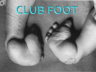
Club foot
1.
2. CLUBFOOT
VAGUETERM USEDTO DESCRIBE A
NUMBER OF DIFFERENTABNORMALITIES
INTHE SHAPE OFTHE FOOT
NOW IT HAS COMETO BE SYNONYMOUS
WITH THE COMMONEST CONGENITAL
FOOTABNORMALITY i.e., CTEV
7. ANATOMY
LIGAMENTS
DELTOID L. : MEDIAL COLLATERAL LIG. OF ANKLE
SPRING L. : CALCANIUM – NAVICULAR
CAPSULAR L. :T – N , N – C , C – M
PLANTAR L. :LONGITUDINAL ARCH OF FOOT
8. NOMENCLATURE
Planus: flatfoot
Cavus: highly arched foot
Varus: heal going towards
the midline
Valgus: heel going away
from the midline
Adduction: forefoot going
towards the midline
Abduction: forefoot going away
From the midline
10. CLUB FOOT
Definitions
Talipes: Talus = ankle
Pes = foot
Equinus: (Latin = horse)
Foot that is in a position of
planter flexion at the ankle,
looks like that of the horse.
Calcaneus: Full dorsiflexion at the ankle
14. CLASSIFICATION
EXTRINSIC
FLEXIBLEWITH ABNORMAL BONE RELATION
WITHOUT MARKED FIBROSIS
CONSERVATIVETREATMENT
INTRINSIC
RIGIDWITH ABNORMAL BONE RELATION
MARKED FIBROSIS
OPERATIVETREATMENT
15. THEORIES OF CTEV
TURCO’S : medial displacement of navicular and
calcaneous around talus
BROCKMAN’S : congenital atresia of theT – N joint
Mc- KAY’s :3-D bony deformity of the subtalar complex
INTRAUTERINE:compression by malpositon of fetus in utero
Germ plasm theory
Soft tissue theory
Prenatal muscle imbalance theory
16. PATHO-ANATOMY
BONES AND JOINTS
CALCANEUS : INVARUS POSITION
TALUS : DISPLACED MEDIAL AND PLANTARWARDS
NAVICULAR : MEDIALLY DISPLACED AND ROTATED
CUBOID : DISPLACED MEDIALLY AND ARTICULATES
WITHTHE NON-ARTICULAR SURFACE
OF CALCANEUM ( CUBOID SIGN /
LOCKED CUBOID )
METATARSALS : DEVIATES MEDIALLY ATT-M JOINTS
DISLOCATION OFTALOCALCANEAL ARTICULATION
TIBIA – MEDIALTORSION
21. PATHO-ANATOMY
SKIN
Adapts shortening on the medial side
Deep creases on the medial side
Dimples on the lateral aspect
SECONDARY CHANGES
Occurs when the child starts walking-exaggerates the
deformity
Callosities and bursae
22. CLINICAL FEATURES
COMMON PRESENTATIONS
Detected at birth
Infancy and early child hood
Late childhood
23. CLINICAL FEATURES
Short Achilles tendon
High and small heel
No creases behind Heel
Abnormal crease in middle of the foot
Foot is smaller in unilateral affection
Callosities at abnormal pressure areas
Internal torsion of the leg
Calf muscles wasting
Deformities don’t prevent walking
24. CLINICAL FEATURES
Seek a detailed family history of clubfoot or
neuromuscular disorders, and perform a general
examination to identify any other abnormalities.
Similar deformities are seen with myelomeningocele
and arthrogryposis.Therefore, always examine for
these associated conditions.
25. CLINICAL FEATURES
DORSIFLEXION TEST :
PLUMBLINETEST : tibial torsion
child is made to sit on a table with both LL hanging from the
edge.
Line drawn from the centre of the patella to the tibial tubercle
when extended down should cut the foot at 1st or 2nd
intermetatarsal space normally.- PLUMBLINE
In CTEV , with medial rotation of tibia it cuts through 4th or 5th
space
SCRATCHTEST – INFANTS
MEDIAL SCRACTHTEST : FOOT EVERTS - PERONEALS
LATERAL SCRACTHTEST: FOOT INVERTS - INVERTORS
26. INVESTIGATIONS
RADIOGRAPHY
APVIEW :angle formed b/w
talus and calcaneum ( NORMAL 30-35) REDUCED
Talus and metatarsals ( NORMAL 5 -15 ) -VE
Helps to asses angle of varus and forefoot adduction
27. RADIOGRAPHY
LATERALVIEW - ANGLE FORMED B/W
TIBIA AND CALCANEUM ( NORMAL 5- 15 ) -VE
TALUS AND CALCANEUM ( NORMAL 20- 50)
TO KNOWTHE EXTENTOF EQINUSANDVARUS DEFORMITY
CT , MRI , ARTHROGRAPHY
28. MANAGEMENT
The goal of treatment for clubfoot is to obtain a plantigrade foot
that is functional, painless, and stable
A cosmetically pleasing appearance is also an important goal
CONSERVATIVE
SURGICAL
EXTERNAL FIXATORS
29. CONSERVATIVE
INFANTS (< 6 MONTHS)
1ST 6WEEKS : SERIAL MANIPULATION AND CASTING
Corrective casting
First correction of adductus of midfoot
Folowed by correction of inversion
Finally correction of the equinus
30. Conservative management
Weekly serial manipulation and casting
Every weekly for 1st 6 week
Fortnightly till 6 months
Correction acheived Correction not achieved
Splint
day time
Phelp’s Brace
night time
Dennis Brown Splint
For 6 – 18 mothhs
CTEV shoes
( upto 4 years )
SURGERY EXT. FIXATOR
<4YRS STR
>4YRS STR+BONY
PROCEDURE
31. SURGICAL TRATMENT
Indications
Late presentation, after 6 months of age
Complementary to conservative treatment
Failure of conservative treatment
Residual deformities after conservative treatment
Recurrence after conservative treatment
32. SURGICAL TREATMENT
Soft tissue operations
Release of contractures
Tendon elongation
Tendon transfer
Restoration of normal bony relationship
Bony operations
Usually accompanied with soft tissue operation
Types:
- Osteotomy, to correct foot deformity or int. tibial torsion
- Wedge excision
- Arthrodesis (usually after bone maturity)
one or several joints
- Salvage operation to restore shape
33. P M S R
POSTERO MEDIAL SOFTTISSUE RELEASE ( 6-12 months )
TURCO’S PROCEDURE
On the posterior Side
Z- plasty of tendo – Achilles
Posterior capsulotomy of ankle and subtalar jnt
Release of posterior talo-fibular and calcaneo-fibular lig
On the medial side
Lenghtening ofTP , FHL, FDL
Release of talonavicular lig., spring lig., superficial part of
deltoid lig.
Release of interossious talocalcaneal lig, capsules of naviculo-
cuniform and 1st metatarsao-cuniform jnts
34. P M S R
On the plantar side
Plantar fascia release
Release of AH , FDB
Post – op regimen
Change cast at 2 weeks
Remove K wire at 6 weeks
Long leg cast for 3 months
Ankle foot orthoses for 6- 9 months
35. LIMITED SOFT TISSUE RELEASE
When only one component present
Equinus – posterior release
Adduction – medial release
Cavus – plantar release
36. CIRCUMFERENTIAL RELEASE
McKAY’s
All structures on PMSR + lateral structures
Superior peroneal retinaculam
Inferior extensor retinaculam
Dorsal calcaneo-cuboid lig.
12 – 36 months
Passively correctable deformity resulting from muscle
imbalance
37. RESISTANT CLUBFOOT
• >5YR. METATARSAL OSTEOTOMY
METATARSUS
ADDUCTUS
• <2- 3YR .modified McKey”s procedure
• 3- 10 yr
• Dwyer osteotomy
• Dilwyn –Evans operation
• 10-12 yr tripple arthrodesis
HIND FOOT
VARUS
• TendoAchillus lengthening + posterior capsulotomy sub
talar and ankle joint
• Lambrunidis triple arthrodesis
EQINUS
38. OPERATIONS
TRIPPLE ARTHRODESIS(>10YRS)
Lateral closed wedge osteotomy through subtalar and
midtarsal joints is done to fuse
SUBTALAR
TALONAVICULAR
CALCANEOCUBOID
TALECTOMY
Severe uncorrected club-foot
SURGERY FOR CORRECTION OFTIBIALTORSION
>15deg should be corrected by derotation osteotomy
39. DILWYN-EVANS OPERATION
Soft tissue release and calcaneocuboid fusion
1st three stages : extensive soft tissue release
Finally calcaneaocuboid wedge is excised
Neglected or recurred foot in children of 4-8 yrs
40. EXTERNAL FIXATORS
ILIZAROV’S EXTERNAL FIXATOR FRAME
JOSHI’S EXTERNAL FIXATOR FRAME
Allows gradual distraction
Transfixing wires through
Tibia, calcaneum ad metatarsals
Distractors positioned
Posteriorly, medially and laterally
Frame completed by
interconnecting the components
41. TREATMENT IN ADULT PATIENT
CUNIFORMTARSECTOMY
Vertical wedge of bone , with its base laterally is
removed from
Calcaneus – behind the metatarsal joints
Cuboid – infront of the joint
Curved wedge , with its base upwards and laterally
from head and neck of talus
42. RETENSION OF CTEV CORRECTION
DENIS BROWN SPLINT
PHELP’S BRACE
BELOW KNEEWALKING CALIPERS
CTEV SHOES