
Recomendados
Mais conteúdo relacionado
Mais procurados
Mais procurados (20)
Destaque
Destaque (20)
Semelhante a Ocular Emergency
Semelhante a Ocular Emergency (20)
Mais de Narenthorn EMS Center
Mais de Narenthorn EMS Center (20)
Último
Último (20)
Ocular Emergency
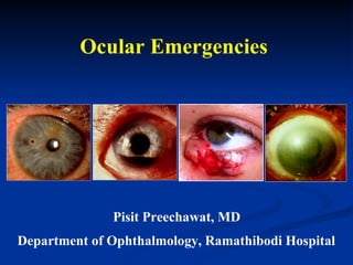
- 1. Ocular Emergencies Pisit Preechawat, MD Department of Ophthalmology, Ramathibodi Hospital
- 3. 1. Frontal bone 2. Zygomatic bone 3. Maxillary bone 4. Sphenoid bone 5. Ethmoid bone 6. Lacrimal bone 7. Palatine bone 1 2 3 4 5 6 7 Bony Components of Orbit Size 30 x 40 x 45 mm
- 5. Ocular Anatomy Orbicularis Oculi
- 10. Optic Nerve
- 11. Venous System
- 12. Ocular Emergencies Trauma Non - trauma Blunt trauma Penetrating trauma
- 13. Retinal arterial Perforation Orbital cellulitis occlusion Ruptured Orbital injury Chemical burns Acute glaucoma Corneal ulcer Sudden congestion Corneal abrasion proptosis Hyphema Intraocular FB Retinal detachment Macular edema ( Immediately ) ( Within a few hours ) ( Within one day ) Acute Eye Conditions Emergency Very Urgent Urgent
- 14. Ocular condiitons requiring immediate treatment Acute Angle-Closure Glaucoma Central Retinal Artery Occlusion Orbital Cellulitis Cavernous Sinus Thrombosis Endophthalmitis Retinal Detachment Toxic Causes of blindness Nontraumatic Ocular Emergencies Acute Dacryocystitis Acute Dacryoadenitis Acute Hordeolum Preseptal cellulitis Spontaneous subconjunctival hemorrhage Conjunctivitis Bacterial corneal ulcer Viral keratoconjunctivitis Acute hydrops of the cornea Hyphema Uveitis ( iritis & iridocyclitis ) Vitreous hemorrhage Retinal hemorrhage Central retinal vein occlusion Optic neuritis Ocular Emergencies
- 15. Ocular burns and trauma Ocular Burn Alkali Burns Acid Burns Thermal Burns Burns Due to Ultraviolet Radiation Mechanical Trauma to the Eye Penetrating or Perforating injuries Blunt Trauma to the Eye, Adnexa,& Orbit 1. Ecchymosis of the Eyelids 2. Lacerations of the Eyelids 3. Orbital hemorrhage 4. Fracture of the Ethmoid bone 5. Blowout Fractures of the Floor of the Orbit 6. Corneal Abrasions 7. Corneal & Conjunctival Foreign Bodies Ocular Emergencies
- 16. Eye Examination Visual acuity External Eye : orbit, periorbital skin, eyelids Confrontation visual fields Ocular motility
- 17. Anterior Segment Conjunctiva Cornea Anterior chamber Iris Lens Pupils : RAPD Eye Examination
- 18. A dilated pupil makes it easier to see the optic nerve, macula, and retina - 1% tropicamide ( Mydriacyl ) - 2.5% phenylephrine ( Neo-Synephrine ) Fundus Examination PanOptic Ophthalmoscope Indirect Ophthalmoscope
- 19. Digital palpation Schiotz tonometer Intraocular Pressure Measurement
- 20. Ocular Trauma Closed Globe Open Globe Burn Contusion Laceration Laceration Penetrating Perforating Rupture
- 22. Pain , photophobia , FB sensation, tearing Conjunctival injection, swollen eyelid Corneal Abrasion Epithelial staining defect with fluorescein
- 24. Corneal Ulcer Hypopyon Eye Shield No patching Topical antibiotics Ophthalmologist referral
- 26. Corneal foreign body with rust ring Rust ring Corneal Foreign Bodies
- 28. Corneal Foreign Body Removal
- 30. Traumatic Hyphema : Classification Total IV 1/2 to less than total III 1/3 to 1/2 II Less than 1/3 I No layered blood circulating red blood cells only 0 Size of Hyphema Grade
- 35. Full Thickness Lid Lacerations - Gray line - Lash line - Mucocutaneous junction Tear lid margin
- 36. Laceration of lower eyelid margin Post-operative result following a primary repair Lid Margin Repair
- 38. Lid Lacerations with tear canaliculi
- 42. Irregular pupil
- 43. Penetrating / Ruptured Globe
- 44. Ruptured globe caused by golf ball Penetrating / Ruptured Globe
- 46. Intraocular or Intraorbital Foreign Bodies
- 47. Ocular Trauma Traumatic cataract Traumatic mydriasis Traumatic lens subluxation Traumatic lens subluxation
- 50. Irrigation in case of chemical injury
- 52. Chemical Ocular Injury : Classification Grade I Grade II Grade III Grade IV
- 54. Bilateral Alkali Injuries Chemical Ocular Injury
- 55. Chemical Ocular Injury : Management Corneal Transplantation Keratoprosthesis
- 59. The woman suddenly experienced nausea, vomiting, and extreme pain in the left eye while in a movie theater. Her vision has worsened since that time and the eye has become very red. A 55-year-old woman with a red eye, blurred vision with halos, nausea, and vomiting
- 60. VA - HM Conjunctival injection Hazy cornea Shallow anterior chamber Fixed mid-dilated pupil A 55-year-old woman with a red eye, blurred vision with halos, nausea, and vomiting Acute Angle Closure Glaucoma IOP 56 mmHg
- 62. Reduce the intraocular pressure O.5% Timolol 1 drop 2-4 % Pilocarpine 1 drop every 15 minutes 20% Mannitol 250-500 ml IV drip Acetazolamide 500 mg oral 100% Glycerin 1 cc/kg Consult ophthalmologist Acute Angle Closure Glaucoma
- 63. A 60-year-old woman with acute, painless loss of vision in the right eye Visual acuity CF – LP in 90% of cases Opaque white retina and attenuated vessels Central Retinal Artery Occlusion
- 64. Treatment must be initiated immediately. Ocular massage Inhaled carbogen ( 95% O2 and 5% CO2 ) Reduced intraocular pressure Central Retinal Artery Occlusion Consult ophthalmologist immediately Anterior chamber paracentesis Direct infusion of t-PA or urokinase in the ophthalmic artery
- 65. A 40-year-old man with left eyelid edema and pain ( worse on eye movement )
- 67. Broad spectrum intravenous antibiotics CT scan orbit Ophthalmology & ENT consultation Orbital Cellulitis Subperiosteal abscess
- 69. Endophthalmitis
- 71. A 36-year-old-woman with subacute visual loss in right eye and pain on eye movement VA 20/200, 20/25 RAPD +ve OD VF central scotoma OD Retrobulbar optic neuritis
- 72. A 55-year-old man with HT and acute visual loss in RE VA 20/100, 20/20 RAPD +ve RE Nonarteritic anterior ischemic optic neuropathy ESR 10 mm/hr
- 73. A 73-year-old woman with acute visual loss of right eye, headache, anorexia and weight loss VA 10/200, 20/25 RAPD + ve RE ESR 94 mm/hr, high level of C - reactive protein Arteritic anterior ischemic optic neuropathy
- 74. Pathology : Giant Cell ( Temporal ) Arteritis
- 75. A 35-year-old man with left painful third nerve palsy VA 20/25, 20/30 Dilated, nonreactive pupil LE
- 76. A 35-year-old man with a suspicious of aneurysmal third nerve palsy Conventional CT scan or MRI are not the procedure of choice High false negative rate 12 – 40 % Magnetic resonance angiography (MRA) Computed tomography angiography (CTA) Overall sensitivity up to 97 %
- 77. A 35-year-old man with a suspicious of aneurysmal third nerve palsy
- 78. A 40-year-old woman with sudden onset of left third nerve palsy, visual loss and severe headache What is the diagnosis? VA 20/30, LP +ve RAPD LE
- 79. Pituitary Apoplexy Characterized by sudden visual loss, headache, and ophthalmoplegia secondary to rapid expansion of pituitary macroadenoma into the suprasellar space and/or cavernous sinus Commonly results from hemorrhage into a pre-existing pituitary mass
- 80. A 17-year-old man with right blured vision after minor blunt trauma. VA 20/32, 20/20 + ve RAPD RE Normal fundi RE LE
- 81. A 16-year-old man with head injury and left blured vision after falls from height VA 20/30, LP + ve RAPD LE Normal fundi
- 82. Traumatic Optic Neuropathy : Classification and Mechanisms Direct injury - Penetrating injury from knife, projectile - Injury from fractured bone - Avulsion, transection Indirect injury - Contusion with transmission of force through bone - Compression secondary to orbital hemorrhage or intrasheath hemorrhage
- 83. Clinical Features of Traumatic Optic Neuropathy Most commonly unilateral May be overlooked in setting of significant globe or maxillofacial trauma Reduced visual acuity ( NLP to 20/20 ) Visual field defect : No pathognomonic defect Normal optic disc with development of optic atrophy
- 84. Medical Management Options Steroids : Controversial - Thought to limit free-radical amplification of the injury response - Dosages ( low, high, mega) - May be harmful Observation : 57% of untreated patients shown to have 3 lines or more acuity improvement
- 85. Surgical Management Options Lateral canthotomy and cantholysis for orbital hemorrhage Surgical decompression of the optic nerve within its canal There is no defined standard protocol of treatment for indirect optic nerve injury .
- 86. Thank you for your attention