SlideShare uma empresa Scribd logo
1 de 8
Baixar para ler offline
P O S I T I O N

S T A T E M E N T

Diagnosis and Classification of Diabetes
Mellitus
AMERICAN DIABETES ASSOCIATION

DEFINITION AND
DESCRIPTION OF DIABETES
MELLITUSdDiabetes is a group of
metabolic diseases characterized by hyperglycemia resulting from defects in insulin secretion, insulin action, or both.
The chronic hyperglycemia of diabetes is
associated with long-term damage, dysfunction, and failure of different organs,
especially the eyes, kidneys, nerves, heart,
and blood vessels.
Several pathogenic processes are involved in the development of diabetes.
These range from autoimmune destruction of the b-cells of the pancreas with
consequent insulin deficiency to abnormalities that result in resistance to insulin
action. The basis of the abnormalities in
carbohydrate, fat, and protein metabolism in diabetes is deficient action of insulin on target tissues. Deficient insulin
action results from inadequate insulin secretion and/or diminished tissue responses to insulin at one or more points in the
complex pathways of hormone action.
Impairment of insulin secretion and defects in insulin action frequently coexist in
the same patient, and it is often unclear
which abnormality, if either alone, is the
primary cause of the hyperglycemia.
Symptoms of marked hyperglycemia include polyuria, polydipsia,weight loss,sometimes with polyphagia, and blurred vision.
Impairment of growth and susceptibility to
certain infections may also accompany
chronic hyperglycemia. Acute, life-threatening consequences of uncontrolled diabetes
are hyperglycemia with ketoacidosis or the
nonketotic hyperosmolar syndrome.
Long-term complications of diabetes
include retinopathy with potential loss
of vision; nephropathy leading to renal
failure; peripheral neuropathy with risk
of foot ulcers, amputations, and Charcot
joints; and autonomic neuropathy causing gastrointestinal, genitourinary, and

cardiovascular symptoms and sexual dysfunction. Patients with diabetes have an
increased incidence of atherosclerotic cardiovascular, peripheral arterial, and cerebrovascular disease. Hypertension and
abnormalities of lipoprotein metabolism
are often found in people with diabetes.
The vast majority of cases of diabetes
fall into two broad etiopathogenetic categories (discussed in greater detail below).
In one category, type 1 diabetes, the cause
is an absolute deficiency of insulin secretion. Individuals at increased risk of developing this type of diabetes can often be
identified by serological evidence of an
autoimmune pathologic process occurring
in the pancreatic islets and by genetic
markers. In the other, much more prevalent category, type 2 diabetes, the cause is a
combination of resistance to insulin action
and an inadequate compensatory insulin
secretory response. In the latter category, a
degree of hyperglycemia sufficient to cause
pathologic and functional changes in various target tissues, but without clinical
symptoms, may be present for a long
period of time before diabetes is detected.
During this asymptomatic period, it is
possible to demonstrate an abnormality in
carbohydrate metabolism by measurement
of plasma glucose in the fasting state or
after a challenge with an oral glucose load.
The degree of hyperglycemia (if any)
may change over time, depending on the
extent of the underlying disease process
(Fig. 1). A disease process may be present
but may not have progressed far enough
to cause hyperglycemia. The same disease
process can cause impaired fasting glucose (IFG) and/or impaired glucose tolerance (IGT) without fulfilling the criteria
for the diagnosis of diabetes. In some individuals with diabetes, adequate glycemic control can be achieved with weight
reduction, exercise, and/or oral glucoselowering agents. These individuals

c c c c c c c c c c c c c c c c c c c c c c c c c c c c c c c c c c c c c c c c c c c c c c c c c

Section on gestational diabetes diagnosis revised Fall 2010.
DOI: 10.2337/dc12-s064
© 2012 by the American Diabetes Association. Readers may use this article as long as the work is properly
cited, the use is educational and not for profit, and the work is not altered. See http://creativecommons.org/
licenses/by-nc-nd/3.0/ for details.

S64

DIABETES CARE, VOLUME 35, SUPPLEMENT 1, JANUARY 2012

therefore do not require insulin. Other
individuals who have some residual insulin secretion but require exogenous insulin for adequate glycemic control can
survive without it. Individuals with extensive b-cell destruction and therefore
no residual insulin secretion require insulin for survival. The severity of the metabolic abnormality can progress, regress,
or stay the same. Thus, the degree of hyperglycemia reflects the severity of the
underlying metabolic process and its
treatment more than the nature of the
process itself.
CLASSIFICATION OF
DIABETES MELLITUS AND
OTHER CATEGORIES
OF GLUCOSE
REGULATIONdAssigning a type of
diabetes to an individual often depends
on the circumstances present at the time
of diagnosis, and many diabetic individuals do not easily fit into a single class. For
example, a person with gestational diabetes mellitus (GDM) may continue to
be hyperglycemic after delivery and may
be determined to have, in fact, type 2
diabetes. Alternatively, a person who
acquires diabetes because of large doses
of exogenous steroids may become normoglycemic once the glucocorticoids are
discontinued, but then may develop diabetes many years later after recurrent
episodes of pancreatitis. Another example
would be a person treated with thiazides
who develops diabetes years later. Because
thiazides in themselves seldom cause severe
hyperglycemia, such individuals probably
have type 2 diabetes that is exacerbated by
the drug. Thus, for the clinician and patient,
it is less important to label the particular
type of diabetes than it is to understand the
pathogenesis of the hyperglycemia and to
treat it effectively.
Type 1 diabetes (b-cell destruction,
usually leading to absolute insulin
deficiency)
Immune-mediated diabetes. This form
of diabetes, which accounts for only
5–10% of those with diabetes, previously
encompassed by the terms insulindependent diabetes, type 1 diabetes, or
juvenile-onset diabetes, results from a cellular-mediated autoimmune destruction
care.diabetesjournals.org
Position Statement

Figure 1dDisorders of glycemia: etiologic types and stages. *Even after presenting in ketoacidosis, these patients can briefly return to normoglycemia without requiring continuous therapy (i.e., “honeymoon” remission); **in rare instances, patients in these categories (e.g., Vacor toxicity,
type 1 diabetes presenting in pregnancy) may require insulin for survival.

of the b-cells of the pancreas. Markers of
the immune destruction of the b-cell include islet cell autoantibodies, autoantibodies to insulin, autoantibodies to GAD
(GAD65), and autoantibodies to the tyrosine phosphatases IA-2 and IA-2b.
One and usually more of these autoantibodies are present in 85–90% of individuals when fasting hyperglycemia is
initially detected. Also, the disease has
strong HLA associations, with linkage to
the DQA and DQB genes, and it is influenced by the DRB genes. These HLA-DR/
DQ alleles can be either predisposing or
protective.
In this form of diabetes, the rate of
b-cell destruction is quite variable, being
rapid in some individuals (mainly infants
and children) and slow in others (mainly
adults). Some patients, particularly children and adolescents, may present with
ketoacidosis as the first manifestation of
the disease. Others have modest fasting
hyperglycemia that can rapidly change
to severe hyperglycemia and/or ketoacidosis in the presence of infection or other
stress. Still others, particularly adults,
may retain residual b-cell function sufficient to prevent ketoacidosis for many
years; such individuals eventually become dependent on insulin for survival
and are at risk for ketoacidosis. At this
latter stage of the disease, there is little
or no insulin secretion, as manifested by
low or undetectable levels of plasma
care.diabetesjournals.org

C-peptide. Immune-mediated diabetes
commonly occurs in childhood and adolescence, but it can occur at any age, even
in the 8th and 9th decades of life.
Autoimmune destruction of b-cells
has multiple genetic predispositions and
is also related to environmental factors
that are still poorly defined. Although patients are rarely obese when they present
with this type of diabetes, the presence of
obesity is not incompatible with the diagnosis. These patients are also prone to
other autoimmune disorders such as
Graves’ disease, Hashimoto’s thyroiditis,
Addison’s disease, vitiligo, celiac sprue,
autoimmune hepatitis, myasthenia gravis,
and pernicious anemia.
Idiopathic diabetes. Some forms of type
1 diabetes have no known etiologies.
Some of these patients have permanent
insulinopenia and are prone to ketoacidosis, but have no evidence of autoimmunity. Although only a minority of
patients with type 1 diabetes fall into
this category, of those who do, most are of
African or Asian ancestry. Individuals
with this form of diabetes suffer from
episodic ketoacidosis and exhibit varying
degrees of insulin deficiency between
episodes. This form of diabetes is strongly
inherited, lacks immunological evidence
for b-cell autoimmunity, and is not HLA
associated. An absolute requirement for
insulin replacement therapy in affected
patients may come and go.

Type 2 diabetes (ranging from
predominantly insulin resistance
with relative insulin deficiency to
predominantly an insulin secretory
defect with insulin resistance)
This form of diabetes, which accounts for
;90–95% of those with diabetes, previously referred to as non–insulin-dependent diabetes, type 2 diabetes, or adultonset diabetes, encompasses individuals
who have insulin resistance and usually
have relative (rather than absolute) insulin deficiency At least initially, and often
throughout their lifetime, these individuals do not need insulin treatment to survive. There are probably many different
causes of this form of diabetes. Although
the specific etiologies are not known, autoimmune destruction of b-cells does not
occur, and patients do not have any of the
other causes of diabetes listed above or
below.
Most patients with this form of diabetes are obese, and obesity itself causes
some degree of insulin resistance. Patients
who are not obese by traditional weight
criteria may have an increased percentage
of body fat distributed predominantly in
the abdominal region. Ketoacidosis seldom occurs spontaneously in this type of
diabetes; when seen, it usually arises in
association with the stress of another
illness such as infection. This form of
diabetes frequently goes undiagnosed for
many years because the hyperglycemia

DIABETES CARE, VOLUME 35, SUPPLEMENT 1, JANUARY 2012

S65
Diagnosis and Classification
develops gradually and at earlier stages is
often not severe enough for the patient to
notice any of the classic symptoms of
diabetes. Nevertheless, such patients are
at increased risk of developing macrovascular and microvascular complications. Whereas patients with this form of
diabetes may have insulin levels that
appear normal or elevated, the higher
blood glucose levels in these diabetic
patients would be expected to result in
even higher insulin values had their b-cell
function been normal. Thus, insulin secretion is defective in these patients and
insufficient to compensate for insulin resistance. Insulin resistance may improve
with weight reduction and/or pharmacological treatment of hyperglycemia but is
seldom restored to normal. The risk of
developing this form of diabetes increases
with age, obesity, and lack of physical activity. It occurs more frequently in women
with prior GDM and in individuals with
hypertension or dyslipidemia, and its frequency varies in different racial/ethnic subgroups. It is often associated with a strong
genetic predisposition, more so than is the
autoimmune form of type 1 diabetes. However, the genetics of this form of diabetes
are complex and not clearly defined.
Other specific types of diabetes
Genetic defects of the b-cell. Several
forms of diabetes are associated with
monogenetic defects in b-cell function.
These forms of diabetes are frequently
characterized by onset of hyperglycemia
at an early age (generally before age 25
years). They are referred to as maturityonset diabetes of the young (MODY) and
are characterized by impaired insulin secretion with minimal or no defects in insulin action. They are inherited in an
autosomal dominant pattern. Abnormalities at six genetic loci on different chromosomes have been identified to date.
The most common form is associated
with mutations on chromosome 12 in a
hepatic transcription factor referred to as
hepatocyte nuclear factor (HNF)-1a. A
second form is associated with mutations
in the glucokinase gene on chromosome
7p and results in a defective glucokinase
molecule. Glucokinase converts glucose
to glucose-6-phosphate, the metabolism
of which, in turn, stimulates insulin secretion by the b-cell. Thus, glucokinase
serves as the “glucose sensor” for the
b-cell. Because of defects in the glucokinase gene, increased plasma levels of glucose are necessary to elicit normal levels
of insulin secretion. The less common
S66

forms result from mutations in other transcription factors, including HNF-4a,
HNF-1b, insulin promoter factor (IPF)1, and NeuroD1.
Point mutations in mitochondrial
DNA have been found to be associated
with diabetes and deafness The most
common mutation occurs at position
3,243 in the tRNA leucine gene, leading
to an A-to-G transition. An identical
lesion occurs in the MELAS syndrome
(mitochondrial myopathy, encephalopathy, lactic acidosis, and stroke-like syndrome); however, diabetes is not part of
this syndrome, suggesting different phenotypic expressions of this genetic lesion.
Genetic abnormalities that result in
the inability to convert proinsulin to insulin have been identified in a few families, and such traits are inherited in an
autosomal dominant pattern. The resultant glucose intolerance is mild. Similarly,
the production of mutant insulin molecules with resultant impaired receptor
binding has also been identified in a few
families and is associated with an autosomal inheritance and only mildly impaired
or even normal glucose metabolism.
Genetic defects in insulin action. There
are unusual causes of diabetes that result
from genetically determined abnormalities of insulin action. The metabolic abnormalities associated with mutations of
the insulin receptor may range from
hyperinsulinemia and modest hyperglycemia to severe diabetes. Some individuals
with these mutations may have acanthosis
nigricans. Women may be virilized and
have enlarged, cystic ovaries. In the past,
this syndrome was termed type A insulin
resistance. Leprechaunism and the RabsonMendenhall syndrome are two pediatric
syndromes that have mutations in the
insulin receptor gene with subsequent
alterations in insulin receptor function
and extreme insulin resistance. The former
has characteristic facial features and is
usually fatal in infancy, while the latter is
associated with abnormalities of teeth and
nails and pineal gland hyperplasia.
Alterations in the structure and function of the insulin receptor cannot be
demonstrated in patients with insulinresistant lipoatrophic diabetes. Therefore,
it is assumed that the lesion(s) must reside
in the postreceptor signal transduction
pathways.
Diseases of the exocrine pancreas. Any
process that diffusely injures the pancreas
can cause diabetes. Acquired processes
include pancreatitis, trauma, infection, pancreatectomy, and pancreatic carcinoma.

DIABETES CARE, VOLUME 35, SUPPLEMENT 1, JANUARY 2012

With the exception of that caused by
cancer, damage to the pancreas must be
extensive for diabetes to occur; adrenocarcinomas that involve only a small
portion of the pancreas have been associated with diabetes. This implies a mechanism other than simple reduction in
b-cell mass. If extensive enough, cystic fibrosis and hemochromatosis will also
damage b-cells and impair insulin secretion. Fibrocalculous pancreatopathy may
be accompanied by abdominal pain radiating to the back and pancreatic calcifications identified on X-ray examination.
Pancreatic fibrosis and calcium stones
in the exocrine ducts have been found at
autopsy.
Endocrinopathies. Several hormones
(e.g., growth hormone, cortisol, glucagon, epinephrine) antagonize insulin action. Excess amounts of these hormones
(e.g., acromegaly, Cushing’s syndrome,
glucagonoma, pheochromocytoma, respectively) can cause diabetes. This generally occurs in individuals with
preexisting defects in insulin secretion,
and hyperglycemia typically resolves
when the hormone excess is resolved.
Somatostatinoma- and aldosteronoma-induced hypokalemia can cause
diabetes, at least in part, by inhibiting
insulin secretion. Hyperglycemia generally resolves after successful removal of
the tumor.
Drug- or chemical-induced diabetes.
Many drugs can impair insulin secretion.
These drugs may not cause diabetes by
themselves, but they may precipitate diabetes in individuals with insulin resistance. In such cases, the classification is
unclear because the sequence or relative
importance of b-cell dysfunction and insulin resistance is unknown. Certain toxins such as Vacor (a rat poison) and
intravenous pentamidine can permanently destroy pancreatic b-cells. Such
drug reactions fortunately are rare. There
are also many drugs and hormones that
can impair insulin action. Examples include nicotinic acid and glucocorticoids.
Patients receiving a-interferon have been
reported to develop diabetes associated
with islet cell antibodies and, in certain
instances, severe insulin deficiency. The
list shown in Table 1 is not all-inclusive,
but reflects the more commonly recognized drug-, hormone-, or toxin-induced
forms of diabetes.
Infections. Certain viruses have been
associated with b-cell destruction. Diabetes occurs in patients with congenital rubella, although most of these patients
care.diabetesjournals.org
Position Statement
Table 1dEtiologic classification of diabetes mellitus
I. Type 1 diabetes (b-cell destruction, usually leading to absolute insulin deficiency)
A. Immune mediated
B. Idiopathic
II. Type 2 diabetes (may range from predominantly insulin resistance with relative insulin deficiency
to a predominantly secretory defect with insulin resistance)
III. Other specific types
A. Genetic defects of b-cell function
1. Chromosome 12, HNF-1a (MODY3)
2. Chromosome 7, glucokinase (MODY2)
3. Chromosome 20, HNF-4a (MODY1)
4. Chromosome 13, insulin promoter factor-1 (IPF-1; MODY4)
5. Chromosome 17, HNF-1b (MODY5)
6. Chromosome 2, NeuroD1 (MODY6)
7. Mitochondrial DNA
8. Others
B. Genetic defects in insulin action
1. Type A insulin resistance
2. Leprechaunism
3. Rabson-Mendenhall syndrome
4. Lipoatrophic diabetes
5. Others
C. Diseases of the exocrine pancreas
1. Pancreatitis
2. Trauma/pancreatectomy
3. Neoplasia
4. Cystic fibrosis
5. Hemochromatosis
6. Fibrocalculous pancreatopathy
7. Others
D. Endocrinopathies
1. Acromegaly
2. Cushing’s syndrome
3. Glucagonoma
4. Pheochromocytoma
5. Hyperthyroidism
6. Somatostatinoma
7. Aldosteronoma
8. Others
E. Drug or chemical induced
1. Vacor
2. Pentamidine
3. Nicotinic acid
4. Glucocorticoids
5. Thyroid hormone
6. Diazoxide
7. b-adrenergic agonists
8. Thiazides
9. Dilantin
10. g-Interferon
11. Others
F. Infections
1. Congenital rubella
2. Cytomegalovirus
3. Others
G. Uncommon forms of immune-mediated diabetes
1. “Stiff-man” syndrome
2. Anti-insulin receptor antibodies
3. Others
H. Other genetic syndromes sometimes associated with diabetes
1. Down syndrome
2. Klinefelter syndrome
3. Turner syndrome
4. Wolfram syndrome
5. Friedreich ataxia
6. Huntington chorea
7. Laurence-Moon-Biedl syndrome
8. Myotonic dystrophy
9. Porphyria
10. Prader-Willi syndrome
11. Others
IV. Gestational diabetes mellitus
Patients with any form of diabetes may require insulin treatment at some stage of their disease. Such use of
insulin does not, of itself, classify the patient.

care.diabetesjournals.org

have HLA and immune markers characteristic of type 1 diabetes. In addition,
coxsackievirus B, cytomegalovirus, adenovirus, and mumps have been implicated in inducing certain cases of the
disease.
Uncommon forms of immune-mediated
diabetes. In this category, there are two
known conditions, and others are likely
to occur. The stiff-man syndrome is an
autoimmune disorder of the central nervous system characterized by stiffness of
the axial muscles with painful spasms.
Patients usually have high titers of the
GAD autoantibodies, and approximately
one-third will develop diabetes.
Anti-insulin receptor antibodies can
cause diabetes by binding to the insulin
receptor, thereby blocking the binding of
insulin to its receptor in target tissues.
However, in some cases, these antibodies
can act as an insulin agonist after binding
to the receptor and can thereby cause
hypoglycemia. Anti-insulin receptor antibodies are occasionally found in patients
with systemic lupus erythematosus and
other autoimmune diseases. As in other
states of extreme insulin resistance, patients with anti-insulin receptor antibodies often have acanthosis nigricans. In the
past, this syndrome was termed type B
insulin resistance.
Other genetic syndromes sometimes
associated with diabetes. Many genetic
syndromes are accompanied by an increased incidence of diabetes. These include the chromosomal abnormalities of
Down syndrome, Klinefelter syndrome,
and Turner syndrome. Wolfram’s syndrome is an autosomal recessive disorder
characterized by insulin-deficient diabetes and the absence of b-cells at autopsy.
Additional manifestations include diabetes
insipidus, hypogonadism, optic atrophy,
and neural deafness. Other syndromes are
listed in Table 1.
Gestational diabetes mellitus
For many years, GDM has been defined as
any degree of glucose intolerance with
onset or first recognition during pregnancy. Although most cases resolve with
delivery, the definition applied whether
or not the condition persisted after pregnancy and did not exclude the possibility
that unrecognized glucose intolerance
may have antedated or begun concomitantly with the pregnancy. This definition
facilitated a uniform strategy for detection
and classification of GDM, but its limitations were recognized for many years.
As the ongoing epidemic of obesity and

DIABETES CARE, VOLUME 35, SUPPLEMENT 1, JANUARY 2012

S67
Diagnosis and Classification
Table 2dCategories of increased risk for
diabetes*
FPG 100 mg/dl (5.6 mmol/l) to 125 mg/dl (6.9
mmol/l) [IFG]
2-h PG in the 75-g OGTT 140 mg/dl (7.8
mmol/l) to 199 mg/dl (11.0 mmol/l) [IGT]
A1C 5.7–6.4%
*For all three tests, risk is continuous, extending
below the lower limit of the range and becoming
disproportionately greater at higher ends of the
range.

diabetes has led to more type 2 diabetes in
women of childbearing age, the number of
pregnant women with undiagnosed type 2
diabetes has increased.
After deliberations in 2008–2009, the
International Association of Diabetes and
Pregnancy Study Groups (IADPSG), an
international consensus group with representatives from multiple obstetrical and
diabetes organizations, including the
American Diabetes Association (ADA),
recommended that high-risk women
found to have diabetes at their initial prenatal visit, using standard criteria (Table
3), receive a diagnosis of overt, not gestational, diabetes. Approximately 7% of all
pregnancies (ranging from 1 to 14%, depending on the population studied and
the diagnostic tests employed) are complicated by GDM, resulting in more than
200,000 cases annually.
CATEGORIES OF INCREASED
RISK FOR DIABETESdIn 1997 and
2003, The Expert Committee on Diagnosis and Classification of Diabetes Mellitus
(1,2) recognized an intermediate group of
individuals whose glucose levels do not
meet criteria for diabetes, yet are higher
than those considered normal. These people were defined as having impaired fasting glucose (IFG) [fasting plasma glucose
(FPG) levels 100 mg/dl (5.6 mmol/l) to
125 mg/dl (6.9 mmol/l)], or impaired glucose tolerance (IGT) [2-h values in the
oral glucose tolerance test (OGTT) of
140 mg/dl (7.8 mmol/l) to 199 mg/dl
(11.0 mmol/l)].
Individuals with IFG and/or IGT have
been referred to as having pre-diabetes,
indicating the relatively high risk for the
future development of diabetes. IFG and
IGT should not be viewed as clinical
entities in their own right but rather risk
factors for diabetes as well as cardiovascular disease. They can be observed as
intermediate stages in any of the disease
processes listed in Table 1. IFG and IGT
are associated with obesity (especially
S68

abdominal or visceral obesity), dyslipidemia with high triglycerides and/or low
HDL cholesterol, and hypertension.
Structured lifestyle intervention, aimed
at increasing physical activity and producing 5–10% loss of body weight, and
certain pharmacological agents have been
demonstrated to prevent or delay the development of diabetes in people with IGT;
the potential impact of such interventions
to reduce mortality or the incidence of
cardiovascular disease has not been demonstrated to date. It should be noted that
the 2003 ADA Expert Committee report
reduced the lower FPG cut point to define
IFG from 110 mg/dl (6.1 mmol/l) to 100
mg/dl (5.6 mmol/l), in part to ensure that
prevalence of IFG was similar to that of
IGT. However, the World Health Organization (WHO) and many other diabetes
organizations did not adopt this change in
the definition of IFG.
As A1C is used more commonly to
diagnose diabetes in individuals with risk
factors, it will also identify those at higher
risk for developing diabetes in the future.
When recommending the use of the A1C
to diagnose diabetes in its 2009 report,
the International Expert Committee (3)
stressed the continuum of risk for diabetes with all glycemic measures and did not
formally identify an equivalent intermediate category for A1C. The group did note
that those with A1C levels above the laboratory “normal” range but below the diagnostic cut point for diabetes (6.0 to
,6.5%) are at very high risk of developing diabetes. Indeed, incidence of diabetes in people with A1C levels in this range
is more than 10 times that of people with
lower levels (4–7). However, the 6.0 to
,6.5% range fails to identify a substantial
number of patients who have IFG and/or
IGT. Prospective studies indicate that
people within the A1C range of 5.5–
6.0% have a 5-year cumulative incidence
of diabetes that ranges from 12 to 25%
(4–7), which is appreciably (three- to
eightfold) higher than incidence in the
U.S. population as a whole (8). Analyses
of nationally representative data from the
National Health and Nutrition Examination Survey (NHANES) indicate that the
A1C value that most accurately identifies
people with IFG or IGT falls between 5.5
and 6.0%. In addition, linear regression
analyses of these data indicate that among
the nondiabetic adult population, an FPG
of 110 mg/dl (6.1 mmol/l) corresponds
to an A1C of 5.6%, while an FPG of 100
mg/dl (5.6 mmol/l) corresponds to an
A1C of 5.4% (R.T. Ackerman, personal

DIABETES CARE, VOLUME 35, SUPPLEMENT 1, JANUARY 2012

communication). Finally, evidence from
the Diabetes Prevention Program (DPP),
wherein the mean A1C was 5.9% (SD
0.5%), indicates that preventive interventions are effective in groups of people with
A1C levels both below and above 5.9%
(9). For these reasons, the most appropriate A1C level above which to initiate preventive interventions is likely to be
somewhere in the range of 5.5–6%.
As was the case with FPG and 2-h PG,
defining a lower limit of an intermediate
category of A1C is somewhat arbitrary,
as the risk of diabetes with any measure
or surrogate of glycemia is a continuum,
extending well into the normal ranges. To
maximize equity and efficiency of preventive interventions, such an A1C cut
point should balance the costs of “false
negatives” (failing to identify those who
are going to develop diabetes) against
the costs of “false positives” (falsely identifying and then spending intervention resources on those who were not going to
develop diabetes anyway).
Compared to the fasting glucose cutpoint of 100 mg/dl (5.6 mmol/l), an A1C
cutpoint of 5.7% is less sensitive but more
specific and has a higher positive predictive value to identify people at risk for
later development of diabetes. A large
prospective study found that a 5.7% cutpoint has a sensitivity of 66% and specificity of 88% for the identification of
subsequent 6-year diabetes incidence
(10). Receiver operating curve analyses
of nationally representative U.S. data
(NHANES 1999-2006) indicate that an
A1C value of 5.7% has modest sensitivity
(39-45%) but high specificity (81-91%)
to identify cases of IFP (FPG .100 mg/
dl) (5.6 mmol/l) or IGT (2-h glucose .
140 mg/dl) (R.T. Ackerman, personal
communication). Other analyses suggest
that an A1C of 5.7% is associated with
diabetes risk similar to the high-risk participants in the DPP (R.T. Ackerman, personal communication). Hence, it is
reasonable to consider an A1C range of
5.7 to 6.4% as identifying individuals
with high risk for future diabetes and to
whom the term pre-diabetes may be applied if desired.
Individuals with an A1C of 5.7–6.4%
should be informed of their increased risk
for diabetes as well as cardiovascular disease and counseled about effective strategies, such as weight loss and physical
activity, to lower their risks. As with glucose measurements, the continuum of risk
is curvilinear, so that as A1C rises, the risk
of diabetes rises disproportionately.
care.diabetesjournals.org
Position Statement
Accordingly, interventions should be
most intensive and follow-up should be
particularly vigilant for those with A1C
levels above 6.0%, who should be considered to be at very high risk. However, just
as an individual with a fasting glucose of
98 mg/dl (5.4 mmol/l) may not be at negligible risk for diabetes, individuals with
A1C levels below 5.7% may still be at
risk, depending on level of A1C and presence of other risk factors, such as obesity
and family history.
Table 2 summarizes the categories of
increased risk for diabetes. Evaluation of
patients at risk should incorporate a
global risk factor assessment for both diabetes and cardiovascular disease. Screening for and counseling about risk of
diabetes should always be in the pragmatic context of the patient’s comorbidities, life expectancy, personal capacity to
engage in lifestyle change, and overall
health goals.
DIAGNOSTIC CRITERIA FOR
DIABETES MELLITUSdFor decades, the diagnosis of diabetes has been
based on glucose criteria, either the FPG
or the 75-g OGTT. In 1997, the first
Expert Committee on the Diagnosis and
Classification of Diabetes Mellitus revised
the diagnostic criteria, using the observed
association between FPG levels and presence of retinopathy as the key factor with
which to identify threshold glucose level.
The Committee examined data from three
cross-sectional epidemiologic studies that
assessed retinopathy with fundus photography or direct ophthalmoscopy and
measured glycemia as FPG, 2-h PG, and
A1C. These studies demonstrated glycemic levels below which there was little
prevalent retinopathy and above which
the prevalence of retinopathy increased in
an apparently linear fashion. The deciles
of the three measures at which retinopathy began to increase were the same for
each measure within each population.
Moreover, the glycemic values above
which retinopathy increased were similar
among the populations. These analyses
helped to inform a new diagnostic cut
point of $126 mg/dl (7.0 mmol/l) for
FPG and confirmed the long-standing diagnostic 2-h PG value of $200 mg/dl
(11.1 mmol/l).
A1C is a widely used marker of
chronic glycemia, reflecting average
blood glucose levels over a 2- to 3-month
period of time. The test plays a critical role
in the management of the patient with
diabetes, since it correlates well with both
care.diabetesjournals.org

Table 3dCriteria for the diagnosis of diabetes
A1C $6.5%. The test should be performed in a laboratory using a method that is NGSP certified
and standardized to the DCCT assay.*
OR
FPG $126 mg/dl (7.0 mmol/l). Fasting is defined as no caloric intake for at least 8 h.*
OR
2-h plasma glucose $200 mg/dl (11.1 mmol/l) during an OGTT. The test should be performed as
described by the World Health Organization, using a glucose load containing the equivalent of
75 g anhydrous glucose dissolved in water.*
OR
In a patient with classic symptoms of hyperglycemia or hyperglycemic crisis, a random plasma
glucose $200 mg/dl (11.1 mmol/l).
*In the absence of unequivocal hyperglycemia, criteria 1–3 should be confirmed by repeat testing.

microvascular and, to a lesser extent,
macrovascular complications and is
widely used as the standard biomarker
for the adequacy of glycemic management. Prior Expert Committees have not
recommended use of the A1C for diagnosis of diabetes, in part due to lack of
standardization of the assay. However,
A1C assays are now highly standardized
so that their results can be uniformly
applied both temporally and across populations. In their recent report (3), an International Expert Committee, after an
extensive review of both established and
emerging epidemiological evidence, recommended the use of the A1C test to diagnose diabetes, with a threshold of
$6.5%, and ADA affirms this decision.
The diagnostic A1C cut point of 6.5% is
associated with an inflection point for retinopathy prevalence, as are the diagnostic
thresholds for FPG and 2-h PG (3). The
diagnostic test should be performed
using a method that is certified by the National Glycohemoglobin Standardization
Program (NGSP) and standardized or
traceable to the Diabetes Control and
Complications Trial reference assay.
Point-of-care A1C assays are not sufficiently accurate at this time to use for diagnostic purposes.
There is an inherent logic to using a
more chronic versus an acute marker of
dysglycemia, particularly since the A1C is
already widely familiar to clinicians as a
marker of glycemic control. Moreover,
the A1C has several advantages to the
FPG, including greater convenience,
since fasting is not required, evidence to
suggest greater preanalytical stability, and
less day-to-day perturbations during periods of stress and illness. These advantages, however, must be balanced by
greater cost, the limited availability of
A1C testing in certain regions of the
developing world, and the incomplete
correlation between A1C and average

glucose in certain individuals. In addition, the A1C can be misleading in patients with certain forms of anemia and
hemoglobinopathies, which may also
have unique ethnic or geographic distributions. For patients with a hemoglobinopathy but normal red cell turnover, such
as sickle cell trait, an A1C assay without
interference from abnormal hemoglobins
should be used (an updated list is available at www.ngsp.org/prog/index3.
html). For conditions with abnormal red
cell turnover, such as anemias from hemolysis and iron deficiency, the diagnosis
of diabetes must employ glucose criteria
exclusively.
The established glucose criteria for
the diagnosis of diabetes remain valid.
These include the FPG and 2-h PG.
Additionally, patients with severe hyperglycemia such as those who present with
severe classic hyperglycemic symptoms
or hyperglycemic crisis can continue to be
diagnosed when a random (or casual)
plasma glucose of $200 mg/dl (11.1
mmol/l) is found. It is likely that in such
cases the health care professional would
also measure an A1C test as part of the
initial assessment of the severity of the diabetes and that it would (in most cases) be
above the diagnostic cut point for diabetes. However, in rapidly evolving diabetes, such as the development of type 1
diabetes in some children, A1C may not
be significantly elevated despite frank
diabetes.
Just as there is less than 100% concordance between the FPG and 2-h PG
tests, there is not full concordance between A1C and either glucose-based
test. Analyses of NHANES data indicate
that, assuming universal screening of the
undiagnosed, the A1C cut point of
$6.5% identifies one-third fewer cases
of undiagnosed diabetes than a fasting
glucose cut point of $126 mg/dl (7.0
mmol/l) (cdc website tbd). However, in

DIABETES CARE, VOLUME 35, SUPPLEMENT 1, JANUARY 2012

S69
Diagnosis and Classification
practice, a large portion of the population
with type 2 diabetes remains unaware of
their condition. Thus, it is conceivable
that the lower sensitivity of A1C at the
designated cut point will be offset by the
test’s greater practicality, and that wider
application of a more convenient test
(A1C) may actually increase the number
of diagnoses made.
Further research is needed to better
characterize those patients whose glycemic status might be categorized differently by two different tests (e.g., FPG and
A1C), obtained in close temporal approximation. Such discordance may arise from
measurement variability, change over
time, or because A1C, FPG, and postchallenge glucose each measure different
physiological processes. In the setting of
an elevated A1C but “nondiabetic” FPG,
the likelihood of greater postprandial glucose levels or increased glycation rates
for a given degree of hyperglycemia may
be present. In the opposite scenario (high
FPG yet A1C below the diabetes cut
point), augmented hepatic glucose production or reduced glycation rates may
be present.
As with most diagnostic tests, a test
result diagnostic of diabetes should be
repeated to rule out laboratory error,
unless the diagnosis is clear on clinical
grounds, such as a patient with classic
symptoms of hyperglycemia or hyperglycemic crisis. It is preferable that the same
test be repeated for confirmation, since
there will be a greater likelihood of concurrence in this case. For example, if the
A1C is 7.0% and a repeat result is 6.8%,
the diagnosis of diabetes is confirmed.
However, there are scenarios in which results of two different tests (e.g., FPG and
A1C) are available for the same patient. In
this situation, if the two different tests are
both above the diagnostic thresholds, the
diagnosis of diabetes is confirmed.
On the other hand, when two different tests are available in an individual and
the results are discordant, the test whose
result is above the diagnostic cut point
should be repeated, and the diagnosis is
made on the basis of the confirmed test.
That is, if a patient meets the diabetes
criterion of the A1C (two results $6.5%)
but not the FPG (,126 mg/dl or 7.0
mmol/l), or vice versa, that person should
be considered to have diabetes. Admittedly, in most circumstance the “nondiabetic” test is likely to be in a range very
close to the threshold that defines diabetes.
Since there is preanalytic and analytic
variability of all the tests, it is also possible
S70

that when a test whose result was above
the diagnostic threshold is repeated, the
second value will be below the diagnostic
cut point. This is least likely for A1C,
somewhat more likely for FPG, and most
likely for the 2-h PG. Barring a laboratory
error, such patients are likely to have test
results near the margins of the threshold
for a diagnosis. The healthcare professional might opt to follow the patient
closely and repeat the testing in 3–6
months.
The decision about which test to use
to assess a specific patient for diabetes
should be at the discretion of the health
care professional, taking into account the
availability and practicality of testing an
individual patient or groups of patients.
Perhaps more important than which diagnostic test is used, is that the testing for
diabetes be performed when indicated.
There is discouraging evidence indicating
that many at-risk patients still do not receive adequate testing and counseling for
this increasingly common disease, or for its
frequently accompanying cardiovascular
risk factors. The current diagnostic criteria
for diabetes are summarized in Table 3.
Diagnosis of gestational diabetes
GDM carries risks for the mother and
neonate. The Hyperglycemia and Adverse
Pregnancy Outcomes (HAPO) study
(11), a large-scale (;25,000 pregnant
women) multinational epidemiologic
study, demonstrated that risk of adverse
maternal, fetal, and neonatal outcomes
continuously increased as a function of
maternal glycemia at 24-28 weeks, even
within ranges previously considered normal for pregnancy. For most complications, there was no threshold for risk.
These results have led to careful reconsideration of the diagnostic criteria for GDM.
After deliberations in 2008-2009, the
IADPSG, an international consensus
group with representatives from multiple
obstetrical and diabetes organizations, including ADA, developed revised recommendations for diagnosing GDM. The
group recommended that all women not
known to have diabetes undergo a 75-g
OGTT at 24-28 weeks of gestation. Additionally, the group developed diagnostic
cutpoints for the fasting, 1-h, and 2-h
plasma glucose measurements that conveyed an odds ratio for adverse outcomes
of at least 1.75 compared with women
with mean glucose levels in the HAPO
study. Current screening and diagnostic
strategies, based on the IADPSG statement (12), are outlined in Table 4.

DIABETES CARE, VOLUME 35, SUPPLEMENT 1, JANUARY 2012

Table 4dScreening for and diagnosis of
GDM
Perform a 75-g OGTT, with plasma glucose
measurement fasting and at 1 and 2 h, at
24-28 of weeks gestation in women not
previously diagnosed with overt diabetes.
The OGTT should be performed in the
morning after an overnight fast of at least
8 h.
The diagnosis of GDM is made when any of the
following plasma glucose values are
exceeded
c Fasting: $92 mg/dl (5.1 mmol/l)
c 1 h: $180 mg/dl (10.0 mmol/l)
c 2 h: $153 mg/dl (8.5 mmol/l)

These new criteria will significantly
increase the prevalence of GDM, primarily because only one abnormal value, not
two, is sufficient to make the diagnosis.
The ADA recognizes the anticipated significant increase in the incidence of GDM
to be diagnosed by these criteria and is
sensitive to concerns about the “medicalization” of pregnancies previously categorized as normal. These diagnostic criteria
changes are being made in the context of
worrisome worldwide increases in obesity and diabetes rates, with the intent of
optimizing gestational outcomes for
women and their babies.
Admittedly, there are few data from
randomized clinical trials regarding therapeutic interventions in women who will
now be diagnosed with GDM based on
only one blood glucose value above the
specified cutpoints (in contrast to the
older criteria that stipulated at least two
abnormal values). Expected benefits to
their pregnancies and offspring is inferred
from intervention trials that focused on
women with more mild hyperglycemia
than identified using older GDM diagnostic criteria and that found modest
benefits (13,14). The frequency of their
follow-up and blood glucose monitoring
is not yet clear but likely to be less intensive than women diagnosed by the older
criteria. Additional well-designed clinical
studies are needed to determine the optimal intensity of monitoring and treatment of women with GDM diagnosed
by the new criteria (that would not
have met the prior definition of GDM).
It is important to note that 80-90% of
women in both of the mild GDM studies
(whose glucose values overlapped with
the thresholds recommended herein)
could be managed with lifestyle therapy
alone.
care.diabetesjournals.org
Position Statement
References
1. Expert Committee on the Diagnosis and
Classification of Diabetes Mellitus. Report
of the Expert Committee on the Diagnosis
and Classification of Diabetes Mellitus.
Diabetes Care 1997;20:1183–1197
2. Genuth S, Alberti KG, Bennett P, Buse J,
Defronzo R, Kahn R, Kitzmiller J, Knowler
WC, Lebovitz H, Lernmark A, Nathan D,
Palmer J, Rizza R, Saudek C, Shaw J,
Steffes M, Stern M, Tuomilehto J, Zimmet
P, Expert Committee on the Diagnosis and
Classification of Diabetes Mellitus2, the
Expert Committee on the Diagnosis and
Classification of Diabetes Mellitus. Follow-up report on the diagnosis of diabetes
mellitus. Diabetes Care 2003;26:3160–
3167
3. International Expert Committee. International Expert Committee report on the
role of the A1C assay in the diagnosis of
diabetes. Diabetes Care 2009;32:1327–
1334
4. Edelman D, Olsen MK, Dudley TK, Harris
AC, Oddone EZ. Utility of hemoglobin
A1c in predicting diabetes risk. J Gen Intern Med 2004;19:1175–1180
5. Pradhan AD, Rifai N, Buring JE, Ridker
PM. Hemoglobin A1c predicts diabetes
but not cardiovascular disease in nondiabetic women. Am J Med 2007;120:
720–727

care.diabetesjournals.org

6. Sato KK, Hayashi T, Harita N, Yoneda T,
Nakamura Y, Endo G, Kambe H. Combined measurement of fasting plasma
glucose and A1C is effective for the prediction of type 2 diabetes: the Kansai
Healthcare Study. Diabetes Care 2009;32:
644–646
7. Shimazaki T, Kadowaki T, Ohyama Y,
Ohe K, Kubota K. Hemoglobin A1c
(HbA1c) predicts future drug treatment
for diabetes mellitus: a follow-up study
using routine clinical data in a Japanese
university hospital. Translational Research 2007;149:196–204
8. Geiss LS, Pan L, Cadwell B, Gregg EW,
Benjamin SM, Engelgau MM. Changes in
incidence of diabetes in U.S. adults,
1997–2003. Am J Prev Med 2006;30:
371–377
9. Knowler WC, Barrett-Connor E, Fowler
SE, Hamman RF, Lachin JM, Walker EA,
Nathan DM, Diabetes Prevention Program
Research Group. Reduction in the incidence of type 2 diabetes with lifestyle
intervention or metformin. N Engl J Med
2002;346:393–403
10. Droumaguet C, Balkau B, Simon D, Caces
E, Tichet J, Charles MA, Eschwege E, the
DESIR Study Group. Use of HbA1c in
predicting progression to diabetes in
French men and women: data from an
Epidemiological Study on the Insulin

11.

12.

13.

14.

Resistance Syndrome (DESIR) Diabetes
Care 2006;29:1619–1625.
Metzger BE, Lowe LP, Dyer AR, Trimble
ER, Chaovarindr U, Coustan DR, Hadden
DR, McCance DR, Hod M, McIntyre HD,
Oats JJ, Persson B, Rogers MS, Sacks DA.
Hyperglycemia and adverse pregnancy
outcomes. N Engl J Med 2008;358:1991–
2002
Metzger BE, Gabbe SG, Persson B,
Buchanan TA, Catalano PA, Damm P,
Dyer AR, Leiva A, Hod M, Kitzmiler JL,
Lowe LP, McIntyre HD, Oats JJ, Omori Y,
Schmidt MI. International Association of
Diabetes and Pregnancy Study Groups
recommendations on the diagnosis and
classification of hyperglycemia in pregnancy. Diabetes Care 2010;33:676–682
Landon MB, Spong CY, Thom E,
Carpenter MW, Ramin SM, Casey B,
Wapner RJ, Varner MW, Rouse DJ, Thorp
JM, Jr., Sciscione A, Catalano P, Harper M,
Saade G, Lain KY, Sorokin Y, Peaceman
AM, Tolosa JE, Anderson GB. A multicenter, randomized trial of treatment for
mild gestational diabetes. N Engl J Med
2009;361:1339–1348
Crowther CA, Hiller JE, Moss JR, McPhee
AJ, Jeffries WS, Robinson JS. Effect of
treatment of gestational diabetes mellitus
on pregnancy outcomes. N Engl J Med
2005;352:2477–2486

DIABETES CARE, VOLUME 35, SUPPLEMENT 1, JANUARY 2012

S71

Mais conteúdo relacionado

Mais procurados

Eating Disorders and Type 1 Diabetes Mellitus
Eating Disorders and Type 1 Diabetes MellitusEating Disorders and Type 1 Diabetes Mellitus
Eating Disorders and Type 1 Diabetes MellitusYasir Hameed
 
Epidemiology of diabetes mellitus
Epidemiology of diabetes mellitusEpidemiology of diabetes mellitus
Epidemiology of diabetes mellitusmgmcricommunitymed
 
Case Study - Type 2 Diabetes
Case Study - Type 2 DiabetesCase Study - Type 2 Diabetes
Case Study - Type 2 DiabetesTrey Vilhauer
 
Idf course module 2 overview of diabetes management
Idf course module 2 overview of diabetes managementIdf course module 2 overview of diabetes management
Idf course module 2 overview of diabetes managementDiabetes for all
 
Outpatient Diabetes Education - Haiti Symposia
Outpatient Diabetes Education - Haiti SymposiaOutpatient Diabetes Education - Haiti Symposia
Outpatient Diabetes Education - Haiti SymposiaThe CRUDEM Foundation
 
Diabetes mellitus Type 2
Diabetes mellitus Type 2Diabetes mellitus Type 2
Diabetes mellitus Type 2Jack Frost
 
Diagnosis of Diabetes Mellitus
Diagnosis of Diabetes MellitusDiagnosis of Diabetes Mellitus
Diagnosis of Diabetes Mellitustpgmedical
 
Diabetes Poster Presentation
Diabetes Poster PresentationDiabetes Poster Presentation
Diabetes Poster PresentationDonna Rauschuber
 
Diabetes Mellitus Type 2
Diabetes Mellitus Type 2Diabetes Mellitus Type 2
Diabetes Mellitus Type 2Jack Frost
 
DIABETES MELLITUS
DIABETES MELLITUS DIABETES MELLITUS
DIABETES MELLITUS arunmtin
 

Mais procurados (20)

Eating Disorders and Type 1 Diabetes Mellitus
Eating Disorders and Type 1 Diabetes MellitusEating Disorders and Type 1 Diabetes Mellitus
Eating Disorders and Type 1 Diabetes Mellitus
 
Epidemiology of diabetes mellitus
Epidemiology of diabetes mellitusEpidemiology of diabetes mellitus
Epidemiology of diabetes mellitus
 
Case Study - Type 2 Diabetes
Case Study - Type 2 DiabetesCase Study - Type 2 Diabetes
Case Study - Type 2 Diabetes
 
APPROACH TO DIABETES
APPROACH TO DIABETESAPPROACH TO DIABETES
APPROACH TO DIABETES
 
Idf course module 2 overview of diabetes management
Idf course module 2 overview of diabetes managementIdf course module 2 overview of diabetes management
Idf course module 2 overview of diabetes management
 
Diabetes Mellitus (DM)
Diabetes Mellitus (DM)Diabetes Mellitus (DM)
Diabetes Mellitus (DM)
 
DIABETES MELLITUS
DIABETES MELLITUSDIABETES MELLITUS
DIABETES MELLITUS
 
diabetes mellitus project
diabetes mellitus projectdiabetes mellitus project
diabetes mellitus project
 
Outpatient Diabetes Education - Haiti Symposia
Outpatient Diabetes Education - Haiti SymposiaOutpatient Diabetes Education - Haiti Symposia
Outpatient Diabetes Education - Haiti Symposia
 
Pathophysiology and Classification of diabetes by Dr Selim
Pathophysiology and Classification of diabetes by Dr SelimPathophysiology and Classification of diabetes by Dr Selim
Pathophysiology and Classification of diabetes by Dr Selim
 
Diabetes mellitus Type 2
Diabetes mellitus Type 2Diabetes mellitus Type 2
Diabetes mellitus Type 2
 
Diabetes mellitus by dr shahjada selim
Diabetes mellitus by dr shahjada selimDiabetes mellitus by dr shahjada selim
Diabetes mellitus by dr shahjada selim
 
Diabetes mellitus 2010
Diabetes mellitus 2010Diabetes mellitus 2010
Diabetes mellitus 2010
 
Diabetes mellitus
Diabetes mellitus Diabetes mellitus
Diabetes mellitus
 
Diagnosis of Diabetes Mellitus
Diagnosis of Diabetes MellitusDiagnosis of Diabetes Mellitus
Diagnosis of Diabetes Mellitus
 
Diabetes Poster Presentation
Diabetes Poster PresentationDiabetes Poster Presentation
Diabetes Poster Presentation
 
Diabetes
DiabetesDiabetes
Diabetes
 
Type 1 Diabetes
Type 1 Diabetes Type 1 Diabetes
Type 1 Diabetes
 
Diabetes Mellitus Type 2
Diabetes Mellitus Type 2Diabetes Mellitus Type 2
Diabetes Mellitus Type 2
 
DIABETES MELLITUS
DIABETES MELLITUS DIABETES MELLITUS
DIABETES MELLITUS
 

Semelhante a 2 diagnosis and class of dm - ada

Criterios de clasificacion de Diabetes ADA
Criterios de clasificacion de Diabetes ADACriterios de clasificacion de Diabetes ADA
Criterios de clasificacion de Diabetes ADAFiorella Lr
 
Ada diganosis and clasification dm .full
Ada diganosis and clasification dm .fullAda diganosis and clasification dm .full
Ada diganosis and clasification dm .fullyepez1987
 
Harrison book based diabetes-Rupak1-3.pptx
Harrison book based diabetes-Rupak1-3.pptxHarrison book based diabetes-Rupak1-3.pptx
Harrison book based diabetes-Rupak1-3.pptxManishShah102081
 
diabetes-180101201516 (1).pdf
diabetes-180101201516 (1).pdfdiabetes-180101201516 (1).pdf
diabetes-180101201516 (1).pdfIna Aguilar
 
Pathophysiology of Diabetes Mellitus (Harrison’s Principles of Internal Medic...
Pathophysiology of Diabetes Mellitus (Harrison’s Principles of Internal Medic...Pathophysiology of Diabetes Mellitus (Harrison’s Principles of Internal Medic...
Pathophysiology of Diabetes Mellitus (Harrison’s Principles of Internal Medic...Batoul Ghosn
 
Diabetes Mellitus Types Diet Maintenance and Exercise
Diabetes Mellitus Types  Diet Maintenance and ExerciseDiabetes Mellitus Types  Diet Maintenance and Exercise
Diabetes Mellitus Types Diet Maintenance and Exerciseshama shabbir
 
Diabetes and its Complication
Diabetes and its ComplicationDiabetes and its Complication
Diabetes and its ComplicationRajan Kumar
 
Diabetes mellitus part-1
Diabetes mellitus part-1Diabetes mellitus part-1
Diabetes mellitus part-1Namrata Chhabra
 
Diabetes Mellitus
Diabetes MellitusDiabetes Mellitus
Diabetes MellitusDrManuSS
 
Diabetes Mellitus
Diabetes Mellitus Diabetes Mellitus
Diabetes Mellitus Vivek Kumar
 

Semelhante a 2 diagnosis and class of dm - ada (20)

Criterios de clasificacion de Diabetes ADA
Criterios de clasificacion de Diabetes ADACriterios de clasificacion de Diabetes ADA
Criterios de clasificacion de Diabetes ADA
 
Diabetes care
Diabetes careDiabetes care
Diabetes care
 
Dia care 2009--s62-7
Dia care 2009--s62-7Dia care 2009--s62-7
Dia care 2009--s62-7
 
Ada diganosis and clasification dm .full
Ada diganosis and clasification dm .fullAda diganosis and clasification dm .full
Ada diganosis and clasification dm .full
 
Harrison book based diabetes-Rupak1-3.pptx
Harrison book based diabetes-Rupak1-3.pptxHarrison book based diabetes-Rupak1-3.pptx
Harrison book based diabetes-Rupak1-3.pptx
 
Different forms of diabetes
Different forms of diabetesDifferent forms of diabetes
Different forms of diabetes
 
diabetes-180101201516 (1).pdf
diabetes-180101201516 (1).pdfdiabetes-180101201516 (1).pdf
diabetes-180101201516 (1).pdf
 
Pathophysiology of Diabetes Mellitus (Harrison’s Principles of Internal Medic...
Pathophysiology of Diabetes Mellitus (Harrison’s Principles of Internal Medic...Pathophysiology of Diabetes Mellitus (Harrison’s Principles of Internal Medic...
Pathophysiology of Diabetes Mellitus (Harrison’s Principles of Internal Medic...
 
Diabetes revised march 2013
Diabetes revised march 2013Diabetes revised march 2013
Diabetes revised march 2013
 
2
22
2
 
Diabetes Mellitus Types Diet Maintenance and Exercise
Diabetes Mellitus Types  Diet Maintenance and ExerciseDiabetes Mellitus Types  Diet Maintenance and Exercise
Diabetes Mellitus Types Diet Maintenance and Exercise
 
Diabetes and its Complication
Diabetes and its ComplicationDiabetes and its Complication
Diabetes and its Complication
 
Diabetes mellitus
Diabetes mellitusDiabetes mellitus
Diabetes mellitus
 
Diabetes mellitus part-1
Diabetes mellitus part-1Diabetes mellitus part-1
Diabetes mellitus part-1
 
Diabetes
Diabetes Diabetes
Diabetes
 
36831.ppt
36831.ppt36831.ppt
36831.ppt
 
Diabetes Mellitus
Diabetes MellitusDiabetes Mellitus
Diabetes Mellitus
 
36831
3683136831
36831
 
Diabetes Mellitus
Diabetes Mellitus Diabetes Mellitus
Diabetes Mellitus
 
Diabetes mellitus.pptx
Diabetes mellitus.pptxDiabetes mellitus.pptx
Diabetes mellitus.pptx
 

2 diagnosis and class of dm - ada

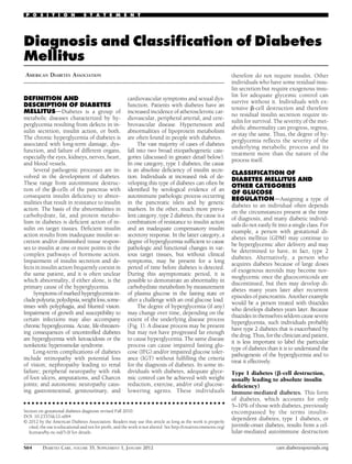
  • 1. P O S I T I O N S T A T E M E N T Diagnosis and Classification of Diabetes Mellitus AMERICAN DIABETES ASSOCIATION DEFINITION AND DESCRIPTION OF DIABETES MELLITUSdDiabetes is a group of metabolic diseases characterized by hyperglycemia resulting from defects in insulin secretion, insulin action, or both. The chronic hyperglycemia of diabetes is associated with long-term damage, dysfunction, and failure of different organs, especially the eyes, kidneys, nerves, heart, and blood vessels. Several pathogenic processes are involved in the development of diabetes. These range from autoimmune destruction of the b-cells of the pancreas with consequent insulin deficiency to abnormalities that result in resistance to insulin action. The basis of the abnormalities in carbohydrate, fat, and protein metabolism in diabetes is deficient action of insulin on target tissues. Deficient insulin action results from inadequate insulin secretion and/or diminished tissue responses to insulin at one or more points in the complex pathways of hormone action. Impairment of insulin secretion and defects in insulin action frequently coexist in the same patient, and it is often unclear which abnormality, if either alone, is the primary cause of the hyperglycemia. Symptoms of marked hyperglycemia include polyuria, polydipsia,weight loss,sometimes with polyphagia, and blurred vision. Impairment of growth and susceptibility to certain infections may also accompany chronic hyperglycemia. Acute, life-threatening consequences of uncontrolled diabetes are hyperglycemia with ketoacidosis or the nonketotic hyperosmolar syndrome. Long-term complications of diabetes include retinopathy with potential loss of vision; nephropathy leading to renal failure; peripheral neuropathy with risk of foot ulcers, amputations, and Charcot joints; and autonomic neuropathy causing gastrointestinal, genitourinary, and cardiovascular symptoms and sexual dysfunction. Patients with diabetes have an increased incidence of atherosclerotic cardiovascular, peripheral arterial, and cerebrovascular disease. Hypertension and abnormalities of lipoprotein metabolism are often found in people with diabetes. The vast majority of cases of diabetes fall into two broad etiopathogenetic categories (discussed in greater detail below). In one category, type 1 diabetes, the cause is an absolute deficiency of insulin secretion. Individuals at increased risk of developing this type of diabetes can often be identified by serological evidence of an autoimmune pathologic process occurring in the pancreatic islets and by genetic markers. In the other, much more prevalent category, type 2 diabetes, the cause is a combination of resistance to insulin action and an inadequate compensatory insulin secretory response. In the latter category, a degree of hyperglycemia sufficient to cause pathologic and functional changes in various target tissues, but without clinical symptoms, may be present for a long period of time before diabetes is detected. During this asymptomatic period, it is possible to demonstrate an abnormality in carbohydrate metabolism by measurement of plasma glucose in the fasting state or after a challenge with an oral glucose load. The degree of hyperglycemia (if any) may change over time, depending on the extent of the underlying disease process (Fig. 1). A disease process may be present but may not have progressed far enough to cause hyperglycemia. The same disease process can cause impaired fasting glucose (IFG) and/or impaired glucose tolerance (IGT) without fulfilling the criteria for the diagnosis of diabetes. In some individuals with diabetes, adequate glycemic control can be achieved with weight reduction, exercise, and/or oral glucoselowering agents. These individuals c c c c c c c c c c c c c c c c c c c c c c c c c c c c c c c c c c c c c c c c c c c c c c c c c Section on gestational diabetes diagnosis revised Fall 2010. DOI: 10.2337/dc12-s064 © 2012 by the American Diabetes Association. Readers may use this article as long as the work is properly cited, the use is educational and not for profit, and the work is not altered. See http://creativecommons.org/ licenses/by-nc-nd/3.0/ for details. S64 DIABETES CARE, VOLUME 35, SUPPLEMENT 1, JANUARY 2012 therefore do not require insulin. Other individuals who have some residual insulin secretion but require exogenous insulin for adequate glycemic control can survive without it. Individuals with extensive b-cell destruction and therefore no residual insulin secretion require insulin for survival. The severity of the metabolic abnormality can progress, regress, or stay the same. Thus, the degree of hyperglycemia reflects the severity of the underlying metabolic process and its treatment more than the nature of the process itself. CLASSIFICATION OF DIABETES MELLITUS AND OTHER CATEGORIES OF GLUCOSE REGULATIONdAssigning a type of diabetes to an individual often depends on the circumstances present at the time of diagnosis, and many diabetic individuals do not easily fit into a single class. For example, a person with gestational diabetes mellitus (GDM) may continue to be hyperglycemic after delivery and may be determined to have, in fact, type 2 diabetes. Alternatively, a person who acquires diabetes because of large doses of exogenous steroids may become normoglycemic once the glucocorticoids are discontinued, but then may develop diabetes many years later after recurrent episodes of pancreatitis. Another example would be a person treated with thiazides who develops diabetes years later. Because thiazides in themselves seldom cause severe hyperglycemia, such individuals probably have type 2 diabetes that is exacerbated by the drug. Thus, for the clinician and patient, it is less important to label the particular type of diabetes than it is to understand the pathogenesis of the hyperglycemia and to treat it effectively. Type 1 diabetes (b-cell destruction, usually leading to absolute insulin deficiency) Immune-mediated diabetes. This form of diabetes, which accounts for only 5–10% of those with diabetes, previously encompassed by the terms insulindependent diabetes, type 1 diabetes, or juvenile-onset diabetes, results from a cellular-mediated autoimmune destruction care.diabetesjournals.org
  • 2. Position Statement Figure 1dDisorders of glycemia: etiologic types and stages. *Even after presenting in ketoacidosis, these patients can briefly return to normoglycemia without requiring continuous therapy (i.e., “honeymoon” remission); **in rare instances, patients in these categories (e.g., Vacor toxicity, type 1 diabetes presenting in pregnancy) may require insulin for survival. of the b-cells of the pancreas. Markers of the immune destruction of the b-cell include islet cell autoantibodies, autoantibodies to insulin, autoantibodies to GAD (GAD65), and autoantibodies to the tyrosine phosphatases IA-2 and IA-2b. One and usually more of these autoantibodies are present in 85–90% of individuals when fasting hyperglycemia is initially detected. Also, the disease has strong HLA associations, with linkage to the DQA and DQB genes, and it is influenced by the DRB genes. These HLA-DR/ DQ alleles can be either predisposing or protective. In this form of diabetes, the rate of b-cell destruction is quite variable, being rapid in some individuals (mainly infants and children) and slow in others (mainly adults). Some patients, particularly children and adolescents, may present with ketoacidosis as the first manifestation of the disease. Others have modest fasting hyperglycemia that can rapidly change to severe hyperglycemia and/or ketoacidosis in the presence of infection or other stress. Still others, particularly adults, may retain residual b-cell function sufficient to prevent ketoacidosis for many years; such individuals eventually become dependent on insulin for survival and are at risk for ketoacidosis. At this latter stage of the disease, there is little or no insulin secretion, as manifested by low or undetectable levels of plasma care.diabetesjournals.org C-peptide. Immune-mediated diabetes commonly occurs in childhood and adolescence, but it can occur at any age, even in the 8th and 9th decades of life. Autoimmune destruction of b-cells has multiple genetic predispositions and is also related to environmental factors that are still poorly defined. Although patients are rarely obese when they present with this type of diabetes, the presence of obesity is not incompatible with the diagnosis. These patients are also prone to other autoimmune disorders such as Graves’ disease, Hashimoto’s thyroiditis, Addison’s disease, vitiligo, celiac sprue, autoimmune hepatitis, myasthenia gravis, and pernicious anemia. Idiopathic diabetes. Some forms of type 1 diabetes have no known etiologies. Some of these patients have permanent insulinopenia and are prone to ketoacidosis, but have no evidence of autoimmunity. Although only a minority of patients with type 1 diabetes fall into this category, of those who do, most are of African or Asian ancestry. Individuals with this form of diabetes suffer from episodic ketoacidosis and exhibit varying degrees of insulin deficiency between episodes. This form of diabetes is strongly inherited, lacks immunological evidence for b-cell autoimmunity, and is not HLA associated. An absolute requirement for insulin replacement therapy in affected patients may come and go. Type 2 diabetes (ranging from predominantly insulin resistance with relative insulin deficiency to predominantly an insulin secretory defect with insulin resistance) This form of diabetes, which accounts for ;90–95% of those with diabetes, previously referred to as non–insulin-dependent diabetes, type 2 diabetes, or adultonset diabetes, encompasses individuals who have insulin resistance and usually have relative (rather than absolute) insulin deficiency At least initially, and often throughout their lifetime, these individuals do not need insulin treatment to survive. There are probably many different causes of this form of diabetes. Although the specific etiologies are not known, autoimmune destruction of b-cells does not occur, and patients do not have any of the other causes of diabetes listed above or below. Most patients with this form of diabetes are obese, and obesity itself causes some degree of insulin resistance. Patients who are not obese by traditional weight criteria may have an increased percentage of body fat distributed predominantly in the abdominal region. Ketoacidosis seldom occurs spontaneously in this type of diabetes; when seen, it usually arises in association with the stress of another illness such as infection. This form of diabetes frequently goes undiagnosed for many years because the hyperglycemia DIABETES CARE, VOLUME 35, SUPPLEMENT 1, JANUARY 2012 S65
  • 3. Diagnosis and Classification develops gradually and at earlier stages is often not severe enough for the patient to notice any of the classic symptoms of diabetes. Nevertheless, such patients are at increased risk of developing macrovascular and microvascular complications. Whereas patients with this form of diabetes may have insulin levels that appear normal or elevated, the higher blood glucose levels in these diabetic patients would be expected to result in even higher insulin values had their b-cell function been normal. Thus, insulin secretion is defective in these patients and insufficient to compensate for insulin resistance. Insulin resistance may improve with weight reduction and/or pharmacological treatment of hyperglycemia but is seldom restored to normal. The risk of developing this form of diabetes increases with age, obesity, and lack of physical activity. It occurs more frequently in women with prior GDM and in individuals with hypertension or dyslipidemia, and its frequency varies in different racial/ethnic subgroups. It is often associated with a strong genetic predisposition, more so than is the autoimmune form of type 1 diabetes. However, the genetics of this form of diabetes are complex and not clearly defined. Other specific types of diabetes Genetic defects of the b-cell. Several forms of diabetes are associated with monogenetic defects in b-cell function. These forms of diabetes are frequently characterized by onset of hyperglycemia at an early age (generally before age 25 years). They are referred to as maturityonset diabetes of the young (MODY) and are characterized by impaired insulin secretion with minimal or no defects in insulin action. They are inherited in an autosomal dominant pattern. Abnormalities at six genetic loci on different chromosomes have been identified to date. The most common form is associated with mutations on chromosome 12 in a hepatic transcription factor referred to as hepatocyte nuclear factor (HNF)-1a. A second form is associated with mutations in the glucokinase gene on chromosome 7p and results in a defective glucokinase molecule. Glucokinase converts glucose to glucose-6-phosphate, the metabolism of which, in turn, stimulates insulin secretion by the b-cell. Thus, glucokinase serves as the “glucose sensor” for the b-cell. Because of defects in the glucokinase gene, increased plasma levels of glucose are necessary to elicit normal levels of insulin secretion. The less common S66 forms result from mutations in other transcription factors, including HNF-4a, HNF-1b, insulin promoter factor (IPF)1, and NeuroD1. Point mutations in mitochondrial DNA have been found to be associated with diabetes and deafness The most common mutation occurs at position 3,243 in the tRNA leucine gene, leading to an A-to-G transition. An identical lesion occurs in the MELAS syndrome (mitochondrial myopathy, encephalopathy, lactic acidosis, and stroke-like syndrome); however, diabetes is not part of this syndrome, suggesting different phenotypic expressions of this genetic lesion. Genetic abnormalities that result in the inability to convert proinsulin to insulin have been identified in a few families, and such traits are inherited in an autosomal dominant pattern. The resultant glucose intolerance is mild. Similarly, the production of mutant insulin molecules with resultant impaired receptor binding has also been identified in a few families and is associated with an autosomal inheritance and only mildly impaired or even normal glucose metabolism. Genetic defects in insulin action. There are unusual causes of diabetes that result from genetically determined abnormalities of insulin action. The metabolic abnormalities associated with mutations of the insulin receptor may range from hyperinsulinemia and modest hyperglycemia to severe diabetes. Some individuals with these mutations may have acanthosis nigricans. Women may be virilized and have enlarged, cystic ovaries. In the past, this syndrome was termed type A insulin resistance. Leprechaunism and the RabsonMendenhall syndrome are two pediatric syndromes that have mutations in the insulin receptor gene with subsequent alterations in insulin receptor function and extreme insulin resistance. The former has characteristic facial features and is usually fatal in infancy, while the latter is associated with abnormalities of teeth and nails and pineal gland hyperplasia. Alterations in the structure and function of the insulin receptor cannot be demonstrated in patients with insulinresistant lipoatrophic diabetes. Therefore, it is assumed that the lesion(s) must reside in the postreceptor signal transduction pathways. Diseases of the exocrine pancreas. Any process that diffusely injures the pancreas can cause diabetes. Acquired processes include pancreatitis, trauma, infection, pancreatectomy, and pancreatic carcinoma. DIABETES CARE, VOLUME 35, SUPPLEMENT 1, JANUARY 2012 With the exception of that caused by cancer, damage to the pancreas must be extensive for diabetes to occur; adrenocarcinomas that involve only a small portion of the pancreas have been associated with diabetes. This implies a mechanism other than simple reduction in b-cell mass. If extensive enough, cystic fibrosis and hemochromatosis will also damage b-cells and impair insulin secretion. Fibrocalculous pancreatopathy may be accompanied by abdominal pain radiating to the back and pancreatic calcifications identified on X-ray examination. Pancreatic fibrosis and calcium stones in the exocrine ducts have been found at autopsy. Endocrinopathies. Several hormones (e.g., growth hormone, cortisol, glucagon, epinephrine) antagonize insulin action. Excess amounts of these hormones (e.g., acromegaly, Cushing’s syndrome, glucagonoma, pheochromocytoma, respectively) can cause diabetes. This generally occurs in individuals with preexisting defects in insulin secretion, and hyperglycemia typically resolves when the hormone excess is resolved. Somatostatinoma- and aldosteronoma-induced hypokalemia can cause diabetes, at least in part, by inhibiting insulin secretion. Hyperglycemia generally resolves after successful removal of the tumor. Drug- or chemical-induced diabetes. Many drugs can impair insulin secretion. These drugs may not cause diabetes by themselves, but they may precipitate diabetes in individuals with insulin resistance. In such cases, the classification is unclear because the sequence or relative importance of b-cell dysfunction and insulin resistance is unknown. Certain toxins such as Vacor (a rat poison) and intravenous pentamidine can permanently destroy pancreatic b-cells. Such drug reactions fortunately are rare. There are also many drugs and hormones that can impair insulin action. Examples include nicotinic acid and glucocorticoids. Patients receiving a-interferon have been reported to develop diabetes associated with islet cell antibodies and, in certain instances, severe insulin deficiency. The list shown in Table 1 is not all-inclusive, but reflects the more commonly recognized drug-, hormone-, or toxin-induced forms of diabetes. Infections. Certain viruses have been associated with b-cell destruction. Diabetes occurs in patients with congenital rubella, although most of these patients care.diabetesjournals.org
  • 4. Position Statement Table 1dEtiologic classification of diabetes mellitus I. Type 1 diabetes (b-cell destruction, usually leading to absolute insulin deficiency) A. Immune mediated B. Idiopathic II. Type 2 diabetes (may range from predominantly insulin resistance with relative insulin deficiency to a predominantly secretory defect with insulin resistance) III. Other specific types A. Genetic defects of b-cell function 1. Chromosome 12, HNF-1a (MODY3) 2. Chromosome 7, glucokinase (MODY2) 3. Chromosome 20, HNF-4a (MODY1) 4. Chromosome 13, insulin promoter factor-1 (IPF-1; MODY4) 5. Chromosome 17, HNF-1b (MODY5) 6. Chromosome 2, NeuroD1 (MODY6) 7. Mitochondrial DNA 8. Others B. Genetic defects in insulin action 1. Type A insulin resistance 2. Leprechaunism 3. Rabson-Mendenhall syndrome 4. Lipoatrophic diabetes 5. Others C. Diseases of the exocrine pancreas 1. Pancreatitis 2. Trauma/pancreatectomy 3. Neoplasia 4. Cystic fibrosis 5. Hemochromatosis 6. Fibrocalculous pancreatopathy 7. Others D. Endocrinopathies 1. Acromegaly 2. Cushing’s syndrome 3. Glucagonoma 4. Pheochromocytoma 5. Hyperthyroidism 6. Somatostatinoma 7. Aldosteronoma 8. Others E. Drug or chemical induced 1. Vacor 2. Pentamidine 3. Nicotinic acid 4. Glucocorticoids 5. Thyroid hormone 6. Diazoxide 7. b-adrenergic agonists 8. Thiazides 9. Dilantin 10. g-Interferon 11. Others F. Infections 1. Congenital rubella 2. Cytomegalovirus 3. Others G. Uncommon forms of immune-mediated diabetes 1. “Stiff-man” syndrome 2. Anti-insulin receptor antibodies 3. Others H. Other genetic syndromes sometimes associated with diabetes 1. Down syndrome 2. Klinefelter syndrome 3. Turner syndrome 4. Wolfram syndrome 5. Friedreich ataxia 6. Huntington chorea 7. Laurence-Moon-Biedl syndrome 8. Myotonic dystrophy 9. Porphyria 10. Prader-Willi syndrome 11. Others IV. Gestational diabetes mellitus Patients with any form of diabetes may require insulin treatment at some stage of their disease. Such use of insulin does not, of itself, classify the patient. care.diabetesjournals.org have HLA and immune markers characteristic of type 1 diabetes. In addition, coxsackievirus B, cytomegalovirus, adenovirus, and mumps have been implicated in inducing certain cases of the disease. Uncommon forms of immune-mediated diabetes. In this category, there are two known conditions, and others are likely to occur. The stiff-man syndrome is an autoimmune disorder of the central nervous system characterized by stiffness of the axial muscles with painful spasms. Patients usually have high titers of the GAD autoantibodies, and approximately one-third will develop diabetes. Anti-insulin receptor antibodies can cause diabetes by binding to the insulin receptor, thereby blocking the binding of insulin to its receptor in target tissues. However, in some cases, these antibodies can act as an insulin agonist after binding to the receptor and can thereby cause hypoglycemia. Anti-insulin receptor antibodies are occasionally found in patients with systemic lupus erythematosus and other autoimmune diseases. As in other states of extreme insulin resistance, patients with anti-insulin receptor antibodies often have acanthosis nigricans. In the past, this syndrome was termed type B insulin resistance. Other genetic syndromes sometimes associated with diabetes. Many genetic syndromes are accompanied by an increased incidence of diabetes. These include the chromosomal abnormalities of Down syndrome, Klinefelter syndrome, and Turner syndrome. Wolfram’s syndrome is an autosomal recessive disorder characterized by insulin-deficient diabetes and the absence of b-cells at autopsy. Additional manifestations include diabetes insipidus, hypogonadism, optic atrophy, and neural deafness. Other syndromes are listed in Table 1. Gestational diabetes mellitus For many years, GDM has been defined as any degree of glucose intolerance with onset or first recognition during pregnancy. Although most cases resolve with delivery, the definition applied whether or not the condition persisted after pregnancy and did not exclude the possibility that unrecognized glucose intolerance may have antedated or begun concomitantly with the pregnancy. This definition facilitated a uniform strategy for detection and classification of GDM, but its limitations were recognized for many years. As the ongoing epidemic of obesity and DIABETES CARE, VOLUME 35, SUPPLEMENT 1, JANUARY 2012 S67
  • 5. Diagnosis and Classification Table 2dCategories of increased risk for diabetes* FPG 100 mg/dl (5.6 mmol/l) to 125 mg/dl (6.9 mmol/l) [IFG] 2-h PG in the 75-g OGTT 140 mg/dl (7.8 mmol/l) to 199 mg/dl (11.0 mmol/l) [IGT] A1C 5.7–6.4% *For all three tests, risk is continuous, extending below the lower limit of the range and becoming disproportionately greater at higher ends of the range. diabetes has led to more type 2 diabetes in women of childbearing age, the number of pregnant women with undiagnosed type 2 diabetes has increased. After deliberations in 2008–2009, the International Association of Diabetes and Pregnancy Study Groups (IADPSG), an international consensus group with representatives from multiple obstetrical and diabetes organizations, including the American Diabetes Association (ADA), recommended that high-risk women found to have diabetes at their initial prenatal visit, using standard criteria (Table 3), receive a diagnosis of overt, not gestational, diabetes. Approximately 7% of all pregnancies (ranging from 1 to 14%, depending on the population studied and the diagnostic tests employed) are complicated by GDM, resulting in more than 200,000 cases annually. CATEGORIES OF INCREASED RISK FOR DIABETESdIn 1997 and 2003, The Expert Committee on Diagnosis and Classification of Diabetes Mellitus (1,2) recognized an intermediate group of individuals whose glucose levels do not meet criteria for diabetes, yet are higher than those considered normal. These people were defined as having impaired fasting glucose (IFG) [fasting plasma glucose (FPG) levels 100 mg/dl (5.6 mmol/l) to 125 mg/dl (6.9 mmol/l)], or impaired glucose tolerance (IGT) [2-h values in the oral glucose tolerance test (OGTT) of 140 mg/dl (7.8 mmol/l) to 199 mg/dl (11.0 mmol/l)]. Individuals with IFG and/or IGT have been referred to as having pre-diabetes, indicating the relatively high risk for the future development of diabetes. IFG and IGT should not be viewed as clinical entities in their own right but rather risk factors for diabetes as well as cardiovascular disease. They can be observed as intermediate stages in any of the disease processes listed in Table 1. IFG and IGT are associated with obesity (especially S68 abdominal or visceral obesity), dyslipidemia with high triglycerides and/or low HDL cholesterol, and hypertension. Structured lifestyle intervention, aimed at increasing physical activity and producing 5–10% loss of body weight, and certain pharmacological agents have been demonstrated to prevent or delay the development of diabetes in people with IGT; the potential impact of such interventions to reduce mortality or the incidence of cardiovascular disease has not been demonstrated to date. It should be noted that the 2003 ADA Expert Committee report reduced the lower FPG cut point to define IFG from 110 mg/dl (6.1 mmol/l) to 100 mg/dl (5.6 mmol/l), in part to ensure that prevalence of IFG was similar to that of IGT. However, the World Health Organization (WHO) and many other diabetes organizations did not adopt this change in the definition of IFG. As A1C is used more commonly to diagnose diabetes in individuals with risk factors, it will also identify those at higher risk for developing diabetes in the future. When recommending the use of the A1C to diagnose diabetes in its 2009 report, the International Expert Committee (3) stressed the continuum of risk for diabetes with all glycemic measures and did not formally identify an equivalent intermediate category for A1C. The group did note that those with A1C levels above the laboratory “normal” range but below the diagnostic cut point for diabetes (6.0 to ,6.5%) are at very high risk of developing diabetes. Indeed, incidence of diabetes in people with A1C levels in this range is more than 10 times that of people with lower levels (4–7). However, the 6.0 to ,6.5% range fails to identify a substantial number of patients who have IFG and/or IGT. Prospective studies indicate that people within the A1C range of 5.5– 6.0% have a 5-year cumulative incidence of diabetes that ranges from 12 to 25% (4–7), which is appreciably (three- to eightfold) higher than incidence in the U.S. population as a whole (8). Analyses of nationally representative data from the National Health and Nutrition Examination Survey (NHANES) indicate that the A1C value that most accurately identifies people with IFG or IGT falls between 5.5 and 6.0%. In addition, linear regression analyses of these data indicate that among the nondiabetic adult population, an FPG of 110 mg/dl (6.1 mmol/l) corresponds to an A1C of 5.6%, while an FPG of 100 mg/dl (5.6 mmol/l) corresponds to an A1C of 5.4% (R.T. Ackerman, personal DIABETES CARE, VOLUME 35, SUPPLEMENT 1, JANUARY 2012 communication). Finally, evidence from the Diabetes Prevention Program (DPP), wherein the mean A1C was 5.9% (SD 0.5%), indicates that preventive interventions are effective in groups of people with A1C levels both below and above 5.9% (9). For these reasons, the most appropriate A1C level above which to initiate preventive interventions is likely to be somewhere in the range of 5.5–6%. As was the case with FPG and 2-h PG, defining a lower limit of an intermediate category of A1C is somewhat arbitrary, as the risk of diabetes with any measure or surrogate of glycemia is a continuum, extending well into the normal ranges. To maximize equity and efficiency of preventive interventions, such an A1C cut point should balance the costs of “false negatives” (failing to identify those who are going to develop diabetes) against the costs of “false positives” (falsely identifying and then spending intervention resources on those who were not going to develop diabetes anyway). Compared to the fasting glucose cutpoint of 100 mg/dl (5.6 mmol/l), an A1C cutpoint of 5.7% is less sensitive but more specific and has a higher positive predictive value to identify people at risk for later development of diabetes. A large prospective study found that a 5.7% cutpoint has a sensitivity of 66% and specificity of 88% for the identification of subsequent 6-year diabetes incidence (10). Receiver operating curve analyses of nationally representative U.S. data (NHANES 1999-2006) indicate that an A1C value of 5.7% has modest sensitivity (39-45%) but high specificity (81-91%) to identify cases of IFP (FPG .100 mg/ dl) (5.6 mmol/l) or IGT (2-h glucose . 140 mg/dl) (R.T. Ackerman, personal communication). Other analyses suggest that an A1C of 5.7% is associated with diabetes risk similar to the high-risk participants in the DPP (R.T. Ackerman, personal communication). Hence, it is reasonable to consider an A1C range of 5.7 to 6.4% as identifying individuals with high risk for future diabetes and to whom the term pre-diabetes may be applied if desired. Individuals with an A1C of 5.7–6.4% should be informed of their increased risk for diabetes as well as cardiovascular disease and counseled about effective strategies, such as weight loss and physical activity, to lower their risks. As with glucose measurements, the continuum of risk is curvilinear, so that as A1C rises, the risk of diabetes rises disproportionately. care.diabetesjournals.org
  • 6. Position Statement Accordingly, interventions should be most intensive and follow-up should be particularly vigilant for those with A1C levels above 6.0%, who should be considered to be at very high risk. However, just as an individual with a fasting glucose of 98 mg/dl (5.4 mmol/l) may not be at negligible risk for diabetes, individuals with A1C levels below 5.7% may still be at risk, depending on level of A1C and presence of other risk factors, such as obesity and family history. Table 2 summarizes the categories of increased risk for diabetes. Evaluation of patients at risk should incorporate a global risk factor assessment for both diabetes and cardiovascular disease. Screening for and counseling about risk of diabetes should always be in the pragmatic context of the patient’s comorbidities, life expectancy, personal capacity to engage in lifestyle change, and overall health goals. DIAGNOSTIC CRITERIA FOR DIABETES MELLITUSdFor decades, the diagnosis of diabetes has been based on glucose criteria, either the FPG or the 75-g OGTT. In 1997, the first Expert Committee on the Diagnosis and Classification of Diabetes Mellitus revised the diagnostic criteria, using the observed association between FPG levels and presence of retinopathy as the key factor with which to identify threshold glucose level. The Committee examined data from three cross-sectional epidemiologic studies that assessed retinopathy with fundus photography or direct ophthalmoscopy and measured glycemia as FPG, 2-h PG, and A1C. These studies demonstrated glycemic levels below which there was little prevalent retinopathy and above which the prevalence of retinopathy increased in an apparently linear fashion. The deciles of the three measures at which retinopathy began to increase were the same for each measure within each population. Moreover, the glycemic values above which retinopathy increased were similar among the populations. These analyses helped to inform a new diagnostic cut point of $126 mg/dl (7.0 mmol/l) for FPG and confirmed the long-standing diagnostic 2-h PG value of $200 mg/dl (11.1 mmol/l). A1C is a widely used marker of chronic glycemia, reflecting average blood glucose levels over a 2- to 3-month period of time. The test plays a critical role in the management of the patient with diabetes, since it correlates well with both care.diabetesjournals.org Table 3dCriteria for the diagnosis of diabetes A1C $6.5%. The test should be performed in a laboratory using a method that is NGSP certified and standardized to the DCCT assay.* OR FPG $126 mg/dl (7.0 mmol/l). Fasting is defined as no caloric intake for at least 8 h.* OR 2-h plasma glucose $200 mg/dl (11.1 mmol/l) during an OGTT. The test should be performed as described by the World Health Organization, using a glucose load containing the equivalent of 75 g anhydrous glucose dissolved in water.* OR In a patient with classic symptoms of hyperglycemia or hyperglycemic crisis, a random plasma glucose $200 mg/dl (11.1 mmol/l). *In the absence of unequivocal hyperglycemia, criteria 1–3 should be confirmed by repeat testing. microvascular and, to a lesser extent, macrovascular complications and is widely used as the standard biomarker for the adequacy of glycemic management. Prior Expert Committees have not recommended use of the A1C for diagnosis of diabetes, in part due to lack of standardization of the assay. However, A1C assays are now highly standardized so that their results can be uniformly applied both temporally and across populations. In their recent report (3), an International Expert Committee, after an extensive review of both established and emerging epidemiological evidence, recommended the use of the A1C test to diagnose diabetes, with a threshold of $6.5%, and ADA affirms this decision. The diagnostic A1C cut point of 6.5% is associated with an inflection point for retinopathy prevalence, as are the diagnostic thresholds for FPG and 2-h PG (3). The diagnostic test should be performed using a method that is certified by the National Glycohemoglobin Standardization Program (NGSP) and standardized or traceable to the Diabetes Control and Complications Trial reference assay. Point-of-care A1C assays are not sufficiently accurate at this time to use for diagnostic purposes. There is an inherent logic to using a more chronic versus an acute marker of dysglycemia, particularly since the A1C is already widely familiar to clinicians as a marker of glycemic control. Moreover, the A1C has several advantages to the FPG, including greater convenience, since fasting is not required, evidence to suggest greater preanalytical stability, and less day-to-day perturbations during periods of stress and illness. These advantages, however, must be balanced by greater cost, the limited availability of A1C testing in certain regions of the developing world, and the incomplete correlation between A1C and average glucose in certain individuals. In addition, the A1C can be misleading in patients with certain forms of anemia and hemoglobinopathies, which may also have unique ethnic or geographic distributions. For patients with a hemoglobinopathy but normal red cell turnover, such as sickle cell trait, an A1C assay without interference from abnormal hemoglobins should be used (an updated list is available at www.ngsp.org/prog/index3. html). For conditions with abnormal red cell turnover, such as anemias from hemolysis and iron deficiency, the diagnosis of diabetes must employ glucose criteria exclusively. The established glucose criteria for the diagnosis of diabetes remain valid. These include the FPG and 2-h PG. Additionally, patients with severe hyperglycemia such as those who present with severe classic hyperglycemic symptoms or hyperglycemic crisis can continue to be diagnosed when a random (or casual) plasma glucose of $200 mg/dl (11.1 mmol/l) is found. It is likely that in such cases the health care professional would also measure an A1C test as part of the initial assessment of the severity of the diabetes and that it would (in most cases) be above the diagnostic cut point for diabetes. However, in rapidly evolving diabetes, such as the development of type 1 diabetes in some children, A1C may not be significantly elevated despite frank diabetes. Just as there is less than 100% concordance between the FPG and 2-h PG tests, there is not full concordance between A1C and either glucose-based test. Analyses of NHANES data indicate that, assuming universal screening of the undiagnosed, the A1C cut point of $6.5% identifies one-third fewer cases of undiagnosed diabetes than a fasting glucose cut point of $126 mg/dl (7.0 mmol/l) (cdc website tbd). However, in DIABETES CARE, VOLUME 35, SUPPLEMENT 1, JANUARY 2012 S69
  • 7. Diagnosis and Classification practice, a large portion of the population with type 2 diabetes remains unaware of their condition. Thus, it is conceivable that the lower sensitivity of A1C at the designated cut point will be offset by the test’s greater practicality, and that wider application of a more convenient test (A1C) may actually increase the number of diagnoses made. Further research is needed to better characterize those patients whose glycemic status might be categorized differently by two different tests (e.g., FPG and A1C), obtained in close temporal approximation. Such discordance may arise from measurement variability, change over time, or because A1C, FPG, and postchallenge glucose each measure different physiological processes. In the setting of an elevated A1C but “nondiabetic” FPG, the likelihood of greater postprandial glucose levels or increased glycation rates for a given degree of hyperglycemia may be present. In the opposite scenario (high FPG yet A1C below the diabetes cut point), augmented hepatic glucose production or reduced glycation rates may be present. As with most diagnostic tests, a test result diagnostic of diabetes should be repeated to rule out laboratory error, unless the diagnosis is clear on clinical grounds, such as a patient with classic symptoms of hyperglycemia or hyperglycemic crisis. It is preferable that the same test be repeated for confirmation, since there will be a greater likelihood of concurrence in this case. For example, if the A1C is 7.0% and a repeat result is 6.8%, the diagnosis of diabetes is confirmed. However, there are scenarios in which results of two different tests (e.g., FPG and A1C) are available for the same patient. In this situation, if the two different tests are both above the diagnostic thresholds, the diagnosis of diabetes is confirmed. On the other hand, when two different tests are available in an individual and the results are discordant, the test whose result is above the diagnostic cut point should be repeated, and the diagnosis is made on the basis of the confirmed test. That is, if a patient meets the diabetes criterion of the A1C (two results $6.5%) but not the FPG (,126 mg/dl or 7.0 mmol/l), or vice versa, that person should be considered to have diabetes. Admittedly, in most circumstance the “nondiabetic” test is likely to be in a range very close to the threshold that defines diabetes. Since there is preanalytic and analytic variability of all the tests, it is also possible S70 that when a test whose result was above the diagnostic threshold is repeated, the second value will be below the diagnostic cut point. This is least likely for A1C, somewhat more likely for FPG, and most likely for the 2-h PG. Barring a laboratory error, such patients are likely to have test results near the margins of the threshold for a diagnosis. The healthcare professional might opt to follow the patient closely and repeat the testing in 3–6 months. The decision about which test to use to assess a specific patient for diabetes should be at the discretion of the health care professional, taking into account the availability and practicality of testing an individual patient or groups of patients. Perhaps more important than which diagnostic test is used, is that the testing for diabetes be performed when indicated. There is discouraging evidence indicating that many at-risk patients still do not receive adequate testing and counseling for this increasingly common disease, or for its frequently accompanying cardiovascular risk factors. The current diagnostic criteria for diabetes are summarized in Table 3. Diagnosis of gestational diabetes GDM carries risks for the mother and neonate. The Hyperglycemia and Adverse Pregnancy Outcomes (HAPO) study (11), a large-scale (;25,000 pregnant women) multinational epidemiologic study, demonstrated that risk of adverse maternal, fetal, and neonatal outcomes continuously increased as a function of maternal glycemia at 24-28 weeks, even within ranges previously considered normal for pregnancy. For most complications, there was no threshold for risk. These results have led to careful reconsideration of the diagnostic criteria for GDM. After deliberations in 2008-2009, the IADPSG, an international consensus group with representatives from multiple obstetrical and diabetes organizations, including ADA, developed revised recommendations for diagnosing GDM. The group recommended that all women not known to have diabetes undergo a 75-g OGTT at 24-28 weeks of gestation. Additionally, the group developed diagnostic cutpoints for the fasting, 1-h, and 2-h plasma glucose measurements that conveyed an odds ratio for adverse outcomes of at least 1.75 compared with women with mean glucose levels in the HAPO study. Current screening and diagnostic strategies, based on the IADPSG statement (12), are outlined in Table 4. DIABETES CARE, VOLUME 35, SUPPLEMENT 1, JANUARY 2012 Table 4dScreening for and diagnosis of GDM Perform a 75-g OGTT, with plasma glucose measurement fasting and at 1 and 2 h, at 24-28 of weeks gestation in women not previously diagnosed with overt diabetes. The OGTT should be performed in the morning after an overnight fast of at least 8 h. The diagnosis of GDM is made when any of the following plasma glucose values are exceeded c Fasting: $92 mg/dl (5.1 mmol/l) c 1 h: $180 mg/dl (10.0 mmol/l) c 2 h: $153 mg/dl (8.5 mmol/l) These new criteria will significantly increase the prevalence of GDM, primarily because only one abnormal value, not two, is sufficient to make the diagnosis. The ADA recognizes the anticipated significant increase in the incidence of GDM to be diagnosed by these criteria and is sensitive to concerns about the “medicalization” of pregnancies previously categorized as normal. These diagnostic criteria changes are being made in the context of worrisome worldwide increases in obesity and diabetes rates, with the intent of optimizing gestational outcomes for women and their babies. Admittedly, there are few data from randomized clinical trials regarding therapeutic interventions in women who will now be diagnosed with GDM based on only one blood glucose value above the specified cutpoints (in contrast to the older criteria that stipulated at least two abnormal values). Expected benefits to their pregnancies and offspring is inferred from intervention trials that focused on women with more mild hyperglycemia than identified using older GDM diagnostic criteria and that found modest benefits (13,14). The frequency of their follow-up and blood glucose monitoring is not yet clear but likely to be less intensive than women diagnosed by the older criteria. Additional well-designed clinical studies are needed to determine the optimal intensity of monitoring and treatment of women with GDM diagnosed by the new criteria (that would not have met the prior definition of GDM). It is important to note that 80-90% of women in both of the mild GDM studies (whose glucose values overlapped with the thresholds recommended herein) could be managed with lifestyle therapy alone. care.diabetesjournals.org
  • 8. Position Statement References 1. Expert Committee on the Diagnosis and Classification of Diabetes Mellitus. Report of the Expert Committee on the Diagnosis and Classification of Diabetes Mellitus. Diabetes Care 1997;20:1183–1197 2. Genuth S, Alberti KG, Bennett P, Buse J, Defronzo R, Kahn R, Kitzmiller J, Knowler WC, Lebovitz H, Lernmark A, Nathan D, Palmer J, Rizza R, Saudek C, Shaw J, Steffes M, Stern M, Tuomilehto J, Zimmet P, Expert Committee on the Diagnosis and Classification of Diabetes Mellitus2, the Expert Committee on the Diagnosis and Classification of Diabetes Mellitus. Follow-up report on the diagnosis of diabetes mellitus. Diabetes Care 2003;26:3160– 3167 3. International Expert Committee. International Expert Committee report on the role of the A1C assay in the diagnosis of diabetes. Diabetes Care 2009;32:1327– 1334 4. Edelman D, Olsen MK, Dudley TK, Harris AC, Oddone EZ. Utility of hemoglobin A1c in predicting diabetes risk. J Gen Intern Med 2004;19:1175–1180 5. Pradhan AD, Rifai N, Buring JE, Ridker PM. Hemoglobin A1c predicts diabetes but not cardiovascular disease in nondiabetic women. Am J Med 2007;120: 720–727 care.diabetesjournals.org 6. Sato KK, Hayashi T, Harita N, Yoneda T, Nakamura Y, Endo G, Kambe H. Combined measurement of fasting plasma glucose and A1C is effective for the prediction of type 2 diabetes: the Kansai Healthcare Study. Diabetes Care 2009;32: 644–646 7. Shimazaki T, Kadowaki T, Ohyama Y, Ohe K, Kubota K. Hemoglobin A1c (HbA1c) predicts future drug treatment for diabetes mellitus: a follow-up study using routine clinical data in a Japanese university hospital. Translational Research 2007;149:196–204 8. Geiss LS, Pan L, Cadwell B, Gregg EW, Benjamin SM, Engelgau MM. Changes in incidence of diabetes in U.S. adults, 1997–2003. Am J Prev Med 2006;30: 371–377 9. Knowler WC, Barrett-Connor E, Fowler SE, Hamman RF, Lachin JM, Walker EA, Nathan DM, Diabetes Prevention Program Research Group. Reduction in the incidence of type 2 diabetes with lifestyle intervention or metformin. N Engl J Med 2002;346:393–403 10. Droumaguet C, Balkau B, Simon D, Caces E, Tichet J, Charles MA, Eschwege E, the DESIR Study Group. Use of HbA1c in predicting progression to diabetes in French men and women: data from an Epidemiological Study on the Insulin 11. 12. 13. 14. Resistance Syndrome (DESIR) Diabetes Care 2006;29:1619–1625. Metzger BE, Lowe LP, Dyer AR, Trimble ER, Chaovarindr U, Coustan DR, Hadden DR, McCance DR, Hod M, McIntyre HD, Oats JJ, Persson B, Rogers MS, Sacks DA. Hyperglycemia and adverse pregnancy outcomes. N Engl J Med 2008;358:1991– 2002 Metzger BE, Gabbe SG, Persson B, Buchanan TA, Catalano PA, Damm P, Dyer AR, Leiva A, Hod M, Kitzmiler JL, Lowe LP, McIntyre HD, Oats JJ, Omori Y, Schmidt MI. International Association of Diabetes and Pregnancy Study Groups recommendations on the diagnosis and classification of hyperglycemia in pregnancy. Diabetes Care 2010;33:676–682 Landon MB, Spong CY, Thom E, Carpenter MW, Ramin SM, Casey B, Wapner RJ, Varner MW, Rouse DJ, Thorp JM, Jr., Sciscione A, Catalano P, Harper M, Saade G, Lain KY, Sorokin Y, Peaceman AM, Tolosa JE, Anderson GB. A multicenter, randomized trial of treatment for mild gestational diabetes. N Engl J Med 2009;361:1339–1348 Crowther CA, Hiller JE, Moss JR, McPhee AJ, Jeffries WS, Robinson JS. Effect of treatment of gestational diabetes mellitus on pregnancy outcomes. N Engl J Med 2005;352:2477–2486 DIABETES CARE, VOLUME 35, SUPPLEMENT 1, JANUARY 2012 S71