1. Three-dimensional finite element analysis of the effect of different bone
quality on stress distribution in an implant-supported crown
M. Sevimay, DDS, PhD,a F. Turhan, DDS,b M. A. Kilicarslan, PhD,c and G. Eskitascioglu, DDS, PhDd
x
School of Dentistry, University of Selcuk, Konya, Turkey; Baskent Hospital, Adana, Turkey; 75th Year
x
Ankara Dental Hospital, Ankara, Turkey
Statement of problem. Primary implant stability and bone density are variables that are considered essential
to achieve predictable osseointegration and long-term clinical survival of implants. Information about the
influence of bone quality on stress distribution in an implant-supported crown is limited.
Purpose. The purpose of this study was to investigate the effect of 4 different bone qualities on stress
distribution in an implant-supported mandibular crown, using 3-dimensional (3-D) finite element (FE) analysis.
Material and methods. A 3-D FE model of a mandibular section of bone with a missing second premolar
tooth was developed, and an implant to receive a crown was developed. A solid 4.1 3 10-mm screw-type dental
implant system (ITI; solid implant) and a metal-ceramic crown using Co-Cr (Wiron 99) and feldspathic
porcelain were modeled. The model was developed with FE software (Pro/Engineer 2000i program), and 4
types of bone quality (D1, D2, D3, and D4) were prepared. A load of 300 N was applied in a vertical direction
to the buccal cusp and distal fossa of the crowns. Optimal bone quality for an implant-supported crown was
evaluated.
Results. The results demonstrated that von Mises stresses in D3 and D4 bone quality were163 MPa and 180
MPa, respectively, and reached the highest values at the neck of the implant. The von Mises stress values in D1
and D2 bone quality were 150 MPa and 152 MPa, respectively, at the neck of the implant. A more homogenous
stress distribution was seen in the entire bone.
Conclusion. For the bone qualities investigated, stress concentrations in compact bone followed the same
distributions as in the D3 bone model, but because the trabecular bone was weaker and less resistant to
deformation than the other bone qualities modeled, the stress magnitudes were greatest for D3 and D4
bone. (J Prosthet Dent 2005;93:227-34.)
CLINICAL IMPLICATIONS
Placement of implants in bone with greater thickness of the cortical shell and greater density of
the core reduced stress concentration and may result in less micromovement, thereby increasing
the likelihood of implant stabilization and tissue integration. However, long-term clinical trials
are required to determine the effect of different bone quality on stress distribution in dental
implants, in relation to the long-term success of implant treatment.
S ince the late 1960s, when dental implants were in-
troduced for rehabilitation of the completely edentulous
Available bone is particularly important in implant
dentistry and describes the external architecture or vol-
patient,1,2 an awareness and subsequent demand for this ume of the edentulous area considered for implants. In
form of therapy has increased.3 Long-term success rates addition, bone has an internal structure described in
as high as 95% for mandibular implants and 90% for terms of quality or density, which reflects the strength
maxillary implants have been reported.4 Still, implant of the bone.7 The density of available bone in an eden-
failure is a source of frustration and disappointment tulous site is a determining factor in treatment planning,
for both the patient and clinician, and strategies for implant design, surgical approach, healing time, and
prevention of failure are crucial.5,6 initial progressive bone loading during prosthetic
reconstruction.8,9
For osseointegration of endosteal implants to occur,
a
Research Assistant, Department of Prosthodontics, School of not only is adequate bone quantity (height, width,
Dentistry, University of Selcuk. shape) required, but adequate density is also needed.10
b
Private practice, Baskent Hospital.
x
c
Private practice, 75th Year Ankara Dental Hospital.
Zarb and Schmitt11 stated that bone structure is the
d
Chairman and Professor, Department of Prosthodontics, School of most important factor in selecting the most favorable
Dentistry, University of Selcuk. treatment outcome in implant dentistry. Bone quality
MARCH 2005 THE JOURNAL OF PROSTHETIC DENTISTRY 227
2. THE JOURNAL OF PROSTHETIC DENTISTRY SEVIMAY ET AL
The mechanical distribution of stress occurs primarily
where bone is in contact with the implant.7 The density
of bone is directly related to the amount of implant-to-
bone contact.7 The percentage of bone contact is signif-
icantly greater in cortical bone than in trabecular bone.7
The initial bone density not only provides mechanical
immobilization during healing but also permits better
distribution and transmission of stresses from the
implant-bone interface.7,17 Increased clinical failure
rates in poor quality, porous bone, as compared to more
dense bone, have been well documented.18-21 To de-
crease stress, the clinician may elect to increase the num-
ber of implants or use an implant design with greater
surface area.7,22-24
Three-D FE analysis has been widely used for the
quantitative evaluation of stresses on the implant and
its surrounding bone.25-27 Some investigators studied
the influence of the implant design on stress concentra-
tion in the bone during loading and indicated that the
implant design was a significant factor influencing the
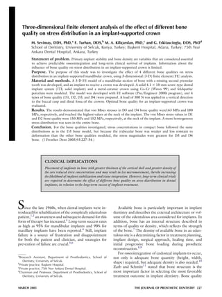
Fig. 1. Schematic presentation of threaded solid dental stress created in the bone.28,29 Others studied the influ-
implant. ence of the bone-implant interface on stress concentra-
tion. These authors demonstrated that when
maximum stress concentration occurs in cortical bone,
Table I. Material properties
it is located in the area of contact with the implant,
Young’s Poisson’s and when the maximum stress concentration occurs in
modulus ratio trabecular bone, it occurs around the apex of the im-
Material (GPa) (v)
plant.30,31 FE analysis was used in the present study to
Titanium implant and abutment 11031 0.35 examine the effect of the bone quality on stress distribu-
Dense trabecular bone (for D1, D2, D3 bone) 1.3732 0.3 tion for an implant-supported crown. The purpose of
Low-density trabecular bone (for D4 bone) 1.1032 0.3 this study was to determine optimal bone quality for
Cortical bone 13.732 0.3
an implant-supported crown.
Co-Cr alloy 21833 0.33
Feldspathic porcelain 82.834 0.35
MATERIAL AND METHODS
A 3-D FE model of a mandibular section of bone with
is a significant factor in determining implant selection, a missing second premolar and an implant to receive
primary stability, and loading time.12 a crown structure was used in this study. The 3-D tetra-
The classification scheme for bone quality proposed hedral structural solid FEs were used to model the bone,
by Lekholm and Zarb13 has since been accepted by implant, framework, and occlusal surface material. The
clinicians and investigators as standard in evaluating pa- simulated crown consisted of framework material and
tients for implant placement. In this system, the sites are porcelain. The length and diameter of the crown were
categorized into 1 of 4 groups on the basis of jawbone 8 mm and 6 mm, respectively. A bone block, 24.2 mm
quality. In Type 1 (D1) bone quality, the entire jaw is in height and 16.3 mm wide, representing the section
comprised of homogenous compact bone. In Type 2 of the mandible in the second premolar region, was
(D2) bone quality, a thick layer (2 mm) of compact modeled. Four distinctly different bone qualities (D1,
bone surrounds a core of dense trabecular bone. In D2, D3, and D4) were used in this model.13 A solid
Type 3 (D3) bone quality, a thin layer (1 mm) of cortical 4.1 3 10-mm screw-type dental implant system (ITI;
bone surrounds a core of dense trabecular bone of favor- Institut Straumann AG, Waldenburg, Switzerland) was
able strength. In Type 4 (D4) bone quality, a thin layer selected for this study. The implant had a threaded helix
(1 mm) of cortical bone surrounds a core of low-density (Fig. 1). Cobalt-chromium (Wiron 99; Bego, Bremen,
trabecular bone.7,14-16 Jaffin and Berman17 reported Germany) was used as the crown framework mate-
that 55% of all failures occurred in D4 bone, with an rial,30,31 and feldspathic porcelain (Ceramco II;
overall 35% failure. To gain insight into the biomechan- Dentsply, Burlington, NJ) was used for the occlusal
ics of oral implants, it is crucial to understand the behav- surface.32,33 The implant, its superstructure, and support-
ior of bone around implants. ing bone were simulated using finite element software
228 VOLUME 93 NUMBER 3
3. SEVIMAY ET AL THE JOURNAL OF PROSTHETIC DENTISTRY
Fig. 2. A, Mathematical model. B, Mesh view of mathematical model.
Fig. 3. A, Values and distribution of load applied to finite element model. B, Boundary conditions.
(Pro/ Engineer 2000i; Parametric Technology Corp, assumed to be fixed, which defined the boundary
Needham, Mass). condition (Fig. 3).
The porcelain thickness used in this study was 2 mm, The geometry of the tooth model has been described
and the metal thickness used was 0.8 mm.10 All materials by Wheeler.41 The applied forces were static. Stress lev-
were presumed to be linear elastic, homogenous, and els were calculated using von Misses stress values.42 Von
isotropic.34 The corresponding elastic properties such Misses stresses are most commonly reported in FE
as Young’s modulus and Poisson ratio were determined analysis studies to summarize the overall stress state
from values obtained from the literature,35-39 and are at a point.24,43-45 The analyses were performed on a per-
summarized in Table I. In total, the model consisted sonal computer (Dell Precision 420 Dual Pentium III;
of 32,083 nodes and 180,884 elements (Fig. 2). An ave- Dell, Austin, Tex) using software (COSMOS/M ver-
rage occlusal force of 300 N was used.40 The total verti- sion 2.5; Structural Research and Analysis Corp, Santa
cal force of 300 N was applied from the buccal cusp Monica, Calif). Boundary conditions, loading, and the
(150 N) and distal fossa (150 N) in centric occlusion. mathematical model were prepared with FE software
The final element on the x-axis for each design was (Pro/Engineer 2000i; Parametric Technology Corp,
MARCH 2005 229
4. THE JOURNAL OF PROSTHETIC DENTISTRY SEVIMAY ET AL
Fig. 4. Cross-section of model simulating different bone qualities.
Fig. 5. Distribution of stresses within main model. Fig. 6. Distribution of stresses within implant and abutment.
A, D1 bone, 150 MPa; B, D2 bone, 152 MPa; C, D3 bone, 163
MPa; D, D4 bone, 180 MPa.
Needham, Mass). The outputs were transferred to 532 MPa at the distal fossa for all bone qualities.
a COSMOS/M program to display stress values and Stresses in cortical bone were almost uniform on the
distributions. Fig. 4 represents a cross-section of buccal and lingual surfaces of the bone for all bone
the model. qualities. Figure 6 represents stress distribution within
the implant and abutment. For D1, D2, or D3 bone
quality, von Mises stresses were concentrated at the
RESULTS
neck of implant. Maximum stresses were: 150 Mpa for
Figure 5 represents stress distribution within the D1 bone quality, 152 Mpa for D2 bone quality, and
main model. Stresses were located on the distal fossa 163 Mpa for D3 bone quality at the neck of the
and buccal cusp, and the maximum stress value was implant. For D4 bone quality, von Mises stresses were
230 VOLUME 93 NUMBER 3
5. SEVIMAY ET AL THE JOURNAL OF PROSTHETIC DENTISTRY
Fig. 7. Distribution of stresses within cortical bone from lingual aspect.
concentrated at the neck of the implant and in the around single-tooth implants as a function of bony sup-
middle of the implant body. Maximum stress was 180 port, prosthesis type, and loading during function. The
Mpa for D4 bone quality. authors concluded that high stresses transmitted
Maximum stresses were located within the cortical through the implant were concentrated at the level of
bone surrounding the implant and within the lingual cortical bone along the facial surface of the implant.
contour of the mandible. There was no stress within The results of the current study are in agreement with
the spongy bone. Maximum stress values within the the findings of these investigators. In the current study,
cortical bone surrounding the implant were 87 Mpa 3-D FE stress analysis was used. Three-D FE analyses are
for D1, 90 Mpa for D2, 113 Mpa for D3, and 146 preferred to 2-D techniques because they are more rep-
Mpa for D4 (Fig. 7). resentative of stress behavior on the supporting bone.45
The FE model created in this study was a multilayered
DISCUSSION
complex structure involving a solid implant and a layered
Micromovement of an endosteal dental implant and specific crown. It is important to note that the stress in
excessive stress at the implant-bone interface have been different bone qualities may be influenced greatly by
suggested as potential causes for peri-implant bone the materials and properties assigned to each layer.5,25
loss and failure of osseointegration.5 In a 3-year longitu- In the application of the FE method to orthopedic
dinal study of successful dental implants, van Steenberge biomechanics, the most common disadvantage is
et al6 reported an average loss of marginal bone of overemphasis on the precise stress values in a model.
0.4 mm during the first year following implant place- While computer modeling offers many advantages
ment and 0.03 mm per year during the second and third over other methods in considering the complexities
years. A clinical investigation9 has demonstrated that that characterize clinical situations, it should be noted
overload of an implant may result in marginal bone re- that these studies are extremely sensitive to the assump-
sorption. While the correlation of poor bone quality to tions made regarding model parameters such as loading
implant failure has been well established, the precise re- conditions, boundary conditions, and material
lationship between bone quality and stress distribution is properties.5,18,30
not adequately understood. In the present study, an im- Several assumptions were made in the development
plant-bone model was developed to evaluate the effect of the model in the present study. The structures in
of different bone qualities by means of FE analyses.25 the model were all assumed to be homogenous and iso-
There are similar studies reported in the literature. tropic and to possess linear elasticity. The properties of
Holmes and Loftus5 examined the influence of bone the materials modeled in this study, particularly the liv-
quality on the transmission of occlusal forces for endos- ing tissues, however, are different. For instance, it is well
seous implants. The authors concluded that the place- documented that the cortical bone of the mandible is
ment of implants in Type 1 bone quality resulted in transversely isotropic and inhomogenous.8 Cement
less micromotion and reduced stress concentration. thickness layer was also ignored.40-43 All interfaces
Papavasiliou et al24 investigated the stress distribution between the materials were assumed to be bonded or
MARCH 2005 231
6. THE JOURNAL OF PROSTHETIC DENTISTRY SEVIMAY ET AL
osseointegrated.12,25,27,44 The stress distribution pat- tages, including less surgical trauma, primary bone
terns simulated also may be different depending on the stabilization, postsurgical implant stabilization, and bio-
materials and properties assigned to each layer of the compatibility of the implant.1 In the current study,
model and the model used in the experiments. These 1 type of implant design was used, but this study could
are inherent limitations of this study.25 be enriched by evaluating different implant designs. Un-
When applying FE analysis to dental implants, it is derstanding the effects of different designs in different
important to consider not only axial loads and horizon- bone qualities is important in implant selection and
tal forces (moment-causing loads) but also a combined long-term success.7,34
load (oblique occlusal force) because the latter repre- The initial bone density not only provides mechanical
sents more realistic occlusal directions and, for a given immobilization of the implant during healing, but after
force, will result in localized stress in cortical bone.20 healing also permits distribution and transmission of
In the current study, only vertical loads were considered. stresses from the prosthesis to the implant bone inter-
The design of the occlusal surface of the model may face. The mechanical distribution of stress occurs
influence the stress distribution pattern. In the current primarily where bone is in contact with the implant.1
study, the locations for the force application were specif- The smaller the area of bone contacting the implant
ically described as buccal cusp tip and distal fossa. body, the greater the overall stress, when all factors are
However, the geometric form of the tooth surface can equal.5 The bone density influences the amount of
produce a pattern of stress distribution that is specific bone in contact with the implant surface, not only at
for the modeled form. The pattern could be different first-stage surgery, but also at second-stage surgery and
with even moderate changes to the occlusal surface of early prosthetic loading. Cortical bone, having a higher
the crown. The occlusal form used for this model would modulus of elasticity than trabecular bone, is stronger
not be expected to be the same for all premolar teeth. and more resistant to deformation.7 For this reason,
Available chromium-based alloys for casting single cortical bone will bear more load than trabecular bone
and multiple unit fixed restorations offer differing in clinical situations.8,13 Although the results of the
hardness and strength values. Most, however, are harder current study showed lower stresses for D1 and D2
and stronger than their noble metal counterparts. bone quality, stresses increased for D3 and D4 bone
Measured bond strengths of many base metal-porcelain quality. This is likely due to the difference in the moduli
combinations are comparable to those of noble alloy- of elasticity in cortical and spongy bone.
porcelain combinations.31 Co-Cr alloys have high ten- Crestal bone loss and early implant failure after load-
sile strength (552 to 1034 Mpa) and high elastic modu- ing results most often from excess stress at the implant-
lus (200.000 Mpa). The high tensile strength permits bone interface.10 This phenomenon is explained by the
use of thinner metal sections than would be possible if evaluation of FE analysis of stress contours in the
noble metal alloys were used. Co-Cr alloys have the bone. In the current study, all of the bone for the D1
highest elastic moduli of all dental alloys, which de- bone model was modeled as compact bone.
creases flexibility to a significant degree. The flexibility Consequently the stress distribution was more uniform
of a fixed partial denture framework constructed of and von Mises stresses were of a lower magnitude.
cobalt-chromium is less than half that of a framework For the D2 bone model, the elastic modulus of the
of the same dimensions made from a high-gold alloy.31 central core of bone was reduced, but the implant still
The Co-Cr alloy used in the present study was also used engaged cortical bone at both the apical and coronal
by Williams et al.30 These authors investigated the effect regions. Stresses were borne mainly by the compact
of stresses on cantilevered prostheses attached to os- bone, and the available volume of compact bone was
seointegrated implants by FE analysis. The authors less than D1 bone quality. In the D3 bone model, the
stated that Co-Cr alloy reduced the maximal and thickness of the cortical shell was reduced and the im-
effective stresses. The much higher elastic modulus of plant did not engage cortical bone at the apex. Stresses
Co-Cr allowed more uniform distribution of stress were principally concentrated in the compact bone,
within the framework, providing more efficient and and again, the available volume of compact bone was less
durable load transfer. than for both D1 and D2 bone qualities. Von Mises
Porcelain is a commonly used material for occlusal stresses were higher than D1 and D2 bone qualities.
surfaces.32 Cibirka et al,32 in an in vitro simulated study, The D4 bone model had the same cortical bone
compared the force transmitted to human bone by gold, configuration as for D3 bone quality; the only difference
porcelain, and resin occlusal surfaces and found no between these 2 models was the elastic modulus speci-
significant differences in the force absorption quotient fied for the central core of bone. The low-density trabec-
of the occlusal surfaces among these 3 materials. There- ular bone was modeled for D4 bone quality. Stress
fore, porcelain was used for the occlusal surface in the concentrations in compact bone showed the same distri-
current study. In the present study, a 4.1 3 10-mm bution as in the D3 bone model, but the von Mises stress
screw-type dental implant was selected for its advan- values were greatest for D4 bone quality.
232 VOLUME 93 NUMBER 3
7. SEVIMAY ET AL THE JOURNAL OF PROSTHETIC DENTISTRY
In a 5-year analysis of Branemark implants, Jaffin and 7. Misch CE. Density of bone: effect on treatment plans , surgical approach,
healing, and progressive bone loading. Int J Oral Implantol 1990;6:23-31.
Berman17 reported that out of 949 implants placed in 8. Cochran DL. The scientific basis for and clinical experiences with
types 1, 2, and 3 bones, only 3% of the implants were Straumann implants including the ITI dental implant system: a consensus
lost, while out of 105 implants placed in type 4 bones, report. Clin Oral Implants Res 2000;11:33-58.
9. Quirynen M, Naert I, van Steenberghe D. Fixture design and overload
35% failed. Bass and Triplett’s15 study correlating im- influence marginal bone loss and fixture success in the Branemark system.
plant success with jaw anatomy for 1097 Branemark im- Clin Oral Implants Res 1992;3:104-11.
plants also revealed that bone quality 4 exhibited the 10. Misch CE. Contemporary implant dentistry. 2nd ed. St. Louis: Mosby;
1998. p. 109-34, 207-17, 329-43, 595-608.
greatest failure rate. Hutton et al16 likewise reported in 11. Zarb GA, Schmitt A. Implant prosthodontic treatment options for the
a prospective study of 510 Branemark implants retaining edentulous patient. J Oral Rehabil 1995;22:661-71.
overdentures that patients who possessed dental 12. Ashman RB, Van Buskirk WC. The elastic properties of a human mandi-
ble. Adv Dent Res 1987;1:64-7.
arches with bone quality 4 were at highest risk for implant 13. Lekholm U, Zarb GA. Tissue-integrated prostheses. In: Branemark PI,
failure. Zarb GA, Albrektsson T. Tissue-integrated prostheses. Chicago: Quintes-
The results of the current study, using 4 different sence; 1985. p. 199-209.
14. Linkow LI, Rinaldi AW, Weiss WW Jr, Smith GH. Factors influencing
bone qualities (D1, D2, D3, and D4), showed long-term implant success. J Prosthet Dent 1990;63:64-73.
maximum stresses in bone quality D4 at the neck of 15. Bass SL, Triplett RG. The effects of preoperative resorption and jaw anat-
the implant and on the middle of the implant body. omy on implant success. A report of 303 cases. Clin Oral Implants Res
1991;2:193-8.
For bone qualities D1, D2, and D3, maximum stress 16. Hutton JE, Heath MR, Chai JY, Harnett J, Jemt T, Johns RB, et al. Factors
was concentrated at the neck of the implant. A key deter- related to success and failure rates at 3-year follow-up in a multicenter
minant for clinical success is the diagnosis of bone den- study of overdentures supported by Branemark implants. Int J Oral
Maxillofac Implants 1995;10:33-42.
sity around an endosteal implant.10 Factors such as the 17. Jaffin RA, Berman CL. The excessive loss of Branemark fixtures in type IV
amount of bone contact, the modulus of elasticity, and bone: a 5-year analysis. J Periodontol 1991;62:2-4.
axial stress contours around an implant are all affected 18. Sato Y, Wadamoto M, Tsuga K, Teixeira ER. The effectiveness of element
downsizing on a three-dimensional finite element model of bone trabec-
by the density of bone. As a consequence, this may ulae in implant biomechanics. J Oral Rehabil 1999;26:288-91.
influence the maintenance of osseointegration and 19. Ichikawa T, Kanitani H, Wigianto R, Kawamato N, Matsumato N. Influ-
long-term survival of implants. ence of bone quality on the stress distribution. An in vitro experiment.
Clin Oral Implants Res 1997;8:18-22.
20. Holmgren EP, Seckinger RJ, Kilgren LM, Mante F. Evaluating parameters
CONCLUSION of osseointegrated dental implants using finite element analysis–a
two-dimensional comparative study examining the effects of implant
A 3-D FE analysis model was constructed to investi- diameter, implant shape, and load direction. J Oral Implantol 1998;24:
80-8.
gate the effect of different bone qualities on stress distri- 21. Rieger MR, Adams WK, Kinzel GL. A finite element survey of eleven
bution in a single-unit crown. Within the limitations of endosseous implants. J Prosthet Dent 1990;63:457-65.
this study, the following conclusions were drawn: 22. Siegele D, Soltesz U. Numerical investigations of the influence of implant
shape on stress distribution in the jaw bone. J Oral Maxillofac Implants
1. Simulating different bone qualities for an implant- 1989;4:333-40.
23. Sahin S, Cehreli MC, Yalcin E. The influence of functional forces on the
supported crown affected stress distribution and stress biomechanics of implant-supported prostheses– a review. J Dent 2002;
values. 30:271-82.
2. Von Mises stresses in D3 and D4 bone qualities 24. Papavasiliou G, Kamposiora P, Bayne SC, Felton DA. Three-dimensional
finite element analysis of stress-distribution around single tooth implants
reached the highest values at the neck of the implant as a function of bony support, prosthesis type, and loading during func-
and were distributed locally. A more homogenous tion. J Prosthet Dent 1996;76:633-40.
stress distribution was seen in the entire bone for bone 25. Eskitascioglu G, Usumez A, Sevimay M, Soykan E, Unsal E. The influence
of occlusal loading location on stresses transferred to implant-supported
groups D1 and D2, and a similar stress distribution prostheses and supporting bone: A three-dimensional finite element study.
was observed. J Prosthet Dent 2004;91:144-50.
26. Rieger MR, Fareed K, Adam S WK, Tanquist RA. Bone stress distribution
for three endosseous implants. J Prosthet Dent 1989;61:223-8.
27. Yokoyama S, Wakabayashi N, Shiota M, Ohyama T. The influence of
REFERENCES implant location and length on stress distribution for three-unit implant-
1. Schroeder A. Oral implantology: basic, ITI hollow cylinder system. New supported posterior cantilever fixed partial dentures. J Prosthet Dent 2004;
York: Thieme Medical Publishers; 1996. p. 60-5. 91:234-40.
2. Branemark PI, Zarb GA, Albrektsson T. Tissue-integrated prostheses. Chi- 28. Borchers L, Reichart P. Three-dimensional stress distribution around a den-
cago: Quintessence; 1985. p. 175-86. tal implant at different stages of interface development. J Dent Res 1983;
3. Adell R, Eriksson B, Lekholm U, Branemark PI, Jemt T. Long-term follow- 62:155-9.
up study of osseointegrated implants in the treatment of totally edentulous 29. Brunski JB. Biomechanical factors affecting the bone-dental implant inter-
jaws. Int J Oral Maxillofac Implants 1990;5:347-59. face. Clin Mater 1992;10:153-201.
4. Quirynen M, Naert I, van Steenberghe D, Nys L. A study of 589 consec- 30. Williams KR, Watson CJ, Murphy WM, Scott J, Gregory M, Sinobad D.
utive implants supporting complete fixed prostheses. Part I: periodontal Finite element analysis of fixed prostheses attached to osseointegrated
aspects. J Prosthet Dent 1992;68:655-63. implants. Quintessence Int 1990;21:563-70.
5. Holmes DC, Loftus JT. Influence of bone quality on stress distribution for 31. O’Brien WJ. Dental materials and their selection. 2nd ed. Chicago: Quin-
endosseous implants. J Oral Implantol 1997;23:104-11. tessence; 1997. p. 259-72.
6. van Steenberghe D, Klinge B, Linden U, Quirynen M, Herrmann I, 32. Cibirka RM, Razzoog ME, Lang BR, Stohler CS. Determining the force
Garpland C. Periodontal indices around natural and titanium abutments: absorption quotient for restorative materials used in implant occlusal
a longitudinal multicenter study. J Periodontol 1993;64:538-41. surfaces. J Prosthet Dent 1992;67:361-4.
MARCH 2005 233
8. THE JOURNAL OF PROSTHETIC DENTISTRY SEVIMAY ET AL
33. Hojjatie B, Anusavice KJ. Three-dimensional finite element analysis of 43. Rieger MR. Finite element stress analysis of root-form implants. J Oral
glass-ceramic dental crowns. J Biomech 1990;23:1157-66. Implantol 1988;14:472-84.
34. Papavasiliou G, Kamposiora P, Bayne SC, Felton DA. 3D-FEA of osseoin- 44. Iplikcioglu H, Akca K. Comparative evaluation of the effect of diameter,
tegration percentages and patterns on implant-bone interfacial stresses. length and number of implants supporting three-unit fixed partial prosthe-
J Dent 1997;25:485-91. ses on stress distribution in the bone. J Dent 2002;30:41-6.
35. Matsushita Y, Kitoh M, Mizuta K, Ikeda H, Suetsugu T. Two-dimensional 45. Darbar UR, Huggett R, Harrison A. Stress analysis techniques in complete
FEM analysis of hydroxyapatite implants: diameter effects on stress distri- dentures. J Dent 1994;22:259-64.
bution. J Oral Implantol 1990;16:6-11.
36. Lewinstein I, Banks-Sills L, Eliasi R. Finite element analysis of a new sys- Reprint requests to:
tem (IL) for supporting an implant-retained cantilever prosthesis. Int J Oral DR MUJDE SEVIMAY
Maxillofac Implants 1995;10:355-66. SELCUK UNIVERSITY SCHOOL OF DENTISTRY
37. Meijer HJ, Kuiper JH, Starmans FJ, Bosman F. Stress distribution around DEPARTMENT OF PROSTHODONTICS
dental implants: influence of superstructure, length of implants, and ALAADDIN KEYKUBAT CAMPUS
height of mandible. J Prosthet Dent 1992;68:96-102. KONYA, TURKEY
38. Anusavice KJ, Philips RW, editors. Philips’ science of dental materials. 42079
11th ed. St Louis: Elsevier; 2003. p. 378. FAX: 903322410062
39. Peyton FA, Craig RG. Current evaluation of plastics in crown and bridge E-MAIL: msevimay@hotmail.com
prosthesis. J Prosthet Dent 1963;13:743-53.
40. Ismail YH, Pahountis LN, Fleming JF. Comparison of two-dimensional 0022-3913/$30.00
and three-dimensional finite element analysis of a blade implant. J Oral Copyright Ó 2005 by The Editorial Council of The Journal of Prosthetic
Implantol 1987;4:25-31. Dentistry.
41. Wheeler RC. An atlas of tooth form. Toronto: Harcourt Canada; 1969.
p. 68.
42. Timoshenko S, Young DH. Elements of strength of materials. 5th ed.
Florence: Wadsworth; 1968. p. 377. doi:10.1016/j.prosdent.2004.12.019
234 VOLUME 93 NUMBER 3