

Recomendados
Mais conteúdo relacionado
Destaque
Destaque (20)
Semelhante a Mitosis activity worksheet
Semelhante a Mitosis activity worksheet (20)
Mais de M, Michelle Jeannite
Mais de M, Michelle Jeannite (19)
Último
Último (20)
Mitosis activity worksheet
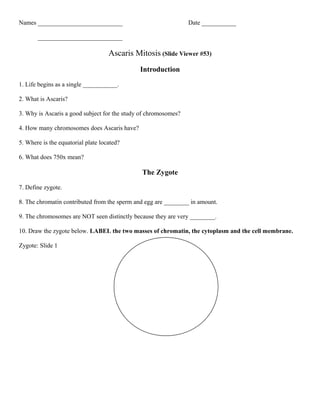
- 1. Names ___________________________ Date ___________ ___________________________ Ascaris Mitosis (Slide Viewer #53) Introduction 1. Life begins as a single ___________. 2. What is Ascaris? 3. Why is Ascaris a good subject for the study of chromosomes? 4. How many chromosomes does Ascaris have? 5. Where is the equatorial plate located? 6. What does 750x mean? The Zygote 7. Define zygote. 8. The chromatin contributed from the sperm and egg are ________ in amount. 9. The chromosomes are NOT seen distinctly because they are very ________. 10. Draw the zygote below. LABEL the two masses of chromatin, the cytoplasm and the cell membrane. Zygote: Slide 1
- 2. Prophase 11. The ____________________ have become much shorter and thicker. 12. How many chromosomes do you see? _______ 13. After fertilization, the _______________ chromosomes and the _______________ chromosomes pair up. 14. Draw prophase. Label the U-shaped and bent chromosomes (Prophase: Slide 2) Metaphase 15. In this slide the chromosomes have moved to the__________________ plate 16. At each pole is a _________________ 17. A starlike structure called an ___________________, radiates from each _________________. 18. Fine _____________ ________________ go from centriole to each chromosome. 19. Chromosomes are held together by a structure called a ________________________. 20. Draw Metaphase. Label: aster, spindle fibers, and chromosomes ( Metaphase: Slide 3)
- 3. Metaphase--Polar View 21. In slide #4, you are looking at the same stage as shown in slide #3, but we are now looking at the cell from the ____________. 22. The four chromosomes are seen as they lie flat on the ___________________ _____________. Early Anaphase 23. How many U's are there in this stage? _________ How many bents? __________ 24. In these eight chromosomes, there is enough hereditary material for two__________. 25. The groups are starting to move toward the ____________ 26. Each chromosome is being pulled by a ____________________, attached to each _______________ 27. Draw early anaphase. Label: centriole, aster, chromosomes, cell membrane (Early anaphase: Slide 5) Anaphase 28. The eight chromosomes are now ___________________ into two groups. 29. Electron microscopes show that spindle fibers are made of structures called _____________________ 30. Why causes the chromosomes to look beaded? 31. Draw anaphase. Anaphase: Slide 6
- 4. Telophase 32. The chromosomes have moved completely ________________________ 33. The cell membrane ________________ inward and the cytoplasm is dividing into two masses 34. As the cell approaches its division into two cells, the chromosomes become less ________________ 35. Draw Telophase. (Telephase: Slide 7) Late Telophase and Cytokinesis 36. The separation is now _________________ 37. Each of these two cells has __________ chromosomes 38. One cell with _______ pairs of chromosomes formed _________ cells. 39. How many chromosomes does a human cell have? _________________ 40. Comparasion of Plant and Animal cell Telophase: Use the slides on plant Mitosis and go to the last slide. Draw late telophase in the animal cell from the ascaris mitosis and late telophase in the plant cell. Name and describe at least three major differences between the two.