SlideShare uma empresa Scribd logo
1 de 2
Baixar para ler offline
Tips from the lab
                          Predictable impressioning
EQUIPMENT


                          By John C. Cranham, DDS, Chesapeake, Va.

                                                                                                                                                           ② Control the soft tissue through preliminary


                          W
                                           ithin a well-run den-    dictability to all indirect esthet-      employed to consistently place the
                                           tal practice, a direct   ic/restorative dental procedures.        margin one-half the depth of the              mouth preparation and chemotherapeutics
                                           relationship exists                                               healthy pocket. If the pocket is 2.5          Starting with a healthy mouth is a
                                           between           pre-   Soft-tissue management                   mm deep, then the margin should               prerequisite of any restorative pro-
                          dictability and positive trends.          From time to time, it is necessary       be placed 1.2 mm underneath the               cedure.        Proper        preliminary
                          These benefits include increased          to control a minor amount of gin-        tissue. This will create an invisible         mouth          preparation          should
                          production, decreased overhead,           gival bleeding during a restorative      margin, and one that exists in a              include any and all procedures to
M AT E R I A L S


                          increased profitability, decreased        procedure. Inadvertently nicking         healthy relationship within the               get the mouth completely free of
                          stress, and increased happiness of        the tissues during margin refine-        periodontal structures.                       disease prior to the final restora-
                          the entire dental team. The better        ment does occur occasionally and                                                       tive phase. Healthy tissue will
                          we become at doing procedures             it is essential to have the ability to   Retraction options                            make our job easier and the final
                          correct the first time, the more we       quickly         control  hemostasis.     There are many excellent ways to              esthetic/restorative result more
                          can experience the attributes pre-        Viscostat from Ultradent dis-            retract tissue if the only goal is to         predictable.
                          viously mentioned. Becoming pre-          pensed from its Dental Infusor is a      simply expose the gingival margin.               Additionally, chemotherapeutic
                          dictable in our work should be the        fast, predictable way to obtain 100      Laser troughing and electrosurgi-             agents such as Viscostat (Ultradent
                          primary focus of every dental             percent control.                         cal procedures are both excellent             Products, figure 2, below) can be
A S S I S TA N T S


                          practice. Without predictability,                                                  ways to expose the restorative mar-
                          there is great uncertainty, and           Start with an impressable preparation    gins. A retraction cord, while more
                          found within uncertainty is a high        Most restorative dentists do not         time-consuming, can be used to
                          degree of stress.                         trim their own dies. While many          more predictably place the margin
                             For a dental practice that does        teaching institutions recommend          in the ideal zone within the gingi-
                          any amount of indirect restorative        doing this, with consistently excel-     val sulcus.
                          dentistry, learning a tried-and-true      lent impressions, it is actually an          A two-cord technique has been
                          technique that consistently cap-          unnecessary step. It is important,       described to accomplish this goal.
                          tures the necessary details of an         therefore, for dentists to prepare       The technique described in this
                          impression is a vital component.          teeth in a way that their margins        article is a modification of this
                          Yet, after speaking to hundreds of        are easily identifiable. We should       process utilizing contemporary
FOCUS


                          laboratory owners over the last sev-      learn to prepare teeth with a con-       materials.
                          eral years, it is obvious that consis-    sistent margin design 360 degrees                                                      used by directly applying it to the
                          tently excellent impressions are a        around the tooth. If a contempo-         A step-by-step approach to an                 tissue to stop any localized bleed-
                          rarity.                                   rary metal ceramic material is your      ideal final impression                        ing. This can also be used to soak
                                                                    restorative choice, then a chamfer       ① Start with an impressable preparation       the retraction cords (figure 3,
                          Start with healthy tissue                 margin with the same general look                                                      below). This chemical agent
                          It is important to understand that,       should be utilized. If you prefer
                          in fabricating an indirect restora-       more of a knife-edge or even a
                          tion, the purpose of the impres-          shoulder, the key is to develop a
                          sion is not to simply capture the         preparation style that your dental
                          details of the hard tissues. The          laboratory gets used to seeing. You
                          goal is to also capture the stable        want to avoid having a margin that
                          relationship of the gingival tissues.     starts as a chamfer, then turns into
                          If we do indirect restorative proce-      a knife-edge, and then ends in a
                          dures in the presence of gingival         shoulder. These become very diffi-       In figure 1 (above), you will see a
                          inflammation, the final restorative       cult to read and trim. Preparing         tooth prepared for a Captek
                          result will likely be inadequate, no      teeth with consistent margin             Crown. The preparation has ade-
                          matter how “good” the impres-             design will add tremendous pre-          quate reduction (as described in              (Ferric Sulfate), not only rapidly
                          sion. After final crown placement,        dictability in the laboratory repro-     Part 1 of this series, printed in             achieves hemostasis, but also fixes
                          when the gingival tissues return to       duction of your preparations.            January) and has been smoothed                the tissue, holding it in the retract-
                          normal size, shape, and contour,                                                   and polished. The margin design               ed position for a longer period of
                          exposure of a margin will occur.          Consistent margin placement              is that of a chamfer, around the              time. This material is an important
                          While the crown may fit fine, the         Consistent margin placement is           entire circumference of the tooth.            component of any restorative den-
                          outcome is likely to be below the         key to providing a restoration that      Employing this type of prepara-               tist’s armamentarium.
                          patient’s esthetic expectations.          will meet the esthetic goals of the      tion guideline will dramatically
                              Beginning with healthy tissues        patient and the optimum health of        increase the readability of your              ③ Placement of first cord
                          will also simplify the impression-        the restorative result. Not prepar-      impressions since your laboratory             Prior to final margin placement,
                          making process. Bleeding tissues          ing far enough into the gingival         support will know exactly what it is          the first cord should be packed. A
                          can be controlled, but should not         sulcus will lead to esthetic prob-       looking for. Whatever margin                  00 Ultradent cord that has been
                          be “the norm.” Taking patients            lems, while going too deep will          design you personally prefer, keep-           soaked in Viscostat is placed into
                          through proper periodontal pro-           invade the biologic width.               ing it consistent will increase the           the depth of the sulcus (figure 4).
                          cedures and completing all neces-            By beginning with healthy tis-        quality and predictability of the             As shown in figure 5, the goal is to
                          sary preliminary mouth prepara-           sue and using normal probing             final restorative result.                     have it perfectly fit once around
                          tion will add tremendous pre-             depths, a technique can be


                     46                                                                                                                        Dental Equipment & Materials • May/June 2003
Comprecap, pushing the Expasyl              way into an otherwise perfect
                                                                                                                       further into the pocket (figure             impression.
EQUIPMENT


                                                                                                                       12). This provides additional
                                                                                                                       retraction by forcing the tissue fur-       ⑨ Visually inspect impression
                                                                                                                       ther away from the tooth. When              The final step is visual inspection
                                                                                                                       you combine this benefit with the           of the impression. Loupes or some
                                                                                                                       additional hemostatic and drying            sort of magnification should be
                                                                           gival sulcus (figure 8, above). It          properties of this material,                used to check the details of the
                                 Figure 4 (above), Figure 5 (below)                                                                                                master impression. As previously
                                                                           has hemostatic agents within its            Comprecap makes for a very pre-
                                                                           body and also has the ability to            dictable method for margin cap-             mentioned, all margins should be
                                                                           absorb crevicular fluid. The most           ture.                                       visible with a minimum of .5 mm
                                                                           positive clinical effect observed is                                                    apical to the marginal tissues (fig-
M AT E R I A L S


                                                                           not just great retraction, but a very       ⑦ Visually inspect retraction objectives    ure 15).
                                                                           dry field. In fact, there seems to be       After washing away the Expasyl                  By combining all of the ele-
                                                                           a complete absence of fluid                 (simply remove by spraying with a           ments of healthy tissue, correct
                                                                           around the preparation — a quali-           three-way syringe) and removing             preparation design, predictable
                                                                           ty that makes for an ideal impres-          the retraction cord, visual inspec-         retraction, and highly esthetic
                          the tooth. Pressure should be                    sion.                                       tion is the final step before taking        porcelain contour and color, we
                          applied so that the 00 cord is sit-                                                          the impression. This is when the            can all achieve excellent clinical
                          ting at the base of the healthy                  ⑥ Apply pressure with Comprecap             doctor should put on his or her             results. The outcome can be seen
                          pocket.                                          After dispensing the appropriate            loupes and, using excellent light-          in this delivered IPS d.SIGN
A S S I S TA N T S


                                                                           amount of Expasyl over the retrac-          ing, visually inspect the retracted         Porcelain to Captek restoration
                          ④ Margin refinement                              tion cord (figure 9) circumferen-           tissue (figure 13, below). All mar-         (figures 16 and 17).
                          Once the tissue has been retracted                                                                                                           Indirect restorative procedures
                          with the first cord, the margin is                                                                                                       can be a highly predictable, highly
                          finished by dropping the margin                                                                                                          profitable component of any den-
                          to the top of the cord (figure 6,                                                                                                        tal practice. To achieve this, how-
                          below). The placement of the cord                                                                                                        ever, the dental team must employ
                                                                                                                                                                   some simple, disciplined steps to
                                                                                                                                                                   achieve consistent results. This
                                                                                                                                                                   article has outlined a step-by-step,
                                                                                Figure 9 (above), Figure 10 (below)                                                highly predictable method of
FOCUS


                                                                                                                       gins should be easily viewable with         obtaining an ideal final impres-
                                                                                                                       a minimum of .5 mm of additional            sion.
                                                                                                                       tooth structure apical to the mar-              Acknowledgment: The author
                                                                                                                       gin. Figure 13 exhibits the ideal           would like to thank the technical staff
                          will protect the biologic width and                                                          retraction goals.                           of Dental Arts Laboratories, Inc. in
                          prevent us from going too far into                                                                                                       Peoria, Ill., for their excellent ceramic
                          the pocket. This is where this tech-                                                         ⑧ Inject with quality impression material   artistry in preparation of this case.
                          nique may have an advantage over                                                             A polyvinyl impression material             Call (800) 227-4142 to contact DAL.
                          laser or electrosurgical retraction.             tially around the tooth, a                  such as Take One from Kerr can                  Note: For complete information on
                          While other techniques can rapid-                Comprecap (figure 10) is placed             be utilized to complete the impres-         Cranham Dental Seminars, visit
                          ly attain exposure of the restora-               over     the   prepared      tooth.         sion procedure. While this materi-          www.cranhamdentalseminars.com or
                          tive margin, there is greater risk of            Comprecap, made by Roeko, look              al has hydrophilic properties,              contact Dr. John Cranham’s office at
                          invading the biologic width or                   like a hollowed-out cotton roll, but        every effort should be made to              (757) 465-8900.
                          leaving the postoperative margin                 are open only on one side and are           keep the field dry. Dry angles, cot-
                          visible.                                         firmer (figure 11). Once placed             ton rolls, and suction should be
                                                                                                                       employed to keep saliva out of the
                          ⑤ Addition of contemporary retraction material                                               field. This is particularly impor-
                          A very popular method of obtain-                                                             tant in the mandibular area. When
                          ing ideal retraction involves the                                                            injecting (figure 14, below), keep
                          placement of two cords on top of
                          one another within the pocket.
                          This technique follows a similar
                          path, but uses a contemporary
                          dental material called Expasyl (fig-                  Figure 11 (above), Figure 12 (below)
                          ure 7, below), recently introduced


                                                                                                                       the tip angled at the margin and
                                                                                                                       burnish the material around the
                                                                                                                       tooth. The thixatropic nature of
                          by Kerr. This material has a clay-                                                           this material will allow it to flow
                          like consistency that is released                over the prepared tooth, the                upon itself, eliminating voids, bub-
                          (under pressure) around the gin-                 patient can bite on the                     bles, and fins that can find their
                                                                                                                                                                         From top to bottom, Figures 15-17

                     48                                                                                                                             Dental Equipment & Materials • May/June 2003

Mais conteúdo relacionado

Mais de Quách Bảo Toàn

Profitable dental (impressions)
Profitable dental (impressions)Profitable dental (impressions)
Profitable dental (impressions)Quách Bảo Toàn
 
An alternative way to use gingival retraction paste
An alternative way to use gingival retraction pasteAn alternative way to use gingival retraction paste
An alternative way to use gingival retraction pasteQuách Bảo Toàn
 
A comparison of gingival inflammation related to retraction cords using bioch...
A comparison of gingival inflammation related to retraction cords using bioch...A comparison of gingival inflammation related to retraction cords using bioch...
A comparison of gingival inflammation related to retraction cords using bioch...Quách Bảo Toàn
 
Fine needle aspiration findings in malignant ameloblastoma a case report and ...
Fine needle aspiration findings in malignant ameloblastoma a case report and ...Fine needle aspiration findings in malignant ameloblastoma a case report and ...
Fine needle aspiration findings in malignant ameloblastoma a case report and ...Quách Bảo Toàn
 
Diagnostic ability of differential diagnosis in ameloblastoma and odontogenic...
Diagnostic ability of differential diagnosis in ameloblastoma and odontogenic...Diagnostic ability of differential diagnosis in ameloblastoma and odontogenic...
Diagnostic ability of differential diagnosis in ameloblastoma and odontogenic...Quách Bảo Toàn
 
Desmoplastic ameloblastoma of maxilla a case report
Desmoplastic ameloblastoma of maxilla  a case reportDesmoplastic ameloblastoma of maxilla  a case report
Desmoplastic ameloblastoma of maxilla a case reportQuách Bảo Toàn
 
Dentigerous cyst over maxillary sinus a case report and literature review
Dentigerous cyst over maxillary sinus  a case report and literature reviewDentigerous cyst over maxillary sinus  a case report and literature review
Dentigerous cyst over maxillary sinus a case report and literature reviewQuách Bảo Toàn
 
Cone beam ct findings in a case of plexiform ameloblatoma
Cone beam ct findings in a case of plexiform ameloblatomaCone beam ct findings in a case of plexiform ameloblatoma
Cone beam ct findings in a case of plexiform ameloblatomaQuách Bảo Toàn
 
Clinical and radiologic behaviour of ameloblastoma in 4 cases
Clinical and radiologic behaviour of ameloblastoma in 4 casesClinical and radiologic behaviour of ameloblastoma in 4 cases
Clinical and radiologic behaviour of ameloblastoma in 4 casesQuách Bảo Toàn
 
Basal cell ameloblastoma a rare case report and review of literature
Basal cell ameloblastoma a rare case report and review of literatureBasal cell ameloblastoma a rare case report and review of literature
Basal cell ameloblastoma a rare case report and review of literatureQuách Bảo Toàn
 
Atypical ameloblastoma – an enigma in diagnosis review of literature and rep...
Atypical ameloblastoma – an enigma in diagnosis  review of literature and rep...Atypical ameloblastoma – an enigma in diagnosis  review of literature and rep...
Atypical ameloblastoma – an enigma in diagnosis review of literature and rep...Quách Bảo Toàn
 

Mais de Quách Bảo Toàn (20)

Profitable dental (impressions)
Profitable dental (impressions)Profitable dental (impressions)
Profitable dental (impressions)
 
Jpd
JpdJpd
Jpd
 
Expasyltriedtrue dpr-2.08
Expasyltriedtrue dpr-2.08Expasyltriedtrue dpr-2.08
Expasyltriedtrue dpr-2.08
 
Expasyl nazarian-ce
Expasyl nazarian-ceExpasyl nazarian-ce
Expasyl nazarian-ce
 
Aquasil ultra
Aquasil ultraAquasil ultra
Aquasil ultra
 
An alternative way to use gingival retraction paste
An alternative way to use gingival retraction pasteAn alternative way to use gingival retraction paste
An alternative way to use gingival retraction paste
 
A comparison of gingival inflammation related to retraction cords using bioch...
A comparison of gingival inflammation related to retraction cords using bioch...A comparison of gingival inflammation related to retraction cords using bioch...
A comparison of gingival inflammation related to retraction cords using bioch...
 
200651850439300
200651850439300200651850439300
200651850439300
 
1447.full
1447.full1447.full
1447.full
 
1354
13541354
1354
 
335 339
335 339335 339
335 339
 
Us4871311
Us4871311Us4871311
Us4871311
 
Fine needle aspiration findings in malignant ameloblastoma a case report and ...
Fine needle aspiration findings in malignant ameloblastoma a case report and ...Fine needle aspiration findings in malignant ameloblastoma a case report and ...
Fine needle aspiration findings in malignant ameloblastoma a case report and ...
 
Diagnostic ability of differential diagnosis in ameloblastoma and odontogenic...
Diagnostic ability of differential diagnosis in ameloblastoma and odontogenic...Diagnostic ability of differential diagnosis in ameloblastoma and odontogenic...
Diagnostic ability of differential diagnosis in ameloblastoma and odontogenic...
 
Desmoplastic ameloblastoma of maxilla a case report
Desmoplastic ameloblastoma of maxilla  a case reportDesmoplastic ameloblastoma of maxilla  a case report
Desmoplastic ameloblastoma of maxilla a case report
 
Dentigerous cyst over maxillary sinus a case report and literature review
Dentigerous cyst over maxillary sinus  a case report and literature reviewDentigerous cyst over maxillary sinus  a case report and literature review
Dentigerous cyst over maxillary sinus a case report and literature review
 
Cone beam ct findings in a case of plexiform ameloblatoma
Cone beam ct findings in a case of plexiform ameloblatomaCone beam ct findings in a case of plexiform ameloblatoma
Cone beam ct findings in a case of plexiform ameloblatoma
 
Clinical and radiologic behaviour of ameloblastoma in 4 cases
Clinical and radiologic behaviour of ameloblastoma in 4 casesClinical and radiologic behaviour of ameloblastoma in 4 cases
Clinical and radiologic behaviour of ameloblastoma in 4 cases
 
Basal cell ameloblastoma a rare case report and review of literature
Basal cell ameloblastoma a rare case report and review of literatureBasal cell ameloblastoma a rare case report and review of literature
Basal cell ameloblastoma a rare case report and review of literature
 
Atypical ameloblastoma – an enigma in diagnosis review of literature and rep...
Atypical ameloblastoma – an enigma in diagnosis  review of literature and rep...Atypical ameloblastoma – an enigma in diagnosis  review of literature and rep...
Atypical ameloblastoma – an enigma in diagnosis review of literature and rep...
 

Tips from the lab predictable impressioning

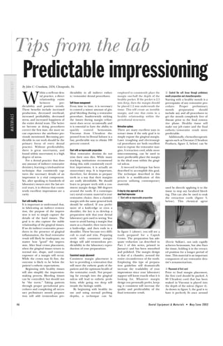
  • 1. Tips from the lab Predictable impressioning EQUIPMENT By John C. Cranham, DDS, Chesapeake, Va. ② Control the soft tissue through preliminary W ithin a well-run den- dictability to all indirect esthet- employed to consistently place the tal practice, a direct ic/restorative dental procedures. margin one-half the depth of the mouth preparation and chemotherapeutics relationship exists healthy pocket. If the pocket is 2.5 Starting with a healthy mouth is a between pre- Soft-tissue management mm deep, then the margin should prerequisite of any restorative pro- dictability and positive trends. From time to time, it is necessary be placed 1.2 mm underneath the cedure. Proper preliminary These benefits include increased to control a minor amount of gin- tissue. This will create an invisible mouth preparation should production, decreased overhead, gival bleeding during a restorative margin, and one that exists in a include any and all procedures to M AT E R I A L S increased profitability, decreased procedure. Inadvertently nicking healthy relationship within the get the mouth completely free of stress, and increased happiness of the tissues during margin refine- periodontal structures. disease prior to the final restora- the entire dental team. The better ment does occur occasionally and tive phase. Healthy tissue will we become at doing procedures it is essential to have the ability to Retraction options make our job easier and the final correct the first time, the more we quickly control hemostasis. There are many excellent ways to esthetic/restorative result more can experience the attributes pre- Viscostat from Ultradent dis- retract tissue if the only goal is to predictable. viously mentioned. Becoming pre- pensed from its Dental Infusor is a simply expose the gingival margin. Additionally, chemotherapeutic dictable in our work should be the fast, predictable way to obtain 100 Laser troughing and electrosurgi- agents such as Viscostat (Ultradent primary focus of every dental percent control. cal procedures are both excellent Products, figure 2, below) can be A S S I S TA N T S practice. Without predictability, ways to expose the restorative mar- there is great uncertainty, and Start with an impressable preparation gins. A retraction cord, while more found within uncertainty is a high Most restorative dentists do not time-consuming, can be used to degree of stress. trim their own dies. While many more predictably place the margin For a dental practice that does teaching institutions recommend in the ideal zone within the gingi- any amount of indirect restorative doing this, with consistently excel- val sulcus. dentistry, learning a tried-and-true lent impressions, it is actually an A two-cord technique has been technique that consistently cap- unnecessary step. It is important, described to accomplish this goal. tures the necessary details of an therefore, for dentists to prepare The technique described in this impression is a vital component. teeth in a way that their margins article is a modification of this Yet, after speaking to hundreds of are easily identifiable. We should process utilizing contemporary FOCUS laboratory owners over the last sev- learn to prepare teeth with a con- materials. eral years, it is obvious that consis- sistent margin design 360 degrees used by directly applying it to the tently excellent impressions are a around the tooth. If a contempo- A step-by-step approach to an tissue to stop any localized bleed- rarity. rary metal ceramic material is your ideal final impression ing. This can also be used to soak restorative choice, then a chamfer ① Start with an impressable preparation the retraction cords (figure 3, Start with healthy tissue margin with the same general look below). This chemical agent It is important to understand that, should be utilized. If you prefer in fabricating an indirect restora- more of a knife-edge or even a tion, the purpose of the impres- shoulder, the key is to develop a sion is not to simply capture the preparation style that your dental details of the hard tissues. The laboratory gets used to seeing. You goal is to also capture the stable want to avoid having a margin that relationship of the gingival tissues. starts as a chamfer, then turns into If we do indirect restorative proce- a knife-edge, and then ends in a dures in the presence of gingival shoulder. These become very diffi- In figure 1 (above), you will see a inflammation, the final restorative cult to read and trim. Preparing tooth prepared for a Captek result will likely be inadequate, no teeth with consistent margin Crown. The preparation has ade- matter how “good” the impres- design will add tremendous pre- quate reduction (as described in (Ferric Sulfate), not only rapidly sion. After final crown placement, dictability in the laboratory repro- Part 1 of this series, printed in achieves hemostasis, but also fixes when the gingival tissues return to duction of your preparations. January) and has been smoothed the tissue, holding it in the retract- normal size, shape, and contour, and polished. The margin design ed position for a longer period of exposure of a margin will occur. Consistent margin placement is that of a chamfer, around the time. This material is an important While the crown may fit fine, the Consistent margin placement is entire circumference of the tooth. component of any restorative den- outcome is likely to be below the key to providing a restoration that Employing this type of prepara- tist’s armamentarium. patient’s esthetic expectations. will meet the esthetic goals of the tion guideline will dramatically Beginning with healthy tissues patient and the optimum health of increase the readability of your ③ Placement of first cord will also simplify the impression- the restorative result. Not prepar- impressions since your laboratory Prior to final margin placement, making process. Bleeding tissues ing far enough into the gingival support will know exactly what it is the first cord should be packed. A can be controlled, but should not sulcus will lead to esthetic prob- looking for. Whatever margin 00 Ultradent cord that has been be “the norm.” Taking patients lems, while going too deep will design you personally prefer, keep- soaked in Viscostat is placed into through proper periodontal pro- invade the biologic width. ing it consistent will increase the the depth of the sulcus (figure 4). cedures and completing all neces- By beginning with healthy tis- quality and predictability of the As shown in figure 5, the goal is to sary preliminary mouth prepara- sue and using normal probing final restorative result. have it perfectly fit once around tion will add tremendous pre- depths, a technique can be 46 Dental Equipment & Materials • May/June 2003
  • 2. Comprecap, pushing the Expasyl way into an otherwise perfect further into the pocket (figure impression. EQUIPMENT 12). This provides additional retraction by forcing the tissue fur- ⑨ Visually inspect impression ther away from the tooth. When The final step is visual inspection you combine this benefit with the of the impression. Loupes or some additional hemostatic and drying sort of magnification should be gival sulcus (figure 8, above). It properties of this material, used to check the details of the Figure 4 (above), Figure 5 (below) master impression. As previously has hemostatic agents within its Comprecap makes for a very pre- body and also has the ability to dictable method for margin cap- mentioned, all margins should be absorb crevicular fluid. The most ture. visible with a minimum of .5 mm positive clinical effect observed is apical to the marginal tissues (fig- M AT E R I A L S not just great retraction, but a very ⑦ Visually inspect retraction objectives ure 15). dry field. In fact, there seems to be After washing away the Expasyl By combining all of the ele- a complete absence of fluid (simply remove by spraying with a ments of healthy tissue, correct around the preparation — a quali- three-way syringe) and removing preparation design, predictable ty that makes for an ideal impres- the retraction cord, visual inspec- retraction, and highly esthetic the tooth. Pressure should be sion. tion is the final step before taking porcelain contour and color, we applied so that the 00 cord is sit- the impression. This is when the can all achieve excellent clinical ting at the base of the healthy ⑥ Apply pressure with Comprecap doctor should put on his or her results. The outcome can be seen pocket. After dispensing the appropriate loupes and, using excellent light- in this delivered IPS d.SIGN A S S I S TA N T S amount of Expasyl over the retrac- ing, visually inspect the retracted Porcelain to Captek restoration ④ Margin refinement tion cord (figure 9) circumferen- tissue (figure 13, below). All mar- (figures 16 and 17). Once the tissue has been retracted Indirect restorative procedures with the first cord, the margin is can be a highly predictable, highly finished by dropping the margin profitable component of any den- to the top of the cord (figure 6, tal practice. To achieve this, how- below). The placement of the cord ever, the dental team must employ some simple, disciplined steps to achieve consistent results. This article has outlined a step-by-step, Figure 9 (above), Figure 10 (below) highly predictable method of FOCUS gins should be easily viewable with obtaining an ideal final impres- a minimum of .5 mm of additional sion. tooth structure apical to the mar- Acknowledgment: The author gin. Figure 13 exhibits the ideal would like to thank the technical staff will protect the biologic width and retraction goals. of Dental Arts Laboratories, Inc. in prevent us from going too far into Peoria, Ill., for their excellent ceramic the pocket. This is where this tech- ⑧ Inject with quality impression material artistry in preparation of this case. nique may have an advantage over A polyvinyl impression material Call (800) 227-4142 to contact DAL. laser or electrosurgical retraction. tially around the tooth, a such as Take One from Kerr can Note: For complete information on While other techniques can rapid- Comprecap (figure 10) is placed be utilized to complete the impres- Cranham Dental Seminars, visit ly attain exposure of the restora- over the prepared tooth. sion procedure. While this materi- www.cranhamdentalseminars.com or tive margin, there is greater risk of Comprecap, made by Roeko, look al has hydrophilic properties, contact Dr. John Cranham’s office at invading the biologic width or like a hollowed-out cotton roll, but every effort should be made to (757) 465-8900. leaving the postoperative margin are open only on one side and are keep the field dry. Dry angles, cot- visible. firmer (figure 11). Once placed ton rolls, and suction should be employed to keep saliva out of the ⑤ Addition of contemporary retraction material field. This is particularly impor- A very popular method of obtain- tant in the mandibular area. When ing ideal retraction involves the injecting (figure 14, below), keep placement of two cords on top of one another within the pocket. This technique follows a similar path, but uses a contemporary dental material called Expasyl (fig- Figure 11 (above), Figure 12 (below) ure 7, below), recently introduced the tip angled at the margin and burnish the material around the tooth. The thixatropic nature of by Kerr. This material has a clay- this material will allow it to flow like consistency that is released over the prepared tooth, the upon itself, eliminating voids, bub- (under pressure) around the gin- patient can bite on the bles, and fins that can find their From top to bottom, Figures 15-17 48 Dental Equipment & Materials • May/June 2003