Chapter 4: Chemical Composition of the Cell
•
9 gostaram•5,264 visualizações

Experiment for biology form 4 chapter 4

Recomendados
Mais conteúdo relacionado
Mais procurados
Mais procurados (20)
Destaque
Destaque (20)
Semelhante a Chapter 4: Chemical Composition of the Cell
Semelhante a Chapter 4: Chemical Composition of the Cell (20)
Último
Último (20)
Chapter 4: Chemical Composition of the Cell
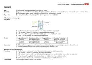
- 1. Biology |Form 4 | Chapter 4: Chemical Composition in Cell 10 Activity 4.1 Aim To differentiate between reducing and non-reducing sugars. Materials 5% glucose solution, 5% fructose solution, 5% galactose solution, 5% maltose solution, 5% lactose solution, 5% sucrose solution, dilute hydrochloric acid, water, sodium hydrogen carbonate powder and Benedict’s solution Apparatus Test tubes, beaker, Bunsen burner, tripod stand, wire gauze and test tube holder A.Testing for reducing sugars Procedure 1. 2 ml of Benedict’s solution are added to 2 ml of glucose solution in a test tube 2. The test tube is placed in a water bath of boiling water for 5 minutes. 3. The colour of the precipitate that is formed (if any) is recorded in the table below. 4. Steps 1 to 3 are repeated for all the other sugar solutions. Results Sugar solution Benedict’s solution Sugar solution Benedict’s solution Glucose Brick-red precipitate Maltose Brick-red precipitate Fructose Brick-red precipitate Lactose Brick-red precipitate Galactose Brick-red precipitate Sucrose Blue solution Discussion 1. Glucose, fructose, galactose, maltose and lactose are reducing sugars because they give a positive Benedict’s test. 2. Sucrose is a non-reducing sugar because it gives a negative Benedict’s test. B.Testing for non-reducing sugar Procedure 1. 2 ml of sucrose solution and 1 ml of dilute hydrochloric acid are added to a test tube 2. The test tube is placed in a water bath and boiled for 5 minutes. 3. The test tube is cooled under a tap and sodium hydrogen carbonate powder is added to neutralize the excess acid. 4. When it stops fizzing, 2 ml of Benedict’s solution are added to the mixture in the test tube。 5. The test tube is placed in the water in the water bath again and boiled for 5 minutes. Observation A brick red precipitate is formed in the test tube.
- 2. Biology |Form 4 | Chapter 4: Chemical Composition in Cell 11 Discussion 1. Sucrose is hydrolysed into glucose and fructose by boiling the sucrose solution with dilute hydrochloride acid. 2. When the solution containing glucose and fructose is boiled with Benedict’s solution, it produces a brick-red precipitate. Conclusion Glucose, fructose, maltose are reducing sugars. Sucrose is non-reducing sugar.
- 3. Biology |Form 4 | Chapter 4: Chemical Composition in Cell 12 Experiment 4.1 Aim To study the effects of temperature on the activity of salivary amylase. Problem statement What are the effects of temperature on the activity of salivary amylase? Hypothesis The optimum temperature for salivary amylase is 37oC. Variables (a) Manipulated variable: Temperature (b) Responding variable: Rate of reaction (c) Fixed variable: Volume and concentration of starch suspension, volume and concentration of saliva suspension, pH of starch and saliva suspensions. Apparatus Beakers, test tubes, tripod stand, wire gauze, Bunsen burner, thermometer, dropper, glass rod, syringes, stopwatch, spotting tile and test tube rack Materials 1% starch starch suspension, saliva, iodine solution, ice cubes and distilled water Technique Recording the time taken for the complete hydrolysis of starch with a stopwatch. The presence of starch is indicated by the iodine test.
- 4. Biology |Form 4 | Chapter 4: Chemical Composition in Cell 13 Procedure 1. The mouth is rinsed with distilled water. 2. The saliva suspension is prepared by spitting into a small beaker and diluting the saliva with an amount of distilled water equal to the amount of saliva. 3. Five water baths are prepared at the following temperatures: 0o C, 20 o C, 37 o C, 50 o C and 60 o C. 4. Two test tubes are labelled P and Q. 5. 4 ml of starch suspension are placed in test tube P and 1 ml of saliva suspension in test tube Q. 6. Both test tubes are placed into the first water bath (at 60 o C) for 5 minutes. 7. A drop of iodine solution is placed into each groove of a clean spotting tile. 8. A drop of suspension from test tube P is poured into the saliva suspension in test tube Q. The stopwatch is started immediately. 9. A drop of suspension is taken out from the test tube Q every minute and tested with iodine solution on the spotting tile. 10. Test tube Q is kept in the water bath thoughout the experiment. 11. The time taken for the complete hydrolysis of starch is recorded in the table, that is, when the iodine solution does not turn blue anymore..
- 5. Biology |Form 4 | Chapter 4: Chemical Composition in Cell 14 12. Steps 4 to 9 are repeated for temperatures 20 o C, 37 o C, 50 o C and 60 o C. 13. The results are recorded in the table below. 14. A graph of rate of reaction against temperature (o C) is plotted. Results Temperature of water bath (o C) Time taken for complete hydrolysis of starch, t (minute) Rate of reaction, (minute-1 ) 0 >30 <0.03 20 8 0.13 37 0.33 3 50 6 0.17 60 >30. <0.03 Discussion 1. The test tubes P and Q are kept in the water bath for 5 minutes to stabilize the temperature of the suspensions in the two test tubes. 2. When the amylase in the saliva has completely hydrolysed the starch, the dark blue colour does not form in the iodine test. 3. At low temperatures, the reaction progresses slowly. As the temperature increases, the rate of reaction increases until the optimum temperature of 37o C is reached. Above the optimum temperature, the rate of reaction falls rapidly. Conclusion The rate of reaction is highest at the temperature 37o C. the hypothesis is accepted.
- 6. Biology |Form 4 | Chapter 4: Chemical Composition in Cell 15 Experiment 4.2 Aim To study the effects of pH on the activity of pepsin. Problem statement What are the effects of pH on the activity of pepsin? Hypothesis The enzyme pepsin works best in acidic conditions. Variables (a) Manipulated variable: pH of medium (b) Responding variable: Rate of reaction (c) Fixed variable: Volume and concentration of egg albumen suspension, volume and concentration of pepsin solution, temperature of medium Materials Egg albumen suspension, dilute hydrochloride acid, dilute sodium hydroxide solution, 1% of pepsin solution, pH paper and distilled water Apparatus Beaker, tripod stand, wire gauze, Bunsen burner, thermometer, dropper, test tubes, test tube rack and syringes Procedure 1. The egg albumen suspension is prepared by adding 1g of dried egg albumen to 100 cm3 of water. The mixture is then heated to 90o C. 2. Three test tubes are labelled P,Q and R. 3. 5 ml of egg albumen suspension and 1 ml of pepsin solution are placed in each test tube. 4. 5 drops of distilled water are added to test tube P. 5. 5 drops of dilute hydrochloric acid are added to test tube Q. 6. 5 drops of dilute sodium hydroxide solution are added to test tube R. 7. All the three test tubes are placed into a water bath at 37o C for 20 minutes. 8. The appearance of the contents in each test tube is recorded at the beginning and the end of the experiment. 9. The contents in each test tube are tested with pH paper. 10. The results are recorded in a table.
- 7. Biology |Form 4 | Chapter 4: Chemical Composition in Cell 16 Results Test tube pH Contents Appearance of contents At the beginning of the experiment At the end of the experiment P 7 Albumen + pepsin + distilled water White suspension White suspension Q 2 Albumen + pepsin + dilute hydrochloric acid White suspension Clear solution R 9 Albumen + pepsin + dilute sodium hydroxide solution White suspension White suspension Discussion 1. The test tubes are kept in a water bath maintained at 37o C because that is the optimum temperature for pepsin. 2. The mixture in test tube Q is clear at the end of the experiment because the albumen has been hydrolysed. 3. Pepsin does not catalyse the hydrolysis of albumen in the neutral and in alkaline conditions. Conclusion Pepsin catalyses the hydrolysis of protein only in acidic conditions. The hypothesis is accepted.
- 8. Biology |Form 4 | Chapter 4: Chemical Composition in Cell 17 Experiment 4.3 Aim To study the effects of substrate concentration on salivary amylase activity. Problem statement What are the effects of substrate concentration on salivary amylase activity? Hypothesis The rate of enzymatic reaction increases with the increase in substrate concentration until it reaches a maximum rate. Variables (a) Manipulated variable: Substrate concentration (b) Responding variable: Rate of concentration (c) Fixed variable: Enzyme concentration, temperature, pH of starch and saliva suspension, volume of starch and saliva suspension Materials 0.1%, 0.2%, 0.3%, 0.4%, 0.5% and 0.6% starch suspensions, saliva, iodine solution and distilled water Apparatus Beakers, test tubes, tripod stand, wire gauze, Bunsen burner, thermometer, dropper, glass rod, syringes, stopwatch, spotting tile and test tube rack Technique Recording the time taken for the complete hydrolysis of starch with a stopwatch. The presence of starch is indicated by the iodine test. Procedure 1. The mouth is rinsed with distilled water. 2. The saliva suspension is prepared by spitting into a small beaker and diluting the saliva with an amount of distilled water is equal to the amount of saliva. 3. Six test tubes are labelled P, Q, R, S, T and Uare filled with the following starch suspensions using different syringe. Test tube Type of starch suspension P 4 ml 0.1% starch suspension Q 4 ml 0.2% starch suspension R 4 ml 0.3% starch suspension S 4 ml 0.4% starch suspension T 4 ml 0.5% starch suspension U 4 ml 0.6% starch suspension 4. 1 ml of saliva suspension is placed in a test tube. 5. A drop of iodine solution is placed into each groove of a clean spotting tile. 6. Test tube P and the test tube containing the saliva suspension are placed into a water bath at 37o C. 7. After 5 minutes, the saliva suspension is poured into the starch suspension in test tube P. the stopwatch is started immediately. 8. A drop of suspension is taken from test tube P every minute and tested with iodine solution on the spotting tile. 9. Test tube P is kept in the water bath throughout the experiment. 10. The time taken for the complete hydrolysis of starch is recorded in the table, that is, when the iodine solution does not turn blue anymore. 11. Steps 4 to 10 are repeated for test tubes Q, R, S, T and U. 12. The results are recorded in the table below. 13. The graph of rate of reaction (% minute-1 ) against concentration of starch suspension (%) is plotted.
- 9. Biology |Form 4 | Chapter 4: Chemical Composition in Cell 18 Results Concentration of starch suspension, C (%) Time taken for the complete hydrolysis of starch,t Rate of reaction = C/t (% minute-1 )(seconds) (minutes) 0.1 300 5.00 0.02 0.2 300 5.00 0.04 0.3 300 5.00 0.06 0.4 300 5.00 0.08 0.5 375 6.25 0.08 0.6 450 7.50 0.08 Discussion 1. The rate of reaction increases with the increase in the concentration of the substrate (starch) concentration, that is from 0.1% to 0.4% starch suspension. 2. The maximum rate is reached at 0.4% starch suspension. At this point all the active sites of the enzymes are filled at anyone one time. This is referred to as saturated point. 3. Beyond the saturation point, the rate of reaction does not increases even through the substrate concentration is increased because the concentration of enzyme becomes a limiting concentration. Conclusion The rate of enzymatic reaction increases with the increase in substrate concentration until it reaches a maximum rate. The hypothesis is accepted.