
Recomendados
Mais conteúdo relacionado
Semelhante a Wound healing dressings and drug delivery systems
Semelhante a Wound healing dressings and drug delivery systems (20)
Último
Último (20)
Wound healing dressings and drug delivery systems
- 1. Wound Healing Dressings and Drug Delivery Systems: A Review JOSHUA S. BOATENG,1 KERR H. MATTHEWS,2 HOWARD N.E. STEVENS,1 GILLIAN M. ECCLESTON1 1 Strathclyde Institute of Pharmacy and Biomedical Sciences, University of Strathclyde, John Arbuthnott Building, 27 Taylor Street, Glasgow G4 0NR, UK 2 School of Pharmacy, The Robert Gordon University, School Hill, Aberdeen AB10 1FR, UK Received 18 June 2007; accepted 17 August 2007 Published online in Wiley InterScience (www.interscience.wiley.com). DOI 10.1002/jps.21210 ABSTRACT: The variety of wound types has resulted in a wide range of wound dressings with new products frequently introduced to target different aspects of the wound healing process. The ideal dressing should achieve rapid healing at reasonable cost with minimal inconvenience to the patient. This article offers a review of the common wound manage- ment dressings and emerging technologies for achieving improved wound healing. It also reviews many of the dressings and novel polymers used for the delivery of drugs to acute, chronic and other types of wound. These include hydrocolloids, alginates, hydro- gels, polyurethane, collagen, chitosan, pectin and hyaluronic acid. There is also a brief section on the use of biological polymers as tissue engineered scaffolds and skin grafts. Pharmacological agents such as antibiotics, vitamins, minerals, growth factors and other wound healing accelerators that take active part in the healing process are discussed. Direct delivery of these agents to the wound site is desirable, particularly when systemic delivery could cause organ damage due to toxicological concerns asso- ciated with the preferred agents. This review concerns the requirement for formulations with improved properties for effective and accurate delivery of the required therapeutic agents. General formulation approaches towards achieving optimum physical properties and controlled delivery characteristics for an active wound healing dosage form are also considered briefly. ß 2007 Wiley-Liss, Inc. and the American Pharmacists Association J Pharm Sci 97:2892–2923, 2008 Keywords: biodegradable polymers; biomaterials; physical characterisation; poly- meric drug delivery systems; wound dressings; wound healing INTRODUCTION wound care market worldwide. In the past, traditional dressings such as natural or synthetic Wound dressings and devices form an important bandages, cotton wool, lint and gauzes all with segment of the medical and pharmaceutical varying degrees of absorbency were used for the management of wounds. Their primary function was to keep the wound dry by allowing evapora- Joshua S. Boateng’s present address is Department of tion of wound exudates and preventing entry of Pharmaceutical, Chemical and Environmental Sciences, harmful bacteria into the wound. It has now been School of Science, Grenville Building, University of Greenwich at Medway, Central Avenue, Chatham Maritime, Kent ME4 shown however, that having a warm moist wound 4TB, UK. environment achieves more rapid and successful Correspondence to: Gillian M. Eccleston (Telephone: þ44- wound healing. The last two decades have 141-548-2510; Fax: þ44-141-552-2562; E-mail: g.m.eccleston@strath.ac.uk) witnessed the introduction of many dressings, Journal of Pharmaceutical Sciences, Vol. 97, 2892–2923 (2008) with new ones becoming available each year. For ß 2007 Wiley-Liss, Inc. and the American Pharmacists Association example, the number of newer dressings available 2892 JOURNAL OF PHARMACEUTICAL SCIENCES, VOL. 97, NO. 8, AUGUST 2008
- 2. WOUND DRESSINGS AND DRUG DELIVERY SYSTEMS 2893 on the Drug Tariff1 in the UK increased from 4 in According to the Wound Healing Society, a wound 1988 to 57 in May 1998 and by February 2007, the is the result of ‘disruption of normal anatomic total number stood at 262. These modern dres- structure and function’.3 Based on the nature of sings are based on the concept of creating an the repair process, wounds can be classified as optimum environment to allow epithelial cells to acute or chronic wounds. Acute wounds are move unimpeded, for the treatment of wounds. usually tissue injuries that heal completely, with Such optimum conditions include a moist envir- minimal scarring, within the expected time frame, onment around the wound, effective oxygen usually 8–12 weeks.4 The primary causes of acute circulation to aid regenerating cells and tissues wounds include mechanical injuries due to and a low bacterial load. Other factors which have external factors such as abrasions and tears contributed to the wide range of wound dressings which are caused by frictional contact between include the different type of wound (e.g. acute, the skin and hard surfaces. Mechanical injuries chronic, exuding and dry wounds, etc.) and the also include penetrating wounds caused by knives fact that no single dressing is suitable for the and gun shots and surgical wounds caused by management of all wounds. In addition, the wound surgical incisions to for example remove tumours. healing process has several different phases that Another category of acute wounds include burns cannot be targeted by any particular dressing. and chemical injuries, which arise from a variety Effective wound management depends on of sources such as radiation, electricity, corrosive understanding a number of different factors such chemicals and thermal sources. The temperatu- as the type of wound being treated, the healing re of the source and the exposure time influence process, patient conditions in terms of health (e.g. the degree of a thermal burn.5 Burns will diabetes), environment and social setting, and the normally require specialist care because of the physical chemical properties of the available associated trauma. dressings.2 It is important therefore, that differ- Chronic wounds on the other hand arise from ent dressings be evaluated and tested in terms of tissue injuries that heal slowly, that is have not their physical properties and clinical performance healed beyond 12 weeks6 and often reoccur. Such for a given type of wound and the stage of wound wounds fail to heal due to repeated tissue insults healing, before being considered for routine use. or underlying physiological conditions7 such as This review discusses the common wound diabetes and malignancies, persistent infections, healing dressings, their key advantages and poor primary treatment and other patient related shortcomings, and the need for dressings with factors. These result in a disruption of the orderly improved properties. The definition and classifi- sequence of events during the wound healing cation of wounds together with the different process (see later). Chronic wounds include decu- stages of wound healing are also briefly described, bitis ulcers (bedsores or pressure sores) and leg as they directly affect the choice of a particular ulcers (venous, ischaemic or of traumatic origin). dressing. In addition, recent advances in dres- Wounds are also classified based on the number sings are discussed within the context of the of skin layers and area of skin affected.8,9 Injury formulations for delivering therapeutic agents to that affects the epidermal skin surface alone is moist wound surfaces. Finally, general physical referred to as a superficial wound, whilst injury characterisation of topical dressings, for applica- involving both the epidermis and the deeper tion to wounds, in terms of their fluid handling, dermal layers, including the blood vessels, sweat moisture vapour permeability, fluid affinity, glands and hair follicles is referred to as partial water uptake, rheological properties (gel and thickness wound. Full thickness wounds occur tensile strength, elasticity), compressive and bio- when the underlying subcutaneous fat or deeper adhesive properties are specifically discussed. tissues are damaged in addition to the epidermis and dermal layers. Ferreira et al.10 have described wounds both acute and chronic that are difficult to heal as WOUNDS ‘complex wounds’ with unique characteristics. The properties of complex wounds from their A wound can be described as a defect or a break review can be summarised as: (a) extensive loss of in the skin, resulting from physical or thermal the integument which comprises skin, hair, and damage or as a result of the presence of an associated glands, (b) infection (e.g. Fournier’s underlying medical or physiological condition. gangrene) which may result in tissue loss, DOI 10.1002/jps JOURNAL OF PHARMACEUTICAL SCIENCES, VOL. 97, NO. 8, AUGUST 2008
- 3. 2894 BOATENG ET AL. (c) tissue death or signs of circulation impairment comprising five overlapping stages that involve and (d) presence of pathology. complex biochemical and cellular processes. These are described as haemostasis, inflammation, migration, proliferation and maturation phases (Fig. 1). In fact, Cooper17 has argued for expand- Wound Healing ing the understanding of wounds beyond the Wound healing is a specific biological process cellular level to a molecular context as well. He related to the general phenomenon of growth and emphasised the need to approach wound healing tissue regeneration. It is not the purpose of this at multiple levels (cellular and molecular) to help paper to review in detail the physiology of wound improve wound treatment and management. healing, but to describe only that which is relevant Wound healing formulations (dressings) and to wound management and the choice of wound novel technologies developed to date focus on dressings. The reader is referred to the biological one or more of these aspects of the natural healing and physiological texts and literature for detailed process18 that are summarised briefly below. scientific expositions.11–14 Wound healing pro- gresses through a series of interdependent and overlapping stages in which a variety of cellular Haemostasis and Inflammation and matrix components act together to reestablish the integrity of damaged tissue and replacement Bleeding usually occurs when the skin is injured of lost tissue.15,16 The wound healing process has and serves to flush out bacteria and/or antigens been reviewed and described by Schultz14 as from the wound. In addition, bleeding activates Figure 1. Schematic representation of the phases of wound healing (a) infiltration of neutrophils into the wound area (b) invasion of wound area by epithelial cells (c) epithelium completely covers the wound (d) many of the capillaries and fibroblasts, formed at early stages have all disappeared (adopted from Gandour—unpublished). JOURNAL OF PHARMACEUTICAL SCIENCES, VOL. 97, NO. 8, AUGUST 2008 DOI 10.1002/jps
- 4. WOUND DRESSINGS AND DRUG DELIVERY SYSTEMS 2895 haemostasis which is initiated by exudate compo- with mature collagen and form aggregates as part nents such as clotting factors. Fibrinogen in the of the clotting mechanism. exudate elicits the clotting mechanism resulting in coagulation of the exudates (blood without cells and platelets) and, together with the formation of Migration a fibrin network, produces a clot in the wound The migration phase involves the movement of causing bleeding to stop. The clot dries to form a epithelial cells and fibroblasts to the injured area scab and provides strength and support to the to replace damaged and lost tissue. These cells injured tissue. Haemostasis therefore, plays a regenerate from the margins, rapidly growing protective role as well as contributing to success- over the wound under the dried scab (clot) ful wound healing.19 accompanied by epithelial thickening. The inflammatory phase occurs almost simul- taneously with haemostasis, sometimes from within a few minutes of injury to 24 h and lasts Proliferation for about 3 days. It involves both cellular and vascular responses. The release of protein-rich The proliferative phase occurs almost simulta- exudate into the wound causes vasodilation neously or just after the migration phase (Day 3 through release of histamine and serotonin, onwards) and basal cell proliferation, which lasts allows phagocytes to enter the wound and engulf for between 2 and 3 days. Granulation tissue is dead cells (necrotic tissue). Necrotic tissue which formed by the in-growth of capillaries and is hard is liquefied by enzymatic action to produce lymphatic vessels into the wound and collagen a yellowish coloured mass described as sloughy is synthesised by fibroblasts giving the skin (Tab. 1). Platelets liberated from damaged blood strength and form. By the fifth day, maximum vessels become activated as they come into contact formation of blood vessels and granulation tissue Table 1. Classification of Wounds Based on the Appearance Wound Type Appearance Stage of Wound Healing Affected Necrotic Often black or olive green due to dead Under favourable conditions, dead devitalised tissue, that is dry, thick tissue in a wound such as a pressure and leathery to touch. Common with sore will usually separate spontaneously pressure sores from the healthy tissue beneath. This occurs as a result of autolysis and presumably involves macrophage activity and the action of proteolytic enzymes which act at the interface of the necrotic and healthy tissue A dry environment prevents the autolytic and proteolytic actions of macrophages and enzymes Sloughy Fluid, moist, loose and stringy Associated with excess exudates during rehydrated necrotic tissue that is inflammatory phase. Slough leads to typically yellow in colour wounds getting stuck in the late inflammatory stage leading resulting in delayed wound healing Granulating Significant quantities of granulation Proliferative phase tissue, generally red or deep pink in colour. May produce excess exudate Epithelialising Pink in colour with formation of new Involves both migratory and proliferative epidermis phases. Final stages of wound healing Infected and malodorous Red, hot inflamed tissue, pus present. Inflammatory response, collagen Infection with anaerobic bacteria synthesis, epithelisation. Infection causes unpleasant odour prolongs the inflammatory process which delays wound healing Each wound type represents the phases that a single wound may go through as it heals.21 DOI 10.1002/jps JOURNAL OF PHARMACEUTICAL SCIENCES, VOL. 97, NO. 8, AUGUST 2008
- 5. 2896 BOATENG ET AL. has occurred. Further epithelial thickening takes Factors Which Impair Wound Healing: place until collagen bridges the wound. The Chronic Wounds fibroblast proliferation and collagen synthesis continues for up to 2 weeks by which time blood Although most wounds will heal uneventfully, vessels decrease and oedema recedes. problems can sometimes occur, that lead to failure of the wound to heal or a prolonged healing time. Failure of a wound to heal within the expected time frame usually results in a chronic wound. A Maturation chronic wound fails to heal because the orderly sequence of events is disrupted at one more of the This phase (also called the ‘remodelling phase’) stages of wound healing. Excessive production of involves the formation of cellular connective exudates can cause maceration of healthy skin tissue and strengthening of the new epithelium tissue around the wound28 and inhibit wound which determines the nature of the final scar. healing. In addition, exudate from chronic wound Cellular granular tissue is changed to an acellular differs from acute wound fluid with relatively mass from several months up to about 2 years. higher levels of tissue destructive proteinase Table 1 describes the appearance of wounds in enzymes29 and therefore more corrosive. The relation to the stages of wound healing. These smell and staining caused by exudate can also descriptions relate not only to different types of have a negative impact on a patient’s general wounds but also to the various stages through health and quality of life.30 which a single wound may pass as it heals.20,21 Foreign bodies introduced deep into the wound at time of injury can cause chronic inflammatory responses delaying healing and sometimes lead- ing to granuloma or abscess formation. Other Wound Exudate problems associated with wound healing include Thomas22 has described wound exudate as: ‘a the formation of keloid (raised) scars resulting generic term given to liquid produced from chronic from excess collagen production in the latter wounds, fistulae or other more acute injuries once part of the wound healing process.19 Pathogenic haemostasis has been achieved’. It is essentially bacteria such as Staphylococcus aureus, Pseudo- blood from which most of the red cells and monas aeruginosa, Streptococcus pyrogenes and platelets have been removed. Exudate is a key some Proteus, Clostridium and Coliform species component in all the stages of wound healing, can be detrimental to the healing process. irrigating the wound continuously and keeping it Inadequate control measures to manage infected moist.23 The maintenance of a moist wound bed is wounds can lead to cellulitis (cell inflammation) widely accepted as the most ideal environment for and ultimately bacteraemia and septicaemia, both effective wound healing.24 Exudate also supplies of which can be fatal. It has been shown that the the wound with nutrients and provides favourable presence of P. aeruginosa and S. aureus signifi- conditions for migration and mitosis of epithelial cantly reduced skin graft healing and also that cells.25 In addition, exudate supplies the wound 94% of ulcers that were slow to heal, or recurred with leucocytes which helps to control bacteria after discharge, contained S. aureus.31 In a review and reduce the incidence of infection at the wound on improving the healing of chronic wounds, surface. Krasner32 outlined a number of factors that In certain conditions such as chronic wounds, needed to be controlled and managed effectively there is excessive amounts of exudates present including preventing infection, optimising exu- which can lead to complications. Excess exudate date control and removing foreign bodies which results from oedema caused by inflammation, could lead to complications. reduced mobility and venous or lymphatic insuf- Poor nutritional status and old age33 also reduce ficiency.26 Increased exudate levels may also be the ability to fight infection. Protein, vitamin (e.g. the result of liquefying hard and eschar-like vitamin C) and mineral deficiencies impair the necrotic tissue to produce a wet and sloughy inflammatory phase and collagen synthesis, mass by a process known as autolytic debride- leading to prolonged healing times.34–36 Under- ment. A key characteristic of modern wound lying diseases such as diabetes37 and anaemia dressings is the removal of excess exudate while delay wound healing because compromised circu- maintaining moisture at the wound bed.27 lation results in the delivery of inadequate JOURNAL OF PHARMACEUTICAL SCIENCES, VOL. 97, NO. 8, AUGUST 2008 DOI 10.1002/jps
- 6. WOUND DRESSINGS AND DRUG DELIVERY SYSTEMS 2897 nutrients, blood cells and oxygen to the wound. wounds, underlying factors such as disease, drug Treatment with drugs such as steroids suppress therapy and patient circumstance must all be the body’s inflammatory responses and thereby reviewed and addressed before a particular wound impede the inflammatory stage of wound healing, dressing is applied. Table 2 describes factors which eventually leads to a compromised immune to be considered in the choice of wound dress- system. Glucocorticoids for example have been ings based on their performance characteristics shown to impair wound healing in both rats and (functions).1,15,56 humans38,39 and Chedid et al.40 investigated the effect of glucorticoids on keratinocyte growth Debridement factor (KGF) and its implications for wound healing inflammation. They suggested that observ- It is important to remove necrotic tissue or foreign ed inhibition of KGF in vitro could have similar material from areas around the wound to increase effects during wound healing. the chances of wound healing and this process is known as wound debridement. Debridment of the wound area is important because the open wound bed cannot be observed and assessed effectively Effective Wound Management with necrotic tissue. The presence of necrotic Several factors apart from the choice of wound tissue or foreign material in a wound also dressings need to be considered to ensure increases the risk of infection and sepsis and also successful wound healing. In the case of chronic prolongs the inflammatory phase, which inhibits Table 2. Functions (Desirable Characteristics) of Wound Dressings after Eccleston21 Desirable Characteristics Clinical Significance to Wound Healing Debridement (wound cleansing) Enhances migration of leucocytes into the wound bed and supports the accumulation of enzymes. Necrotic tissue, foreign bodies and particles prolong the inflammatory phase and serve as a medium for bacterial growth Provide or maintain a moist Prevents desiccation and cell death, enhances wound environment epidermal migration, promotes angiogenesis and connective tissue synthesis and supports autolysis by rehydration of desiccated tissue Absorption. Removal of blood and In chronic wounds, there is excess exudate containing excess exudate tissue degrading enzymes that block the proliferation and activity of cells and break down extracellular matrix materials and growth factors, thus delaying wound healing. Excess exudate can also macerate surrounding skin Gaseous exchange (water vapour and air) Permeability to water vapour controls the management of exudate. Low tissue oxygen levels stimulate angiogenesis. Raised tissue oxygen stimulates epithelialisation and fibroblasts Prevent infection: Protect the wound Infection prolongs the inflammatory phase and delays from bacterial invasion collagen synthesis, inhibits epidermal migration and induces additional tissue damage. Infected wounds can give an unpleasant odour Provision of thermal insulation Normal tissue temperature improves the blood flow to the wound bed and enhances epidermal migration Low adherence. Protects the wound Adherent dressings may be painful and difficult to from trauma remove and cause further tissue damage Cost effective Low frequency of Dressing comparisons based on treatment costs rather dressing change than unit or pack costs should be made (cost-benefit-ratio). Although many dressings are more expensive than traditional materials, the more rapid response to treatment may save considerably on total cost DOI 10.1002/jps JOURNAL OF PHARMACEUTICAL SCIENCES, VOL. 97, NO. 8, AUGUST 2008
- 7. 2898 BOATENG ET AL. wound healing. Several methods are employed for and saline are useful for the initials stages of wound debridement including: surgical removal wound healing for absorbing blood and exudates, using scalpel and scissors, hydrotherapy or wound cleansing and debridement. Other dressings irrigation and autolytic removal by rehydration provide a moist environment during the latter of necrotic tissue, for example using hydrogel stages of wound healing, whilst some medicated dressings (see later), enzymatic removal using dressings and biomaterials can take active part in bacterial derived collagenases or preparations all the stages of wound healing and a detailed such as streptokinase. discussion is found in the ensuing sections. Falabella41 has reviewed the various types of debridement in terms of their advantages and disadvantages as well as the basis for their clinical WOUND DRESSINGS efficacy and safety. Leaper42 has noted in his review of the above debridement methods that, Wound dressings have developed over the years the surgical approach involving continuous tissue from the crude applications of plant herbs, animal debridement using a scalpel should be considered fat and honey to tissue engineered scaffolds. Many the gold standard. However, this approach can traditional medicinal plants used in Africa to only be undertaken by highly skilled and trained treat wounds exhibit antibacterial activity.57 The practitioners. A systemic approach for assessing leaves of Guiera senegalensis used in Senegal and and managing chronic wounds, referred to as Nigeria for treating wounds and inflammatory wound bed preparation has been developed.43 swelling, show antibacterial and anti radical This approach comprises four individual steps effects.58 Ghanaian researchers have reported described with the acronym ‘TIME’: tissue assess- that extracts of Commelina diffusa herb and ment and the management of tissue deficits, Spathodea campanulata bark used traditionally inflammation and infection control, moisture in wound treatment, show antimicrobial and anti- balance and enhancing epithelial advancement oxidant activity against Trichophyton species.59 of wound edges. Other authors have subsequently However, most plants, when applied directly, or as reviewed the scientific and clinical principles the crude extract, would contain microorganisms, underlying wound bed preparation and the reader making them potential sources of infection. Crude is referred to these.44–47 plant extracts also contain other chemicals which There has been a resurrection of the ancient use might be potentially harmful to exposed tissue of maggots for the debridement of wound surfaces and detrimental to the wound healing process. and these insect larvae are now bred under aseptic Recognition of the importance of cleanliness and conditions in the laboratory for such use.48,49 good aseptic practice in medicine and surgery has Maggots debride necrotic and sloughy wounds led to improvements in the quality of wound (see Tab. 1) by dissolving only dead and infected management materials. tissue. This is achieved by the secretion of proteolytic enzymes that liquefy necrotic tissue50 and allows them to absorb the dead tissue in a Classification of Dressings semi-liquid form over the course of several days. In addition to removing necrotic tissue, maggots Dressings are classified in a number of ways disinfect wounds by killing bacteria and also depending on their function in the wound stimulate faster wound healing especially for (debridement, antibacterial, occlusive, absorbent, chronic wounds.51–53 It has been suggested that adherence),60 type of material employed to pro- maggots also stimulate the production of granula- duce the dressing (e.g. hydrocolloid, alginate, tion tissue.54,55 collagen)61 and the physical form of the dressing Thomas56 has noted in a review that, a key (ointment, film, foam, gel)62 and these have been objective in the choice of a dressing is to ‘provide reviewed recently.21 Dressings are further classi- an environment at the surface of the wound in fied into primary, secondary and island dres- which healing could take place at the maximum sings.63 Dressings which make physical contact rate consistent with a completely healed wound, with the wound surface are referred to as primary having an acceptable cosmetic appearance’. In dressings while secondary dressings cover the most cases, a combination of dressings is needed primary dressing. Island dressings possess a in order to achieve complete wound healing in a central absorbent region that is surrounded by reasonable time. Some dressings such as gauze an adhesive portion. Other classification criteria JOURNAL OF PHARMACEUTICAL SCIENCES, VOL. 97, NO. 8, AUGUST 2008 DOI 10.1002/jps
- 8. WOUND DRESSINGS AND DRUG DELIVERY SYSTEMS 2899 include traditional dressings, modern and wounds, semi-solid preparations are not very advanced dressings, skin replacement products effective at remaining on the wound area as they and wound healing devices. There have been rapidly absorb fluid, lose their rheological char- many studies conducted relating to specific acteristics and become mobile. dressings.2,15,56 Classification criteria can be useful in the Traditional Dressings selection of a given dressing but many dressings fit all the criteria.63 For example an occlusive Traditional dressings include cotton wool, natural dressing may also be a hydrocolloid. In this review or synthetic bandages and gauzes. Unlike the dressings are classified according to traditional or topical pharmaceutical formulations, these dres- modern (moist wound environment) dressings. sings are dry and do not provide a moist wound Modern dressings are discussed under the type environment. They may be used as primary or of material (hydrocolloid, alginate, hydrogel) secondary dressings, or form part of a composite of employed to produce the dressing and the physical several dressings with each performing a specific form (film, foam) of the dressing. function. For example Gamgee tissue, comprising a tubular cotton gauze wrap surrounding a layer of absorbent cotton wool is used to absorb exudate Traditional Wound Healing Agents and applied over a primary wound dressing to avoid contaminating the wound with cellulose These were used commonly in the past and though fibres. Bandages are made from natural (cotton now less widely used, they are still of some benefit wool and cellulose) and synthetic (e.g. polyamide) in certain clinical settings for wound treatment. materials which perform different functions. For Traditional wound healing agents include topical example, Cotton Conforming Bandage 198871 is liquid and semi-solid formulations as well as dry used for the retention of light dressings, High traditional dressings. Compression Bandages, are used for the applica- tion of sustained compression in the treatment of Topical Pharmaceutical Formulations venous insufficiency. Short Stretch Compression These formulations are prepared as liquid (solu- Bandage is used for venous leg ulcers and tions, suspensions and emulsions) and semi-solid lymphoedema. Polyamide and Cellulose Contour (ointments and creams) preparations and their Bandage, Knitted BP 198872 is used for dressing use is widespread. Solutions such as povidone- retention. iodine are most effective in the initial stages of Gauze dressings are made from woven and wound healing for reducing bacterial load and as nonwoven fibres of cotton, rayon polyester or a debriding and desloughing agents to prevent combination of both. The history of traditional maceration of healthy tissue by the removal of gauze dressings and problems associated with necrotic tissue from the fresh wound. Antimicro- their use has been reviewed by Jones.73 In this bial agents such as silver, povidone-iodine64 and review, issues are also addressed with reference to polyhexamethylene biguanide65 are sometimes the continued use of traditional gauze dressings incorporated into dressings to control or prevent even with the introduction of more modern ones. infection. Physiological saline solution is used The use of soaked gauze for packing open surgical for wound cleansing to remove dead tissue and and cavity wounds has also been reviewed in the also washing away dissolved polymer dressings light of their known shortcomings in comparison remaining in a wound.66–68 Saline solution is also to the more recent dressings currently available used to irrigate dry wounds during dressing for chronic wounds.74 Sterile gauze pads are used change to aid removal with little or no pain. for packing open wounds to absorb fluid and The major problem with liquid dosage forms, exudates with the fibres in the dressing acting as a however, is short residence times on the wound filter to draw fluid away from the wound. Gauze site, especially where there is a measurable dressings need to be changed regularly to prevent degree of suppuration (exuding) of wound fluid. maceration of the healthy underlying tissue and Semi-solid preparations such as silver sulpha- have been reported to be less cost effective diazine cream69 and silver nitrate ointment70 compared with the more modern dressings.75 used to treat bacterial infection remain on the Though gauze dressings can provide some surface of the wound for a longer period of time bacterial protection, this is lost when the outer compared with solutions. For highly exuding surface of the dressing becomes moistened either DOI 10.1002/jps JOURNAL OF PHARMACEUTICAL SCIENCES, VOL. 97, NO. 8, AUGUST 2008
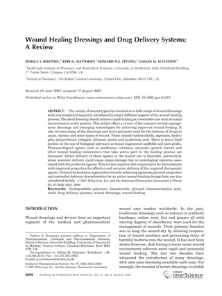
- 9. 2900 BOATENG ET AL. by wound exudate or external fluids. In addition gauze dressings tend to become more adherent to wounds as fluid production diminishes and are painful to remove, thus causing patient discom- fort.76 Gauze dressings also provide little occlu- sion and allow evaporation of moisture resulting in a dehydrated wound bed although gauze impregnated with soft paraffin is occlusive and easier to remove from the skin. It has been suggested that traditional dressings should be employed only for wounds that are clean and dry or used as secondary dressing to absorb exudates and protect the wound.2 Traditional wound healing agents have been largely replaced for chronic wounds and burns by the more recent and advanced dressings because topical liquid and semi-solid formulations do not remain on the wound surface long enough whilst dry traditional dressings do not provide a moist environment for wound healing. Modern Wound Dressings Figure 2. A typical hydrocolloid dressing, (TegasorbTM Thin Hydrocolloid Dressing, 3M Healthcare, Loughbor- Modern dressings have been developed as an ough, UK). The dressing combines’ moisture vapour improvement upon the traditional wound healing permeability with absorbency and conformability, and agents described above. Their essential charac- its transparency allows for wound observation. teristic is to retain and create a moist environ- ment around the wound to facilitate wound healing. The modern dressings are mainly classi- fied according to the materials from which they materials such as alginates. Hydrocolloid dres- are produced including hydrocolloids, alginates sings are useful clinically because unlike other and hydrogels, and generally occur in the form of dressings, they adhere to both moist and dry gels, thin films and foam sheets. sites.78 Hydrocolloid dressings are used for light to moderately exuding wounds (Tab. 1) such as pressure sores, minor burns and traumatic Hydrocolloid Dressings injuries. They are also used to manage leg ulcers Hydrocolloid dressings are among the most widely where they appear to have advantages in the used dressings. The role of hydrocolloid dressings, treatment of wounds that fail to respond to their properties, mechanism of action and the compression therapy alone.79 In their intact state, range of wounds for which they are useful have hydrocolloid dressings are impermeable to water been reviewed.68,77 The term ‘hydrocolloid’ vapour but on absorption of wound exudate, a describes the family of wound management change in physical state occurs with the formation products obtained from colloidal (gel forming of a gel covering the wound. They become agents) materials combined with other materials progressively more permeable to water and air such as elastomers and adhesives. Typical gel as the gel forms.80 As they do not cause pain on forming agents include carboxymethylcellulose removal, they are particularly useful in paediatric (CMC), gelatin and pectin. Examples of hydro- wound care for management of both acute and colloid dressings include GranuflexTM and Aqua- chronic wounds.81 A comparative study conducted celTM (Conva Tec, Hounslow, UK), ComfeelTM to evaluate a hydrocolloid dressing (Comfeel Ulcer (Coloplast, Peterborough, UK) and TegasorbTM DressingTM) combined with compression stocking (3M Healthcare, Loughborough, UK). They occur and a rigid bandage (Unna boot) for treating in the form of thin films and sheets (Fig. 2) or as venous ulcers79 found the hydrocolloid dressing- composite dressings in combination with other compression stocking combination to be superior JOURNAL OF PHARMACEUTICAL SCIENCES, VOL. 97, NO. 8, AUGUST 2008 DOI 10.1002/jps
- 10. WOUND DRESSINGS AND DRUG DELIVERY SYSTEMS 2901 to the rigid bandage. In that study the two groups wounds. The use of alginates as dressings stems of dressings were compared by monitoring time to primarily from their ability to form gels upon complete healing, reduction of wound surface, contact with wound exudates (high absorbency). pain during application and the total duration of The high absorption occurs via strong hydrophilic dressing change. A randomised trial comparing gel formation, which limits wound secretions and paraffin gauze and a hydrocolloid dressing applied minimises bacterial contamination.87 Alginates to skin draft donor sites showed that the hydro- rich in mannuronate, such as SorbsanTM (Maersk, colloid achieves faster healing and is a less painful Suffolk, UK) form soft flexible gels upon hydra- dressing.82 Another study, involving patients with tion whereas those rich in guluronic acid, like lacerations, abrasions and minor operation inci- KaltostatTM (Conva Tec), form firmer gels upon sions, compared a hydrocolloid dressing with a absorbing wound exudate. Some contain calcium nonadherent dressing. While time to heal was alginate fibre such as SorbsanTM and TegagenTM similar for both groups, patients using the (3M Healthcare). Comfeel PlusTM is a hydrocol- hydrocolloid experienced less pain, required less loid/alginate combination dressing. analgesia and were able to carry out their normal When applied to wounds, ions present in the daily activities including bathing or showering alginate fibre are exchanged with those present in without affecting the dressing or the wound.83 exudate and blood to form a protective film of Some possible mechanisms involved in hydrocol- gel.88 This helps to maintain the lesion at an opti- loids’ ability to reduce pain have been discussed.84 mum moisture content and healing temperature. Hoekstra et al.85 found that AquacelTM hydro- The gelling property of the alginates is attributed colloid dressing performed more efficiently com- to the presence of calcium ions which help to form pared with tulle gauze dressing, in terms of the a crosslinked polymeric gel that degrades slowly. acute inflammatory responses observed at the The ability of calcium ions to form crosslinks with initial stages of wound healing, in partial thick- the alginic acid polymer makes calcium alginate ness wounds in rats. Hydrocolloid dressings dressings ideal materials as scaffolds for tissue generally have an occlusive outer cover that engineering.89,90 A comparative study of hydro- prevents water vapour exchange between the colloid dressings and alginates showed that wound and its surroundings. This can be dis- alginates gels remain on the wound for a longer advantageous for infected wounds that require a period than hydrocolloids.91 certain amount of oxygen to heal rapidly. Another Alginate dressings, as well as forming gels, have disadvantage applies to dressings containing a pharmacological function due to the action of the fibres that are deposited in the wound and often calcium ions present in the dressing. The role of have to be removed during dressing change. calcium alginate in the wound healing process Microscopic studies comparing CMC hydrocol- was investigated by Schmidt and Turner92 who loid and alginate dressings for their ability to suggested that it may help in the production of adsorb harmful bacteria showed the CMC dres- mouse fibroblast. This process was further mod- sings to be superior.86 In that study, it was elled in vitro by Doyle et al.93 who showed that demonstrated that the CMC containing wound calcium alginate increased proliferation of fibro- dressing produced a gel upon hydration that was blasts but not their motility. This suggests that effective in encapsulating large numbers of the effects of the dressing may have been P. aeruginosa and S. aureus. The authors also mediated by calcium ions released from the demonstrated the ability of CMC to immobilise alginate and therefore calcium alginate may these bacteria within the swollen fibres unlike the improve some cellular aspects of wound healing alginate gel, which immobilised fewer numbers of but not others. Thomas et al.,94 have reported that these bacteria. some alginate dressings activate human macro- phages to produce tumour necrosis factor-a (TNFa) which initiates inflammatory signals, as Alginate Dressings part of the wound healing process. Lansdown95 Alginate dressings are produced from the calcium has reviewed the potential role of calcium released and sodium salts of alginic acid, a polysaccharide from alginates in the wound healing process. comprising mannuronic and guluronic acid units. Alginate dressings are useful for all stages of Alginate dressings occur either in the form of wound healing described above. Calcium ions freeze-dried porous sheets (foams) or as flexible present in alginate dressings, when released into fibres, the latter indicated for packing cavity the wound, also play a physiological role aiding in DOI 10.1002/jps JOURNAL OF PHARMACEUTICAL SCIENCES, VOL. 97, NO. 8, AUGUST 2008
- 11. 2902 BOATENG ET AL. the clotting mechanism (haemostat) during the so that they physically entrap water. The sheets first stage of wound healing.96–99 The early use of can absorb and retain significant volumes of water alginates as haemostats and wound dressings and upon contact with suppurating wounds. Lay- their apparent lack of toxicity are discussed by Flurrie104 has reviewed the properties of hydro- Blaine100 and later, clinical studies proved their gels by investigating the results from studies into successful use in neurosurgery.101 their efficiency and discussed the wound types Alginate dressings are useful for moderate to that are suited for hydrogel treatment. When heavily exuding wounds. Alginate dressings in the applied to the wound as a gel, hydrogel dressings form of fibres when trapped in a wound are readily usually require a secondary covering such as biodegradable102 and can be rinsed away with gauze and need to be changed frequently.18 The saline irrigation. Subsequent removal therefore, sheets however, do not need a secondary dressing does not destroy granulation tissue, making as a semi-permeable polymer film backing, with or dressing change virtually painless. The ease of without adhesive borders, controls the transmis- biodegradation is exploited in making alginate sion of water vapour through the dressing. In sutures used in surgical wound closures. A study addition the sheets can be cut to fit around the of different brands of alginate dressings showed wound due to their flexible nature. The gels are significant differences in characteristics such as used as primary dressings whereas the hydrogel fluid retention, adherence and dressing resi- films may be used as primary or secondary dues.103 Since alginate dressings require moisture dressings (Fig. 3). Hydrogel dressings contain to function effectively, they cannot be used for dry significant amounts of water (70–90%) and as a wounds and those covered with hard necrotic result they cannot absorb much exudate, thus tissue. This is because it could dehydrate the they are used for light to moderately exuding wound, delaying healing and this is their major wounds. Fluid accumulation can lead to skin disadvantage. maceration and bacterial proliferation which produces a foul smell in infected wounds. In addition, hydrogels have low mechanical strength Hydrogel Dressings and therefore difficult to handle105 and this has Hydrogels are insoluble, swellable hydrophilic been noted to affect patient compliance.106 materials made from synthetic polymers such Hydrogels possess most of the desirable char- as poly(methacrylates) and polyvinylpyrrolidine. acteristics of an ‘ideal dressing’106 (Tab. 2). They Some dressings such as Nu-gelTM (Johnson & are suitable for cleansing of dry, sloughy or Johnson, Ascot, UK) and PurilonTM (Coloplast) necrotic wounds by rehydrating dead tissues are hydrogel/alginate combinations. Hydrogels and enhancing autolytic debridement (Tab. 1). can be applied either as an amorphous gel or as Hydrogel dressings are nonreactive with biologi- elastic, solid sheet or film (Fig. 3). To prepare the cal tissue, permeable to metabolites and are sheets, the polymeric components are crosslinked nonirritant.107 Hydrogels also promote moist healing, are nonadherent and cool the surface of the wound, which may lead to a marked reduction in pain and therefore have high patient accept- ability. In a clinical case study, Moody108 reports the use of a hydrogel gel dressing to treat a chronic leg ulcer for a patient who could not tolerate even reduced compression therapy due to pain, and the hydrogel helped reduce the pain considerably. Hydrogels also leave no residue, are malleable and improve reepithelisation of wounds.18 Morgan2 has stated that hydrogels ‘are suitable for use at all four stages of wound healing with the exception of infected or heavily exuding wounds’. Figure 3. An example of a polymeric hydrogel sheet Semi-Permeable Adhesive Film Dressings wound dressing. Hydrogel sheets do not need a second- ary dressing and due to their flexible nature, can be cut These dressings have been used for a long time to fit around the wound. and their effects on moist wound healing were first JOURNAL OF PHARMACEUTICAL SCIENCES, VOL. 97, NO. 8, AUGUST 2008 DOI 10.1002/jps
- 12. WOUND DRESSINGS AND DRUG DELIVERY SYSTEMS 2903 investigated by Winter109 and Hinman and investigate the efficacy of various dressings Maibach.110 Film dressings were originally including foam and gauze on postoperative made from nylon derivatives supported in an wounds without closure, it was found that foam adhesive polyethylene frame which made them was preferred to gauze in terms of pain reduction, occlusive. The original nylon derived film dres- patient satisfaction and nursing time.117 sings, however, have limited ability to absorb Foam dressings are also indicated for granulat- sufficient quantities of wound exudates which ing wounds where they are reported to help treat results in the accumulation of excess exudates over granulation.118 They are used as primary beneath the dressing.18 This leads to skin mace- wound dressings for absorption and insulation ration and bacterial proliferation and the risk of and a secondary dressing is usually not required infection and therefore require regular changing due to their high absorbency and moisture vapour as well as irrigation of the wound with saline, permeability. Foam dressings are not suitable for making them unsuitable as wound dressings. The dry epithelialising wounds or dry scars as they original nylon dressings are also difficult to apply rely on exudates unlike the polymer films, to and tend to wrinkle on removal from their packs. achieve an optimum wound healing environ- OpsiteTM (Smith and Nephew, Hull, UK) is a thin ment.116 The sheet dressings are not suitable as semi-permeable film made from polyurethane packs for cavity wounds,106 though they may be covered with hypoallergenic acrylic derivatives used as secondary dressings for such wounds. and is more porous and permeable to water vapour Examples of foam dressing include: Lyofoam1 and gases but no liquid from exudates.111 The (Conva Tec) and Allevyn1 (Smith and Nephew). films can be transparent, conform to contours (due to their elastic and flexible nature) such as elbows, Biological Dressings knees and sacral areas and do not require additional taping. However, they are too thin to These dressings are made from biomaterials that be packed into deep or cavity wounds and only play an active part in the wound healing process suitable for relatively shallow wounds. Other and sometimes referred to as ‘bioactive dressings’. available products include CutifilmTM (B.D.F. Bioactive wound healing dressings also include Medical, Milton Keynes, UK), BiooclusiveTM tissue engineered products derived from natural (Johnson & Johnson) and Tegaderm (3M Health- tissues or artificial sources.119 These technologies care). Most of the existing brands differ in terms of usually combine polymers such as collagen,120 vapour permeability, adhesiveness, conformabil- hyaluronic acid,121 chitosan,122 alginates and ity and extensibility.112 elastin. Biomaterials have the advantage of forming part of the natural tissue matrix, are biodegradable and some play an active part in Foam Dressings normal wound healing and new tissue forma- These dressings consist of porous polyurethane tion.123,124 These characteristics make them foam or polyurethane foam film, sometimes with attractive choices from a biocompatibility and adhesive borders.2 Some foam dressings such as toxicological point of view. In some cases they may TielleTM have additional wound contact layers to be incorporated with active compounds such as avoid adherence when the wound is dry and an antimicrobials and growth factors for delivery to occlusive polymeric backing layer to prevent the wound site. excess fluid loss and bacterial contamination.113 Collagen is a natural constituent of connective Foam dressings maintain a moist environment tissue and a major structural protein of any organ. around the wound, provide thermal insulation Its structural, physical, chemical, biological and and are convenient to wear.114 They are highly immunological properties have been discussed absorbent, absorbency being controlled by foam widely in the literature.120,125,126 Collagen is properties such as texture, thickness and pore known to play a vital role in the natural wound size. The open pore structure also gives a high healing process from the induction of clotting to moisture vapour transmission rate (MVTR).115,116 the formation and appearance of the final scar.60 The porous structure of the dressings, make them It stimulates formation of fibroblasts and accel- suitable for partial- or full-thickness wounds with erates the migration of endothelial cells upon minimal or moderate drainage, to highly absor- contact with damaged tissue.127 Schwarzer128 bent structures for heavily exuding wounds.56 In a reported the production of freeze-dried collagen systematic review of clinical trials conducted to biomatrices with the ability to pick up fluid, debris DOI 10.1002/jps JOURNAL OF PHARMACEUTICAL SCIENCES, VOL. 97, NO. 8, AUGUST 2008
- 13. 2904 BOATENG ET AL. and inflammatory cells containing phagocytosed to as acellular and cell containing matrices. bacteria. The matrix can also be medicated, thus Acellular matrices are produced either from serving as a reservoir for drug delivery. The use of synthetic collagen and extracellular matrix com- collagen matrices for delivery of different classes binations such as hyaluronic acid,140 for example of antibiotic drugs have been discussed exten- IntegraTM, or native dermis with the cellular sively.129 components removed but preserving the dermal Hyaluronic acid is a glycoaminoglycan compo- architecture,116 for example AllodermTM. Cell- nent of extracellular matrix with unique physi- containing tissue engineered dressings include cochemical and biological functions such as biodegradable films formed from, for example, lubrication of joints and inflammation processes. collagen and glycosaminoglycans (e.g. ApligrafTM) It is naturally biocompatible, biodegradable and as scaffolds onto which skin cells (patient derived lacks immunogenicity.130 Crosslinked hyaluronic or from recombinant sources) can be seeded for the acid hydrogel films have also been produced for growth of new tissues. These scaffold dressings use as polymeric drug delivery biomaterials.131 possess mechanical properties and anatomic Hyaluronic acid-modified liposomes as bioadhe- characteristics ideally approaching that of the sive carriers for delivering growth factors to tissue (normal dermis) they are to replace.141 wound sites have been studied and reported.132 When introduced into the body they gradually A recent open ended study of hyaluronic acid degrade, leaving behind a matrix of connective based dressing found them to be effective for tissue with the appropriate structural and managing acute wounds particularly in terms of mechanical properties. Some of the developed its safety and efficacy.133 In this study however, no tissue engineered products and skin substitutes standard wound dressing was selected for com- available are summarised in Table 3. parison and the dressing was applied to different Engineered scaffolds either from natural or wound types. Chitosan is known to accelerate synthetic sources are potentially useful for the granulation during the proliferative stage of delivery of additional bioactive materials such as wound healing,101 and its wound healing applica- growth factors and genetic materials to a wound. tion has been reviewed.134 Bioactive dressings are Storie and Mooney142 have reviewed the utility of reported to be more superior to conventional and DNA delivery from polymeric systems in the synthetic dressings such as gauze and hydrogel regeneration of skin tissue in cases such as dressings respectively.135 diabetes foot ulcers. Hoffman143 has described the ideal properties of hydrogels designed to serve different functions as tissue engineered scaffolds such as possessing spores capable of accommo- Tissue Engineered Skin Substitutes dating living cells. Alternatively, they may be Traditional and modern dressings though useful, designed to dissolve or degrade and release cannot replace lost tissue, particularly missing growth factors in the process as well as creating dermis as occurs in severe burns. In more pores into which living cells can penetrate and advanced applications, ‘smart’ polymers from subsequently proliferate, to replace lost or damag- modifications of synthetic and bioactive polymers ed tissue. Ruszczak144 has reviewed the effect of have been developed.136 Advances in the fabrica- collagen on dermal wound healing and noted the tion of biomaterials and the culturing of skin cells advantage of combining collagen with patient have led to the development of a new generation of derived dermal cells, recombinant growth factors, engineered skin substitutes.137 Such polymers act cytokines, living cells and antimicrobial agents. as scaffolds for tissue engineered substrates that This could help speed up formation of granulation replace lost tissue rather than just facilitate tissue and reepithelisation during wound healing. wound healing. The use of ‘smart’ polymers either Though these advanced dressings have great in the natural biological form or semi-synthetic potential for treating chronic wounds and third forms are reported to be able to mimic normal phy- degree burns, they are still limited by the high siologic responses during wound healing.138,139 costs involved, the risk of infection carry over and This can help natural cell and tissue regeneration, antigenicity as well as having to create a second particularly for chronic wounds that are difficult wound in the case of harvesting patient’s own cells to heal. to aid wound healing. These shortcomings in Two major matrices are employed in tissue addition to the legal and ethical issues surround- engineered skin substitutes. These are referred ing stem cell research have probably contributed JOURNAL OF PHARMACEUTICAL SCIENCES, VOL. 97, NO. 8, AUGUST 2008 DOI 10.1002/jps
- 14. WOUND DRESSINGS AND DRUG DELIVERY SYSTEMS 2905 Table 3. Tissue Engineered Skin Substitutes Available Commercially Dressing Type Major Components Manufacturers TM Integra Artificial skin Collagen/chondroitin-6 sulphate Integra LifeScience matrix overlaid with a thin (Plainsborough, NJ) silicone sheet BiobraneTM Biosynthetic skin Silicone, nylon mesh, collagen Dow Hickham/Bertek substitute Pharmaceuticals (Sugar Land, TX) AllodermTM Acellular dermal graft Normal human dermis with all Lifecell Corporation the cellular material removed (Branchberg, NJ) DermagraftTM Dermal skin substitute Cultured human fibroblasts on a Advanced Tissue Sciences biodegradable polyglycolic (LaJolla, CA) acid or polyglactin mesh EpicelTM Epidermal skin Cultured autologous human Genzyme Biosurgery substitute keratinocytes (Cambridge, MA) MyskinTM Epidermal skin Cultured autologous human Celltran Limited (University substitute keratinocytes on medical grade of Sheffield, Sheffield, UK) silicone polymer substrate TranCyteTM Human fibroblast derived Polyglycolic acid/polylactic acid, Advanced Tissue Sciences skin substitute extracellular matrix proteins (synthetic epidermis) derived from allogenic human fibroblasts and collagen ApligrafTM Epidermal and dermal Bovine type I collagen mixed Organogenesis (Canton, MA) skin substitutes with a suspension of dermal fibroblasts Hyalograft 3-DTM Epidermal skin Human fibroblasts on a Fidia Advanced Biopolymers substitute laser-microperforated (Padua, Italy) membrane of benzyl hyaluronate LaserskinTM Epidermal skin Human keratinocytes on a Fidia Advanced Biopolymers substitute laser-microperforated membrane of benzyl hyaluronate BioseedTM Epidermal skin Fibrin sealant and cultured BioTissue Technologies substitute autologous human keratinocytes (Freiburg, Germany) to the slow adoption of these dressings in routine described in previous sections. Traditional dres- clinical practice.145 sings commonly used to deliver drugs include plain gauze and paraffin impregnated gauze (tulle gras). The modern dressings used to deliver active MEDICATED DRESSINGS agents to wounds include hydrocolloids, hydro- FOR DRUG DELIVERY gels, alginates, polyurethane foam/films and silicone gels.146 The incorporated drugs play an The active ingredients used in wound manage- active role in the wound healing process either ment have evolved alongside the pharmaceutical directly or indirectly as cleansing or debriding agents and dressings used to deliver them. The agents for removing necrotic tissue, antimicro- use of topical pharmaceutical agents in the form of bials which prevent or treat infection or growth solutions, creams and ointments to wound sites agents (factors) to aid tissue regeneration. Some of have already been described. For example solu- the commonly used active compounds and the tions such as thymol and hydrogen peroxide106 dressings (and novel polymer systems) used to used commonly for cleansing and debridement, deliver them to wound sites are described below. also possess antiseptic and antibacterial actions. A new generation of medicated dressings Antimicrobials incorporate new chemicals which have therapeu- tic value, and overcome some of the disadvantages The purpose of applying antibiotics and other associated with topical pharmaceutical agents as antibacterials is mainly to prevent or combat DOI 10.1002/jps JOURNAL OF PHARMACEUTICAL SCIENCES, VOL. 97, NO. 8, AUGUST 2008
- 15. 2906 BOATENG ET AL. infections especially for diabetic foot ulcers,147,148 do not necessarily take an active physiological surgical and accident149 wounds where the part in the wound healing process. Growth factors incidence of infections can be high due to reduced are involved with cell division, migration, differ- resistance resulting from extreme trauma. In entiation, protein expression and enzyme produc- some cases, the delivery of certain antibiotics from tion. The wound healing properties of growth paraffin based ointments such as bismuth sub- factors are mediated through the stimulation gallate are known to take active part in the wound of angiogenesis and cellular proliferation, which healing process.150 Common antibiotics incorpo- affects both the production and the degradation of rated into available dressings for delivery to the extracellular matrix and also plays a role wounds include dialkylcarbamoylchloride which in cell inflammation and fibroblast activity.159 is incorporated into Cutisorb1 a highly absorbent Growth factors therefore affect the inflammatory, cotton wool dressing, povidone-iodine used with proliferation and migratory phases of wound fabric dressing and silver used with most of the healing.160 A variety of growth factors have been modern dressings.1 Silver impregnated modern reported which participate in the process of wound dressings available on the UK Drug Tariff include healing including, epidermal growth factor (EGF), Fibrous Hydrocolloid, Poyurethane Foam Film platelet derived growth factor (PDGF), fibroblast and Silicone gels.1 Other antibiotics delivered growth factor (FGF), transforming growth factor to wounds include gentamycin from collagen (TGF-b1), insulin-like growth factor (IGF-1), sponges,151 ofloxacin from silicone gel sheets152,153 human growth hormone and granulocyte-macro- and minocycline from chitosan film dressings.154 phage colony-stimulating factor (GM-CSF).161,162 Some of the reported novel antimicrobial wound In a study of the influence of GM-CSF in full healing dressings reported include, freeze-dried thickness wounds in transgenic mice, Mann fibrin discs for the delivery of tetracycline155 et al.163 suggested that GM-CSF is of fundamental and lactic acid based system for the delivery of importance in the wound healing repair and a ofloxacin and the inhibition of Staphyloccocus deficiency of this growth factor resulted in delayed aureus and P. aeruginosa in split-thickness wound healing and poor quality of newly formed wounds in rats.153 Treatment of dermal depth scar tissue. Lee et al.164 have reported that silver burn wounds using antimicrobial releasing sili- sulphadiazine alone can impair wound healing cone gel sheets which promotes epithelisation of and that EGF helps reverse this impairment when superficial burns has been described by Sawada both are applied together. et al.152 A chitosan-polyurethane film dressing Different dressings have been used to topically incorporating minocycline has also been develop- administer some of the above growth factors ed for treating severe burn wounds.156 to wound sites. These include hydrogel dressings The delivery of antibiotics to local wound sites for delivering transforming growth factor-b1 may be a preferred option to systemic adminis- (TGF-b1),165,166 collagen film for delivering tration for several reasons. Antibiotic doses PDGF167 and human growth hormone,168 alginate needed to achieve sufficient systemic efficiency dressings in the form of beads used to deliver often results in toxic reactions such as the endothelial growth factor,169 polyurethane and cumulative cell and organ toxicity of the amino- collagen film dressings for delivery of EGF.170 glycosides in the ears and kidneys.157,158 The use Park et al.171 showed that a novel porous collagen- of dressings to deliver antibiotics to wound sites hyaluronic acid matrix, containing tobramycin, can provide tissue compatibility, low occurrence basic FGF and platelet-derived growth factor of bacterial resistance and reduced interference significantly enhanced wound healing compared with wound healing.121 The use of lower antibiotic with matrix containing only the antibiotic. It has doses within the dressings also reduces the risk of been reported that EGF when applied to partial systemic toxicity considerably. In addition, local thickness incisions as a cream, stimulated epi- delivery from dressings can overcome the problem dermal regeneration.172 Most of these growth of ineffective systemic antibiotic therapy resulting factors are recombinant proteins and the choice from poor blood circulation at the extremities in of appropriate dressing is critical for effective diabetic foot ulcers. release and action at the wound site. A review of the potential role of growth factors for treating Growth Factors chronic leg ulcers by Khan and Davies173 reported Whilst antibacterial agents prevent or treat encouraging clinical results, though factors such infections and can aid in wound healing, they as small sample size and inconsistent endpoints in JOURNAL OF PHARMACEUTICAL SCIENCES, VOL. 97, NO. 8, AUGUST 2008 DOI 10.1002/jps
- 16. WOUND DRESSINGS AND DRUG DELIVERY SYSTEMS 2907 clinical studies have prevented definite conclu- Keitzman and Braun188 has noted that the low sions being reached. molecular weight protein group, metallothioneins are upregulated around wound margins following topical application of zinc and copper. He sug- gested that the action of these proteins resulted Supplements from the many zinc and copper dependent Another group of active compounds important enzymes required for cell proliferation and to the wound healing process are vitamins and reepithelisation. mineral supplements174 including vitamins A, C, Topical pharmaceutical formulations in the E as well as zinc and copper. The dressings form of liquid emulsions and ointments incorpor- employed for the delivery of vitamins and miner- ating zinc have been applied frequently in the als include oil based liquid emulsions, creams, past and at present. Lansdown189 found that a ointments, gauze and silicone gel sheets. combined formulation of zinc oxide and cod liver Vitamin A is involved with epithelial cell oil emulsion was more effective than vehicle differentiation,175 collagen synthesis and bone only controls as well as those containing a single tissue development.176 It has also been shown to active ingredient. He also found the zinc oxide facilitate normal physiological wound healing as emulsion to be most efficient in rapid healing of well as reversing the corticosteroid induced wounds retarded by corticosteroid treatment. In inhibition of cutaneous wound healing and post another study to investigate the effect of topical operative immune depression.177,178 Vitamin C agents on the healing rate of deep second-degree is an essential compound for the synthesis of burn wounds, application of zinc containing collagen and other organic components of the topical formulation reduced healing times sig- intracellular matrix of tissues such as bones, skin nificantly.190 This reduction in healing time was and other connective tissues.175 It is also involved further improved when basic FGF and EGF were with normal responses to physiological stressors used in combination with the zinc preparation. In such as in accident and surgical trauma and a randomised double-blind placebo controlled the need for ascorbic acid increases during times trial, the effects of topical zinc oxide and mesh of injury.179 In addition, vitamin C aids in on secondary healing pilonidal wounds were improving immune function particularly during compared. Topical zinc was shown to aid faster infection.24,180 The use of vitamins E and C acid wound healing times, decrease Staphyloccocus has been reported to help accelerate wound load in the wound and also no associated cellular healing.181 Vitamin E is also capable of preserving abnormalities.191 However, in another study by important morphological and functional features Cangul et al.192 to evaluate the clinical and of biological membranes159 though its use in histopathological effects of topically applied zinc topical applications has however, been discour- and copper based dressings on open-wound aged due to the problem of contact dermatitis.182 healing in rabbits, it was found that copper In addition, vitamin E its reported to have based topical agents caused wound contraction antioxidant and anti-inflammatory activity183 as and coverage of the wound bed with granulation well as promoting angiogenesis and reduces tissue at a faster rate than zinc based dressings. scarring.184 The delivery of vitamins to wounds from dressings is sparsely reported in the literature; they are mostly administered orally to supple- CONTROLLED DRUG DELIVERY ment body stores but this is outside the scope of TO THE WOUND this review. Vitamin E has been used in combina- tion with silicone gel sheets for the treatment of Controlled release of drugs to a given target hypertrophic and keloid scars.185 Lazovic et al.186 generally involves prolonging the action of the have reported the application of collagen sheet active drug over time by allowing continual dressings wetted with vitamins A and C solutions release from a polymeric dosage form.193 There over burn wounds and showed significant im- is however, little literature on the controlled provements in healing of the wound. Topical zinc delivery of drugs from polymeric wound dressings. can stimulate the healing of leg ulcers through The use of hydrophilic polymers as controlled enhancement of reepithelialisation and also cor- release dressings has great promise because of the rects a local zinc deficiency of the metal.187 potential advantages they offer. DOI 10.1002/jps JOURNAL OF PHARMACEUTICAL SCIENCES, VOL. 97, NO. 8, AUGUST 2008
- 17. 2908 BOATENG ET AL. Advantages of Controlled Drug Delivery and genetic material to wound sites have also been reported.138,142 The modern dressings for drug Controlled delivery dressings can provide an delivery to wounds may be applied in the form of excellent means of delivering drugs to wound gels, films and foams whilst the novel polymeric sites in a consistent and sustained fashion over dressings produced in the form of films and long periods of time without the need for frequent porous sponges such as freeze-dried wafers or dressing change.194 Bioadhesive, synthetic, semi- discs155,222–228 or as tissue engineered polymeric synthetic and naturally derived polymeric dres- scaffolds.155,160 sings are potentially useful in the treatment of local infections where it may be beneficial to have increased local concentrations of antibiotics while avoiding high systemic doses195 thus reducing Mechanism of Controlled Delivery to Wounds patient exposure to an excess of drug beyond that Drug release from polymeric formulations is required at the wound site.196 In addition, they controlled by one or more physical processes are readily biodegradable and therefore can be including (a) hydration of the polymer by fluids easily washed off the wound surface, once they and (b) swelling to form a gel, (c) diffusion of drug have exerted their desired effect.197 Improvement through the swollen gel and (d) eventual erosion of patient compliance is another advantage of the polymer gel.229–236 Although there is little especially in chronic wound management where literature in this area for polymeric wound patients usually undergo long treatments and dressings such as hydrocolloids, alginate, hydro- frequent changing of dressings that can lead to gels and polyurethane, it seems feasible that noncompliance. A dressing that will deliver an swelling, erosion and subsequent drug diffusion active substance to a wound site in a controlled kinetics will play a part in controlled drug release fashion for a sustained period of about a week from these dressings when they come into contact could help solve or minimise this problem. with wound exudate. Upon contact of a dry polymeric dressing with a moist wound surface, wound exudate penetrates into the polymer Polymeric Drug Delivery Dressings matrix. This causes hydration and subsequent Most modern dressings are made from polymers swelling of the dressing to form a gel over the which can serve as vehicles for the release and wound surface.237 Gombotz and Wee238 have delivery of drugs to wound sites. The release of reviewed the controlled release of proteins from drugs from modern polymeric dressings to wounds alginate matrices including dressings to mucosal has been sparsely reported in the literature with tissues such as wounds. They described the few clinical studies carried out to date. The swelling behaviour of the polymer to form a gel polymeric dressings employed for controlled drug which acts as a barrier to drug diffusion. As with delivery to wounds include hydrogels such as all polymers, the swelling observed is due to poly(lactide-co-glycolide)198 poly(vinyl pyrroli- solvation of the polymer chains, which leads to an done),143 poly(vinyl alcohol)188 and poly(hydroxy- increase in the end-to-end distance of the indivi- alkylmethacrylates)199–202 polyurethane-foam,203–207 dual polymer molecules. In certain wound dres- hydrocolloid56 and alginate dressings.169,208–210 sings, the mechanism for drug release has been Other polymeric dressings reported for drug explained by the hydrolytic activity of enzymes delivery to wounds comprise novel formulations present in the wound exudates239 or from bacteria prepared from polymeric biomaterials such as in the case of infected wounds.240 Different hyaluronic acid,131,132 collagen166,168 and chito- techniques have been employed in studies to san.156,211–213 Synthetic polymers employed as characterise the swelling behaviour of hydrophilic swellable dressings for controlled drug delivery polymers upon contact with water.241,242 It has include silicone gel sheets,152 lactic acid.153 Some been shown that in an aqueous medium, the of these novel polymeric dressings for drug polymer also undergoes a relaxation process delivery exist as patents.214–219 Composite dres- resulting in slow, direct erosion (dissolution) of sings comprising both synthetic and naturally the hydrated polymer.243,244. It is possible for occurring polymers have also been reported for both swelling and dissolution to operate simulta- controlled drug delivery to wound sites.220,221 neously in wound dressings with each contribut- Sustained release tissue engineered polymeric ing to the overall release mechanism. Generally, scaffolds for controlled delivery of growth factors however, the rate of release of drug is determined JOURNAL OF PHARMACEUTICAL SCIENCES, VOL. 97, NO. 8, AUGUST 2008 DOI 10.1002/jps