
Recomendados
Mais conteúdo relacionado
Mais procurados
Mais procurados (20)
Destaque
Destaque (8)
Semelhante a Bmj33300181
Semelhante a Bmj33300181 (20)
Último
Último (20)
Bmj33300181
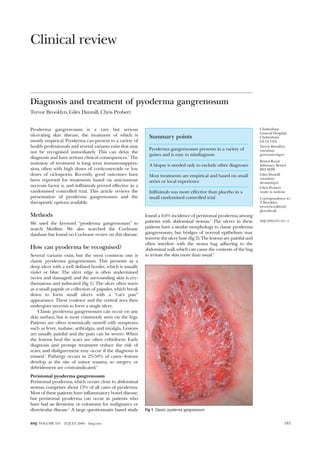
- 1. Clinical review Diagnosis and treatment of pyoderma gangrenosum Trevor Brooklyn, Giles Dunnill, Chris Probert Pyoderma gangrenosum is a rare but serious ulcerating skin disease, the treatment of which is mostly empirical. Pyoderma can present to a variety of health professionals and several variants exist that may not be recognised immediately. This can delay the diagnosis and have serious clinical consequences.1 The mainstay of treatment is long term immunosuppres- sion, often with high doses of corticosteroids or low doses of ciclosporin. Recently, good outcomes have been reported for treatments based on anti-tumour necrosis factor , and infliximab proved effective in a randomised controlled trial. This article reviews the presentation of pyoderma gangrenosum and the therapeutic options available. Methods We used the keyword “pyoderma gangrenosum” to search Medline. We also searched the Cochrane database but found no Cochrane review on this disease. How can pyoderma be recognised? Several variants exist, but the most common one is classic pyoderma gangrenosum. This presents as a deep ulcer with a well defined border, which is usually violet or blue. The ulcer edge is often undermined (worn and damaged) and the surrounding skin is ery- thematous and indurated (fig 1). The ulcer often starts as a small papule or collection of papules, which break down to form small ulcers with a “cat’s paw” appearance. These coalesce and the central area then undergoes necrosis to form a single ulcer. Classic pyoderma gangrenosum can occur on any skin surface, but is most commonly seen on the legs. Patients are often systemically unwell with symptoms such as fever, malaise, arthralgia, and myalgia. Lesions are usually painful and the pain can be severe. When the lesions heal the scars are often cribriform. Early diagnosis and prompt treatment reduce the risk of scars, and disfigurement may occur if the diagnosis is missed.1 Pathergy occurs in 25-50% of cases—lesions develop at the site of minor trauma, so surgery or debridement are contraindicated.2 Peristomal pyoderma gangrenosum Peristomal pyoderma, which occurs close to abdominal stomas, comprises about 15% of all cases of pyoderma. Most of these patients have inflammatory bowel disease, but peristomal pyoderma can occur in patients who have had an ileostomy or colostomy for malignancy or diverticular disease.3 A large questionnaire based study found a 0.6% incidence of peristomal pyoderma among patients with abdominal stomas.4 The ulcers in these patients have a similar morphology to classic pyoderma gangrenosum, but bridges of normal epithelium may traverse the ulcer base (fig 2). The lesions are painful and often interfere with the stoma bag adhering to the abdominal wall, which can cause the contents of the bag to irritate the skin more than usual.5 Summary points Pyoderma gangrenosum presents in a variety of guises and is easy to misdiagnose A biopsy is needed only to exclude other diagnoses Most treatments are empirical and based on small series or local experience Infliximab was more effective than placebo in a small randomised controlled trial Fig 1 Classic pyoderma gangrenosum Cheltenham General Hospital, Cheltenham GL53 7AN Trevor Brooklyn consultant gastroenterologist Bristol Royal Infirmary, Bristol BS2 8HW Giles Dunnill consultant dermatologist Chris Probert reader in medicine Correspondence to: T Brooklyn trevor.brooklyn@ glos.nhs.uk BMJ 2006;333:181–4 181BMJ VOLUME 333 22 JULY 2006 bmj.com
- 2. Pustular pyoderma gangrenosum Pustular pyoderma is a rare superficial variant of the disease. Pyoderma often begins as a pustule or group of pustules that later coalesce and ulcerate. This process stops at the pustular stage in pustular pyoderma, and the patient has a painful pustular lesion that may persist for months (fig 3). Pustular pyoderma seems to be confined to patients with inflammatory bowel disease and tends to occur on the trunk and extensor surfaces of the limbs.6–8 Bullous pyoderma gangrenosum Bullous pyoderma is a superficial variant that affects the upper limbs and face more than the lower limbs. It is associated mostly with haematological conditions. This form of the disease presents as concentric bullous areas that spread rapidly in a concentric pattern. They may break down to form more superficial ulcers than those seen in classic pyoderma, although they still have the blue undermined edge. Prognosis is often poor because of the underlying haematological malignancy.8 9 Vegetative pyoderma gangrenosum Vegetative pyoderma is a superficial form of disease that seems to be less aggressive than other varieties (fig 4). It usually occurs as a single lesion in patients who are otherwise well and may respond to local treatment more readily than other forms of the disease.8 10 What is the histopathology of pyoderma? The histopathology of pyoderma gangrenosum depends on the timing and site of the biopsy.11 Biopsies taken early in the disease and from the advancing, ery- thematous border tend to show an infiltrate of chronic inflammatory cells confined to the dermis (fig 5). They often have features suggestive of vasculitis at the edge of the ulcer, with a perivascular lymphocytic infiltrate and fibrinoid necrosis of the dermal vessel wall. Occa- sionally, extravasation of red blood cells and areas of thrombosis are also seen. Ulceration of the epidermis tends to be secondary to the dermal inflammation. Biopsies taken later in the course of ulceration usually show a polymorphonuclear cell infiltrate with features of ulceration, infarction, and abscess formation.11 Who gets pyoderma gangrenosum? About half of the cases are associated with underlying systemic conditions, such as inflammatory bowel disease, arthritis, and haematological malignancies.2 About 30% of cases occur in patients with inflammatory bowel disease. About 2% of patients with inflammatory bowel disease will develop pyoderma.12 Occasionally, the skin condition presents before the bowel disease. Pyoderma gangrenosum was thought to be associated only with ulcerative colitis, but both Crohn’s disease and ulcerative colitis have a similar incidence.13 Pyoderma gangrenosum is not related to the activity of the inflam- matory bowel disease, and it often occurs in patients whose bowel disease is in clinical remission. About 25% of patients have arthritis, most often seropositive rheumatoid arthritis, although the disease can occur in patients with seronegative arthritis or spondyloarthropathy. As with inflammatory bowel dis- ease, the activity of the arthritis is not related to pyoderma.14 Haematological malignancies are the next most common disorders associated with pyoderma, and these tend to be myeloid rather than lymphoid in origin. Leukaemia is the most frequently reported Fig 2 Peristomal pyoderma gangrenosum Fig 3 Pustular pyoderma gangrenosum Fig 4 Vegetative pyoderma gangrenosum Clinical review 182 BMJ VOLUME 333 22 JULY 2006 bmj.com
- 3. malignancy, usually acute myeloid leukaemia and most commonly the myelocytic or monomyelocytic type.15 What is the differential diagnosis? The diagnosis of pyoderma gangrenosum is based mainly on clinical findings because biopsies show no specific diagnostic features. In many cases, however, a biopsy can help exclude other conditions such as malig- nancy, infections, or cutaneous vasculitis. Swabs should be taken from the ulcer, as pyoderma is treated differently from infection. The differential diagnosis of pyoderma gangrenosum is wide (box). Special mention must be made of Sweet’s syndrome, which is character- ised by sudden onset of fever and an erythematous, papular eruption. Patients have leucocytosis and skin biopsy shows a dense neutrophilic infiltrate. Sweet’s syn- drome and pyoderma can coexist in the same patient as they are both neutrophilic dermatoses.12 How is it treated? No single, specific treatment exists and few controlled trials of treatment have been done.16 Most clinicians use a stepwise approach and both topical and systemic treatments. Immunosuppression is the mainstay of treatment, and the most commonly used drugs are corticosteroids and ciclosporin. Several other immuno- suppressive agents have been used with varied results, but treatment is largely empirical and the choice of treatment often depends on local experience.12 16 Topical treatments Highly potent topical corticosteroids (occasionally underneath occlusive dressings) may be sufficient to induce remission.2 Triamcinolone 40 mg/ml may be injected into the ulcer edge, either alone or as an adjunct to systemic treatment.17 Recently, topical tacrolimus has been shown to be effective in patients with peristomal disease. This is now available as a 0.1% and 0.03% ointment.5 Corticosteroids Most patients need systemic treatment to induce remission and doctors often start patients on oral cor- ticosteroids at an early stage. Prednisolone is the drug of choice and is usually started at high doses (60-120 mg) (level B evidence).8 16 Patients exposed to these doses for a long time are at risk of steroid related side effects and may benefit from the addition of a bone protecting agent. Minocycline 100 mg twice daily may be of some benefit, usually as an adjunct to oral steroids (level C evidence).18 Rapid improvement has been reported in patients with severe disease given intravenous methylprednisolone as pulse therapy of 1 g daily for three to five days (level B evidence), and several series and reviews support this treatment.16 19 Ciclosporin Other immunosuppressive agents may be used—firstly, to reduce the dependence on corticosteroids and, sec- ondly, because pyoderma is often resistant to treatment. When corticosteroids fail, the most widely used alternative is ciclosporin. Several case reports and small case series have demonstrated a good clinical response to low dose ciclosporin (level B evidence). Most patients show clinical improvement within three weeks with a dose of 3-5 mg/kg/day. Ciclosporin has several serious side effects, including nephrotoxicity, hypertension, and increased risk of cancer. Such side Fig 5 A lymphocytic infiltrate within the dermis in pyoderma gangrenosum Differential diagnosis of pyoderma gangrenosum Infections Bacterial Mycobacterial Fungal Viral Parasitic Malignancy Squamous cell carcinoma Cutaneous lymphoma Vascular ulceration Venous or arterial disease Antiphospholipid syndrome Systemic conditions Systemic lupus erythematosis Rheumatoid arthritis Behçet’s disease Wegner’s granulomatosis Sweet’s syndrome Additional educational resources Review article in the Lancet (www.thelancet.com/journals/lancet/article/ PIIS0140673697101878/abstract)—an excellent overview of pyoderma gangrenosum DermAtlas (http://www.dermatlas.org/derm/result.cfm?Diagnosis = 47670916)—images of pyoderma gangrenosum with brief case histories Information for patients emedicine (emedicine.com/derm/topic367.htm )—detailed overview from a leading American based e-learning website (with pictures) Ongoing research When is the optimal time to introduce treatments based on anti-tumour necrosis factor ? If remission is achieved with infliximab, should patients be placed on maintenance infusions? If so, for how long? How does infliximab compare with conventional treatments, such as corticosteroids or ciclosporin? Clinical review 183BMJ VOLUME 333 22 JULY 2006 bmj.com
- 4. effects have not been reported for the low doses used to treat pyoderma.16 20 Other immunosuppressants Azathioprine, used alone or combined with cortico- steroids, has had variable results.16 Tacrolimus has also been used systemically, and two centres where patients received 0.1-0.3 mg/kg have reported sustained responses.21 Anti-tumour necrosis factor agents Pyoderma has been reported to respond to infliximab, a monoclonal antibody against tumour necrosis factor .22 More recently, pyoderma gangrenosum was reported to resolve after treatment with etanercept, a recombinant protein that neutralises the soluble factor. The authors reported a single case where pyoderma gangrenosum healed when etanercept was given in conjunction with oral corticosteroids, although the steroids could not be tapered completely (level C evidence).23 In a randomised controlled trial, we found that infliximab was superior to placebo. We ran- domised 30 patients to infliximab 5 mg/kg or placebo. The response rate between the groups differed by 40% at week 2 (P = 0.025, 95% confidence interval 11% to 70%), giving a number needed to treat of 2.5. Confidence intervals were wide because of the small number of patients. By the end of the study, 20 of 29 patients (69%) had benefited from infliximab, includ- ing six of 29 (21%) who were in remission.24 Two serious adverse effects occurred in the trial. Reports on side effects in inflammatory bowel disease are rare.25 What is our approach to treatment? Morbidity and potential for rapid progression are high in most patients, so once suspected we advise an urgent referral to the dermatology department. We recom- mend oral corticosteroids (with or without mino- cycline) as first line treatment. If patients do not respond promptly, we then use infliximab as this has fewer recognised side effects than ciclosporin and has been used widely in inflammatory bowel disease and rheumatoid arthritis.25 We recommend an induction dose regimen of 5 mg/kg at weeks 0, 2, and 6, followed by further treatments as necessary, depending on response. Another immunosuppressant, such as azathioprine, should be given at the same time as infliximab. This approach is not strictly evidence based but is now accepted practice in other inflammatory conditions, such as Crohn’s disease.25 Contributors: CP had the idea for this review. TB searched the literature and wrote the first draft, which was edited by CP and GD. TB is guarantor. Funding: None. Competing interests: The authors took part in a trial of infliximab for treating pyoderma gangrenosum, supported by a grant from Schering-Plough Ltd. The company played no part in study design; collection, analysis, and interpretation of data; writing of the paper; or the decision to submit the paper for publication. 1 Harris AJ, Regan P, Burge S. Early diagnosis of pyoderma gangrenosum is important to prevent disfigurement. BMJ 1998;316:52-3. 2 Bennett ML, Jackson JM, Jorizzo JL, Fleischer AB Jr, White WL, Callen JP. Pyoderma gangrenosum. A comparison of typical and atypical forms with an emphasis on time to remission. Case review of 86 patients from 2 institutions. Medicine (Baltimore) 2000;79:37-46. 3 Lyon CC, Smith AJ, Beck MH, Wong GA, Griffiths CE. Parastomal pyoderma gangrenosum: clinical features and management. J Am Acad Dermatol 2000;42:992-1002. 4 Lyon CC, Smith AJ, Griffiths CE, Beck MH. The spectrum of skin disor- ders in abdominal stoma patients. Br J Dermatol 2000;143:1248-60. 5 Lyon CC, Stapleton M, Smith AJ, Mendelsohn S, Beck MH, Griffiths CE. Topical tacrolimus in the management of peristomal pyoderma gangrenosum. J Dermatol Treat 2001;12:13-7. 6 Fenske NA, Gern JE, Pierce D, Vasey FB. Vesiculopustular eruption of ulcerative colitis. Arch Dermatol 1983;119:664-9. 7 Callen JP, Woo TY. Vesiculopustular eruption in a patient with ulcerative colitis. Pyoderma gangrenosum. Arch Dermatol 1985;121:399, 402. 8 Powell FC, Collins S. Pyoderma gangrenosum. Clin Dermatol 2000; 18:283-93. 9 Hay CR, Messenger AG, Cotton DW, Bleehen SS, Winfield DA. Atypical bullous pyoderma gangrenosum associated with myeloid malignancies. J Clin Pathol 1987;40:387-92. 10 Wilson-Jones E, Winkelmann RK. Superficial granulomatous pyoderma: a localized vegetative form of pyoderma gangrenosum. J Am Acad Dermatol 1988;18:511-21. 11 Su WP, Schroeter AL, Perry HO, Powell FC. Histopathologic and immu- nopathologic study of pyoderma gangrenosum. J Cutan Pathol 1986; 13:323-30. 12 Callen JP. Pyoderma gangrenosum. Lancet 1998;351:581-5. 13 Thornton JR, Teague RH, Low-Beer TS, Read AE. Pyoderma gangreno- sum and ulcerative colitis. Gut 1980;21:247-8. 14 Von den Driesch P. Pyoderma gangrenosum: a report of 44 cases with follow-up. Br J Dermatol 1997;137:1000-5. 15 Duguid CM, Powell FC. Pyoderma gangrenosum. Clin Dermatol 1993; 11:129-33. 16 Reichrath J, Bens G, Bonowitz A, Tilgen W. Treatment recommendations for pyoderma gangrenosum: an evidence-based review of the literature based on more than 350 patients. J Am Acad Dermatol 2005;53:273-83. 17 Hughes AP, Jackson JM, Callen JP. Clinical features and treatment of peristomal pyoderma gangrenosum. JAMA 2000;284:1546-8. 18 Berth-Jones J, Tan SV, Graham-Brown RAC, Pembroke AC. The success- ful use of minocycline in pyoderma gangrenosum—a report of seven cases and review of the literature. J Dermatol Treat 1989;1:23-5. 19 Johnson RB, Lazarus GS. Pulse therapy. Therapeutic efficacy in the treat- ment of pyoderma gangrenosum. Arch Dermatol 1982;118:76-84. 20 Friedman S, Marion JF, Scherl E, Rubin PH, Present DH. Intravenous cyclosporine in refractory pyoderma gangrenosum complicating inflam- matory bowel disease. Inflamm Bowel Dis 2001;7:1-7. 21 Lyon CC, Kirby B, Griffiths CE. Recalcitrant pyoderma gangrenosum treated with systemic tacrolimus. Br J Dermatol 1999;140:562-4. 22 Regueiro M, Valentine J, Plevy S, Fleisher MR, Lichtenstein GR. Infliximab for treatment of pyoderma gangrenosum associated with inflammatory bowel disease. Am J Gastroenterol 2003;98:1821-6. 23 McGowan JW, Johnson CA, Lynn A. Treatment of pyoderma gangreno- sum with etanercept. J Drugs Dermatol 2004;3:441-4. 24 Brooklyn TN, Dunnill MG, Shetty A, Bowden JJ, Williams JD, Griffiths CE, et al. Infliximab for the treatment of pyoderma gangrenosum: a randomised, double blind, placebo controlled trial. Gut 2006;55:505-9. 25 Hanauer SB, Feagan BG, Lichtenstein GR, Mayer LF, Schreiber S, Colombel JF, et al. Maintenance infliximab for Crohn’s disease: the ACCENT I randomised trial. Lancet 2002;359:1541-9. (Accepted 20 June 2006) GP tips Pyoderma gangrenosum can occur on any skin surface and should be considered in any ulcer or surgical wound that does not heal Pyoderma usually develops rapidly and can progress from a pimple to a crater in 24-48 hours Pyoderma is usually painful and patients may have systemic features such as fever If pyoderma is suspected, the patient should be referred urgently for a specialist opinion A patient’s story I first came across pyoderma about a month after having an ileostomy in December 2001. It started out as a small but sore spot rather like an insect bite, just under the stoma. Over the next 24-48 hours the spot took on a life of its own, growing and erupting into an ulcer with blue and red edges. I occasionally needed morphine for the pain. I was admitted to hospital where I was introduced to a doctor conducting a trial of infliximab, a new treatment for pyoderma. I decided to take up the offer of a place on the trial—I felt I had nothing to lose. The drug was given by infusion and the pyoderma was brought under control within 48 hours. Four years later I am on a maintenance regimen of infusions every eight weeks. I contracted pyoderma on my leg about two years ago, which flared up while I was not being treated with infliximab (during a six month break in treatment). Since being on the maintenance dose I have had no problems. Clinical review 184 BMJ VOLUME 333 22 JULY 2006 bmj.com