SlideShare uma empresa Scribd logo
1 de 5
Baixar para ler offline
Proc. Natl Acad. Sci. USA
Vol. 78, No. 12, pp. 7634-7638, December 1981
Developmental Biology


Isolation of a pluripotent cell line from early mouse embryos
cultured in medium conditioned by teratocarcinoma
stem cells
     (embryonic stem cells/inner cell masses/differentiation in vsitro/embryonal carcinoma cells/growth factors)
GAIL R. MARTIN
Department of Anatomy, University of California, San Francisco, California 94143
Communicated byJ. Michael Bishop, September 14, 1981

ABSTRACT This report describes the establishment directly                             NORMAL MOUSE                                    TERATOMA
from normal preimplantation mouse embryos of a cell line that
forms teratocarcinomas when injected into mice. The pluripotency                       development                                    differentiation
of these embryonic stem cells was demonstrated conclusively by                           in utero                                        in situ
the observation that subclonal cultures, derived from isolated sin-
gle cells, can differentiate into a wide variety of cell types. Such
embryonic stem cells were isolated from inner cell masses of late
blastocysts cultured in medium conditioned by an established ter-                                           transfer to
                                                                                                        extra-uterine site
atocarcinoma stem cell line. This suggests that such conditioned                                                                   TERATOCARCINOMA
medium might contain a growth factor that stimulates the prolif-                        BLASTOCYST     injection into blastocyst
eration or inhibits the differentiation of normal pluripotent em-                                                                           l1
bryonic cells, or both. This method of obtaining embryonic stem
cells makes feasible the isolation of pluripotent cells lines from
various types of noninbred embryo, including those carrying mu-                                                              subcutaneous        culture
tant genes. The availability of such cell lines should make possible                                                           injection         in vitro
new approaches to the study of early mammalian development.
Teratocarcinomas are tumors that arise with relatively high ef-
ficiency when normal 1- to 7.5-day-old inbred mouse embryos
are transplanted to an extra-uterine site in a histocompatible                                                                     TERATOCARCINOMA
host (1, 2). The stem cell lines that are isolated from them are                                                                    STEM CELL LINE
known as embryonal carcinoma cell (ECC) lines. Such cells have
morphological, biochemical, and immunological properties in
common with pluripotent embryonic cells and therefore have                                                                           differentiation
been used extensively as an in vitro model system for the study                                                                         in vitro
of the developing embryo (3-5). In some cases these properties
include the ability to differentiate in vitro in a manner that
closely parallels the normal behavior of the isolated embryonic                                                                     DIFFERENTIATED
                                                                                                                                    CELL CULTURES
inner cell mass (ICM) (6, 7). The most compelling evidence for
the close relationship between the tumor stem cells and normal                          FIG. 1. Relationship between normal embryos and teratocarci-
embryonic cells is the fact that stem cells taken either from ter-                    noma stem cells. Adapted from Martin (10).
atocarcinomas or from embryonal carcinoma cell cultures can
participate in the development of completely normal adult mice                        extra-embryonic site the signals they receive are apparently not
when combined with embryonic cells by the technique of blas-                          conducive to completely normal embryogenesis.
tocyst injection (8, 9).                                                                 It is difficult to study the process by which teratocarcinoma
    These findings, schematically represented in Fig. 1, raise                        stem cells derive from normal embryonic cells as long as the
questions about the origin of the teratocarcinoma stem cell. One                      transforming or selective events occur in vivo. As indicated by
hypothesis is that the tumor stem cells arise as a consequence                        the dotted line in Fig. 1, it has long seemed logical to assume
of a stable, but reversible, epigenetic change in normal pluri-                       that pluripotent stem cells capable of forming teratocarcinomas
potent embryonic cells. Such "transformed" cells presumably                           might be isolated directly from embryos. Recently, Evans and
continue proliferation in the undifferentiated state because neo-                     Kaufman (12) carried out experiments in which pregnant strain
plastic conversion has reduced their efficiency of response to                        129 female mice were subjected to a treatment, involving ova-
the normal signals for differentiation (11). An alternative idea                      riectomy and appropriate hormonal stimulation, that prevents
is that embryonal carcinoma cells are not transformed but rather                      normal implantation of blastocysts. Cells with the properties of
represent a selected population of completely normal embry-                           teratocarcinoma stem cells were isolated from the embryos that
onic cells that are programmed to divide until they receive the                       had been maintained in such "delay" in the reproductive tract.
appropriate signals for differentiation; when the cells are in an                     Other attempts have been made to isolate cells with the prop-
                                                                                      erties of teratocarcinoma stem cells directly from normal early
The publication costs of this article were defrayed in part by page charge
payment. This article must therefore be hereby marked "advertise-                     Abbreviations: ICM, inner cell mass; DME medium, Dulbecco's mod-
ment" in accordance with 18 U. S. C. §1734 solely to indicate this fact.              ified Eagle's medium; ESC, embryonic stem cell(s).
                                                                               7634
Developmental Biology:    Martin                                               Proc. Natl. Acad. Sci. USA 78 (1981)        7635
   mouse   embryos that have not been subjected to alteration in          mycin C-treated STO fibroblasts (6). When colonies with a typ-
   vivo. The approaches   employed included embryo culture in a           ical embryonal carcinoma cell morphology were observed in the
   variety of media, and the use, as a starting material, of normal       dissecting microscope, the culture was washed with phosphate-
   embryos at different stages of development and of giant em-            buffered saline and treated with trypsin/EDTA/saline. By us-
   bryonic cell masses created by embryo aggregation. None of             ing a micropipette, colonies were carefully pulled away from
   these approaches has led to the establishment of pluripotent cell      the feeder cells and transferred, with some disaggregation, to
   cultures, although several differentiated cell lines have been        fresh feeder layers. All colonies that proliferated were subse-
   isolated from early embryos (13).                                     quently passaged in a similar manner at approximately weekly
      This report describes a method, involving culture in condi-        intervals. As the cultures of embryo-derived cells became dens-
  tioned medium, for isolating and establishing pluripotent cell         er, they were passaged to larger dishes of confluent feeder cells.
  lines with the properties of teratocarcinoma stem cells directly       All subsequent handling of the cells, with respect to passage in
  from normal early mouse embryos in vitro. This method should           the undifferentiated state, cloning, and studies of differentia-
  be useful not only for further elucidating the relationship be-        tion in vitro and of tumor formation, was as previously described
  tween teratocarcinoma stem cells and their normal embryonic            for PSA cells (6, 7, 15, 16).
  progenitors but also for generating new, genetically marked
  pluripotent cell lines that -can be used for studying various as-                                     RESULTS
  pects of early mammalian development.                                        Establishment of Embryonic Stem Cell Lines from Isolated
                                                                            Mouse ICMs. The work reported here began with the premise
                  MATERIALS AND METHODS                                    that teratocarcinoma stem -cells are derived from a small pop-
       Embryonal Carcinoma Cell Culture and Preparation of                 ulation of pluripotent stem cells in the peri-implantation em-
    Conditioned Medium. PSA-1 embryonal carcinoma cells were               bryo that proliferate normally as a consequence of the produc-
    used to generate conditioned medium. These cells were main-            tion of an endogenous factor that promotes growth, suppresses
    tained in the undifferentiated state by coculture with fibro-          differentiation, or both. It was assumed that increasing the con-
    blastic STO feeder cells (6) in Dulbecco's modified Eagle's me-        centration of such a factor in the growth medium by providing
    dium (DME medium) supplemented with 10% calf serum                     an exogenous supply might result in an expansion of this stem
    (GIBCO). To prepare conditioned medium the undifferentiated            cell population in normal embryos in vitro. A corollary of this
    PSA-1-feeder cultures were disaggregated by treatment with             idea is that once this population of stem cells is expanded it
   0.05% trypsin and 0.02% EDTA in saline (trypsin/EDTA/sa-                might produce a sufficiently high concentration of the factor to
   line). The resulting single cell suspension was first seeded at         eliminate the need for an exogenous supply. Established tera-
   approximately 4 x 10⁷ cells per 10-cm tissue culture dish and          tocarcinoma stem cell cultures seemed a logical source of such
   incubated for 30 min at 370C. The nonadherent PSA-1 cells              an exogenous supply of growth factor in view of the hypothesis
   were then collected and seeded at approximately 107 cells per          of Todaro and De Larco (17) that the autostimulatory growth
   10-cm tissue culture dish. This "preplating" removes feeder            factor(s) produced by certain tumor cells might be the product
   cells from the PSA-1 culture. After 2 days of growth at 370C the       of a gene normally expressed by embryonic cells.
   serum-containing medium was removed from the PSA-1 cul-                    Medium conditioned by the PSA-1 embryonal carcinoma cell
   tures, and the cells were washed five times with phosphate-            line was prepared and concentrated as described in Materials
  buffered saline and incubated in serum-free DME medium sup-             and Methods. ICMs were isolated from normal late mouse blas-
  plemented with insulin at 10 pug/ml and transferrin at 5 4g/            tocysts, and approximately 30 of these were seeded on a con-
  ml. Forty-eight hours later this conditioned serum-free me-            fluent fibroblastic feeder layer in this conditioned medium.
  dium was collected and the cell debris was removed by low-             Within 1 week, four colonies of cells appeared. These showed
  speed centrifugation. Each 200 ml of conditioned medium was            a remarkable resemblance to those formed when the pluripo-
  dialyzed against 10 liters of 20 mM NH4HCO3 for 2 days, during         tent PSA-1 embryonal carcinoma cell line is cultured on a fi-
  which time the dialysis buffer was changed four to six times. The      broblastic feeder layer (see Fig. 2). These embryo-derived col-
  dialyzed conditioned medium was then lyophilized, resus-               onies were passaged to a fresh feeder layer in conditioned
 pended in 10 ml of serum-free DME medium, sterilized by                 medium, and within 1 week this secondary culture contained
 passage through a 0.2-,um-pore-diameter Millipore filter, and           a very large number of embryonal carcinoma-like colonies. In
 supplemented with 10% calf serum and 0.1 mM 2-mercapto-                 contrast, control cultures of ICM-derived colonies grown and
 ethanol. This conditioned medium, thus concentrated approx-            passaged in the absence ofconditioned medium did not contain
 imately 20-fold, can be stored at -700C without apparent loss          continuously proliferating embryonal carcinoma-like cell colo-
 of activity.                                                           nies. The cells growing in conditioned medium were subse-
     Embryo and Embryonic Stem Cell Culture. Embryos were               quently passaged at approximately weekly intervals until a mass
 obtained by mating superovulated random bred ICR female                culture was obtained. As expected, conditioned medium was
 mice (Simenson Laboratories, Gilroy, CA) with SWR/J males              not required for continued cell proliferation after five passages.
 (The Jackson Laboratory). Early blastocysts were flushed from          These results are reproducible, because mass cultures of similar
 the uterus approximately 76 hr after detection of a copulation         cells have also been obtained in two other experiments, both
plug. Fully expanded late blastocysts were obtained by cultur-         involving embryos of inbred genotypes [(C3H X C57BL/6)Fl
ing the embryos overnight in DME medium supplemented                   hybrids]. In those experiments that have yielded positive re-
with 10% Hyclone fetal calf serum (Sterile Systems, Logan,             sults, individual ICMs apparently gave rise to embryonal car-
UT). ICMs were isolated from these blastocysts by immuno-              cinoma-like colonies with a frequency of approximately 1 in 8.
surgery (14).                                                               As demonstrated below, the cells derived from ICMs' cul-
    All experiments involving ICM-derived cells were carried           tured in conditioned medium have all the essential features of
out in DME medium supplemented with 10% calf serum and                 teratocarcinoma stem cells. Such cells were termed embryonic
0.1 mM 2-mercaptoethanol. When conditioned medium was                  stem cells (ESC) to denote their origin directly from embryos
used, the concentrated material described above was diluted            and to distinguish them from embryonal carcinoma cells (ECC)
1:4 in this culture medium. Isolated ICMs were seeded in a 35-         derived from teratocarcinomas. The specific cell line described
mm tissue culture dish containing a confluent layer of mito-           here was designated ESC-ICR.
7636      Developmental Biology: Martin                                                        Proc. Natl. Acad. Sci. USA 78 (1981)

   The mass cultures of ESC-ICR cells have a growth rate sim-                                       SWR/J PSA-1 ESC-ICR
ilar to that of PSA-1 cells and are morphologically identical to
undifferentiated PSA-1 cells, both in the presence and absence
of fibroblastic feeder cells (see Fig. 2). In addition, like many
embryonal carcinoma cell lines, ESC-ICR cells are diploid.
                                                                                 GPI-la
Preliminary examination of G-banded chromosome spreads in-
dicates that at least some ofthe cells in the culture have a normal              G P1- lb
female karyotype, with the exception of a possible deletion of
the distal portion of one of the two X chromosomes.
   In view of the fact that ESC-ICR cells are morphologically
indistinguishable from the PSA-1 teratocarcinoma stem cells                   FIG. 3. Separation of electrophoretic variants of glucosephosphate
used to condition the medium in which they were grown, it was               isomerase (GPI). Extracts of livers from SWR/J mice (homozygous for
obviously necessary to demonstrate that the former did not arise            Gpi-lb) and of cultures of PSA-1 cells (homozygous for Gpi-la) are
by accidental contamination of the embryo cultures with PSA-                compared with extracts of cultures of ESC-ICR cells. Expression of the
1 cells. The embryos from which the ESC-ICR cells were iso-                 faster electrophoretic form by ESC-ICR cells demonstrates that they
lated were obtained by mating random-bred ICR female mice                   are not derived from PSA-1 cells.
with males ofthe inbred SWR/J strain. Random-bred ICR mice                  5 X 10⁶ ESC-ICR cells. In most cases a typical teratocarcinoma,
segregate the glucosephosphate isomerase alleles Gpi-la and                 containing derivatives of all three primary germ layers, formed
Gpi-lb, whereas SWR/J mice are homozygous for Gpi-lb. (ICR                  within 6 weeks. These results indicated that the ESC-ICR cells
 X SWR/J)Fl hybrids must therefore be either Gpi-lb homo-
zygotes or Gpi-la/b heterozygotes. In contrast, the PSA-1 cell              are similar to embryonal carcinoma cells in their ability to form
line (and all other embryonal carcinoma cell lines currently                tumors and suggested that the embryo-derived cells are plu-
being cultured in this laboratory) are homozygous for Gpi-la.               ripotent. To prove this, single ESC-ICR cells were isolated in
Therefore, if the ESC-ICR cells produce the electrophoretically             microdrops and subelonal cultures were established from them
faster form ofthe enzyme glucosephosphate isomerase encoded                 (19). Although the cloning efficiency of the ESC-ICR cells was
by the Gpi-lb gene, they could not have arisen by contami-                  rather low (approximately 1% as compared with at least 10% for
nation of the embryo cultures with established embryonal car-               PSA-1 cells), four single cell clones were obtained. Each clone
cinoma cells. The results of electrophoresis and staining for glu-          was tested for its ability to differentiate by injection into athymic
cosephosphate isomerase activity (18), shown in Fig. 3, indicate            mice, and each was found to form typical teratocarcinomas (Fig.
that the ESC-ICR cells produce the faster form of the enzyme,               4) containing several differentiated cell types, including en-
thus demonstrating their origin from (ICR X SWR/J) embryos.                 doderm and connective and epithelial tissues, as well as undif-
    Tumorigenicity and Pluripotency of the ESC. To determine                ferentiated stem cells. These results conclusively demonstrate
whether embryonic stem cells are capable of tumor formation,                that individual cells in the ESC culture are pluripotent.
athymic mice were injected subcutaneously with approximately                   Embryonic stem cells and their clonal derivatives were also
                                                                            found to be capable of differentiation in vitro when cultured
                                                                            under conditions appropriate for the differentiation of PSA em-
            PSA-1                           ESC-ICR                         bryonal carcinoma cells (6, 7, 16). When the ESC-ICR cells,
                                                                            which are maintained in the undifferentiated state by coculture
                                                                            with fibroblastic feeder cells, are plated in the absence offeeder
                                                                            cells they form homogeneous rounded aggregates that, when
                                                                            placed in suspension, differentiate an outer layer of endoderm-
                                                                            like cells. These two-layered structures are known as embryoid
                                                                            bodies because of their similarity to the fetus-forming ICM of
                                                                            the normal mouse embryo (6). Further differentiation of the
                                                                            ESC-ICR cells could be obtained by allowing the embryoid


   FIG. 2. Morphological similarity of embryo-derived ESC-ICR cells
to PSA-1 embryonal carcinoma cells. (Upper) Cells growing on a fi-            FIG. 4. Section of a tumor formed by a clonal derivative of the
broblastic feeder layer. (Lower) Mass cultures of the cells seeded in the   ESC-ICR cell line. The tumor contains a variety of differentiated cell
absence of feeder cells. (Phase-contrast microscopy; approximately          types, including cartilage (Ca) and epithelial tissue (Ep). (Stained with
x 250.)                                                                     hematoxylin and eosin; approximately x 100.)
Developmental Biology:        Martin                                                    Proc. Natd Acad. Sci. USA 78 (1981)             7637


   FIG. 5. Differentiation of ESC-ICR cells in vitro. A variety of cell types are apparent during the 6 weeks after the reattachment to tissue culture
 dishes of embryoid bodies formed by ESC-ICR cells. (Upper) Phase-contrast microscopy of live cells. (Approximately x 160). (A) Giant cells, (B)
 neuron-like cells, (C) endodermal cells. (Lower) (D) Section of plastic-embedded culture showing cartilage. (Approximately x 100.) (E) Live cells
 forming tubules. (Approximately x 35.) (F) Section of area shown in E after embedding in plastic. Tubules are filled with a granular, acellular deposit.
 (Approximately x 100.)
 bodies to reattach to a collagen-coated tissue culture substra-                at present about this factor, it seems unlikely that it is commonly
 tum. Within a few days of replating, several morphologically                   produced by other cell types; fibroblastic feeder layers are gen-
 distinct cell types are observed migrating away from the at-                   erally considered potent conditioning agents, but in these ex-
 tached embryoid body cores, and within 6 weeks such cultures                   periments ICMs cultured on STO feeder layers did not give rise
 contain a wide variety of differentiated cell types. Fig. 5 illus-             to ESC cells unless teratocarcinoma-conditioned medium was
 trates some of these, including giant cells, neuron-like cells,                present in the early phases of the culture procedure. Obviously
 endodermal cells, cartilage, and a series of highly structured                 many questions remain to be answered about the teratocarci-
 tubules.                                                                       noma-derived factor. Its purification will be difficult because
                                                                                the biological assay for its activity involves the growth of cells
                          DISCUSSION                                            from isolated mouse embryonic ICMs. It is nevertheless inter-
 The observations described above demonstrate that ICMs iso-                    esting to speculate on its possible relationship to other known
 lated from normal mouse blastocysts and cultured in medium                     tumor-derived growth factors (17, 21). Ultimately the infor-
 conditioned by an established embryonal carcinoma cell line can               mation that is obtained about this factor should help to elucidate
 give rise to cultures of cells with the characteristics of mouse
                                                                               the mechanism by which growth and differentiation are regu-
 teratocarcinoma stem cells. These properties include cell mor-                lated during embryonic development.
 phology, pluripotency, and the ability to form typical terato-                   The culture method described here, as well as the one re-
 carcinomas when injected into mice. In addition, it has been                  cently reported by Evans and Kaufman (12), also has immediate
 found that the embryonic stem cells described here express the                practical value for the isolation of new pluripotent stem cell
 SSEA-1 cell surface antigen (20) common to teratocarcinoma                    lines. It provides a means of circumventing the need for "con-
 stem cells and early embryos but not expressed by -most dif-                  verting" an embryo to a tumor in vivo. This makes feasible the
ferentiated cell types (data not shown).                                       isolation of pluripotent cells from embryos that cannot directly
   By using this culture method, it should now be possible to                  form teratocarcinomas when they are transplanted to an extra-
examine in vitro the way in which normal early embryonic cells                 uterine site. These include all noninbred embryos because, for
give rise to teratocarcinoma stem cells. The results ofsuch stud-              reasons that are not yet understood, stem-cell-containing ter-
ies may ultimately serve to resolve the controversy surrounding               atocarcinomas can be obtained only when an embryo is trans-
the identity of teratocarcinoma stem cell progenitors in the early            planted to a histocompatible host; the alternative approach of
embryo and the question ofwhether the tumor stem cells have                   obtaining stem-cell-containing teratocarcinomas by transplant-
undergone a process of neoplastic transformation.                             ing embryos to immunodeficient athymic mice has not been
   These experiments were undertaken on the premise that                      generally successful (2, 22). Many interesting mutations that
medium conditioned by teratocarcinoma stem cells might con-                   affect early embryonic development are not maintained in
tain a factor, perhaps identical to a normal endogenous embry-                inbred stocks of mice, and it has therefore not been feasible to
onic growth factor, capable of stimulating the proliferation of               obtain teratocarcinomas from embryos carrying these mutant
a small population of pluripotent cells in the normal embryo.                 genes. In this context it is noteworthy that the pluripotent ESC
The success of the approach reported here suggests that this                  line described here was isolated from embryos of a noninbred
working hypothesis has some validity. Although little is known                genotype and thus is derived from an embryo that would not
7638       Developmental Biology: Martin                                                       Proc. Natl. Acad. Sci. USA 78 (1981)

otherwise give rise to a teratocarcinoma. Given these results,                7. Martin, G. R., Wiley, L. M. & Damjanov, I. (1977) Dev. Biol 61,
it seems likely that there will soon be available pluripotent,                   230-244.
embryo-derived cell lines with specific genetic alterations that              8. Mintz, B. & Ilimensee, K. (1975) Proc. Natl Acad. Sci. USA 72,
                                                                                 3585-3589.
should make possible a variety of new approaches to the study                 9. Papaioannou, V. E. (1979) in Cell Lineage, Stem Cells and Cell
of early mammalian development.                                                  Determination, ed. Le Douarin, N. (Elsevier/North-Holland,
                                                                                 New York), pp. 141-155.
Note Added in Proof. The method of ESC isolation described here has          10. Martin, G. R. (1977) in Cell Interactions in Differentiation, eds.
now been  used to establish cell cultures from embryos homozygous for            Karkinen-Jaaskelainen, M., Saxen, L. & Weiss, L. (Academic,
the t"5 mutation of the T/t-complex.                                             New York), pp. 59-75.
                                                                             11. Mintz, B. (1978) Harvey Lect. 71, 193-245.
                                                                             12. Evans, M. J. & Kaufman, M. H. (1981) Nature (London) 292,
   The author is grateful to Max Roth-and Marianne Gallup for technical          154-156.
assistance, to Joanne Fujii and Leslie Lock for technical advice, to David   13. Sherman, M.I. (1975) Cell 5, 343-349.
Akers for his help in the production ofthe figures, and to Dr. G. Steven     14. Solter, D. & Knowles, B. B. (1975) Proc. Nati Acad. Sci. USA 72,
Martin for helpful suggestions during the preparation of this manu-              5099-5102.
script. The work described here was supported by Grant 1 RO1                 15. Martin, G. R. & Evans, M. J. (1975) in Teratomas and Differ-
CA25966 from the National Institutes of Health. The author is a recip-           entiation, eds. Sherman, M. I. & Solter, D. (Academic, New
ient of a Faculty Research Award from. the American Cancer Society.              York), pp. 169-187.
                                                                             16. Martin, G. R. & Evans, M. J. (1975) Cell 6,467-474.
                                                                             17. Tbdaro, G. J. & De Larco, J. E. (1980) in Control Mechanisms in
 1. Stevens, L.C. (1970) Dev. Biol 21, 364-382.                                  Animal Cells, eds. Jimenez de Asua, L., Levi-Montalcini, R.,
 2. Solter, D., Damjanov, I. & Koprowski, H. (1975) in The Early                 Shields, R.. & lacobelli, S. (Raven, New York), pp. 223-243.
    Development of Mammals, eds. Balls, M; & Wild, A. E. (Cam-               18. Fujii, J. T. & Martin, G. R. (1980) Dev. Biol. 74, 239-244.
    bridge Univ. Press, Cambridge, England), pp. 243-264.                    19. Macpherson, I. (1973) in Tissue Culture Methods and Applica-
 3. Hogan, B. L. M. (1977) in Biochemistry of Cell Differentiation               tions, eds. Kruse, P. F. & Patterson, M. K. (Academic, New
    II, ed. Paul, J. (Univ. Park, Baltimore), Vol. 15, pp. 333-376.              York), pp. 241-244.
 4. Solter, D. & Damjanov, I. (1979) Methods Cancer Res. 28,                 20. Solter, D. & Knowles, B. B. (1978) Proc. Nati Acad. Sci. USA 75,
    277-332.                                                                     5565-5569.
 5. Martin, G. R. (1980) Science 209, 768-776.                               21. Todaro, C. J., Fryling, C. & De Larco, J. E. (1980) Proc. Nati
 6. Martin, G. R. & Evans, M. J. (1975) Proc. Natl Acad. Sci. USA                Acad. Sci. USA 77, 5258-5262.
    72, 1441-1445.                                                           22. Solter, D. & Damjanov, I. (1979) Nature (London) 278, 554-555.

Mais conteúdo relacionado

Mais procurados

Stem cell therapy in neurological diseases
Stem cell therapy in neurological diseasesStem cell therapy in neurological diseases
Stem cell therapy in neurological diseases
NeurologyKota
 
Practical 2 07
Practical 2 07Practical 2 07
Practical 2 07
medik.cz
 
Cell proliferation & differentiation
Cell proliferation & differentiationCell proliferation & differentiation
Cell proliferation & differentiation
tamoohy
 
Deriving Mesenchymal Stem Cells from Human Amniotic Fluid – Potential for an ...
Deriving Mesenchymal Stem Cells from Human Amniotic Fluid – Potential for an ...Deriving Mesenchymal Stem Cells from Human Amniotic Fluid – Potential for an ...
Deriving Mesenchymal Stem Cells from Human Amniotic Fluid – Potential for an ...
cordbloodsymposium
 

Mais procurados (20)

Stem cell ppt subhash
Stem cell ppt subhashStem cell ppt subhash
Stem cell ppt subhash
 
Stem Cell
Stem CellStem Cell
Stem Cell
 
Stem cell programming
Stem cell programmingStem cell programming
Stem cell programming
 
STEM CELL CULTURE
STEM CELL CULTURE STEM CELL CULTURE
STEM CELL CULTURE
 
Commitment
Commitment Commitment
Commitment
 
Cancer & Oncogenesis (Tumorigenesis)
Cancer & Oncogenesis (Tumorigenesis)Cancer & Oncogenesis (Tumorigenesis)
Cancer & Oncogenesis (Tumorigenesis)
 
Stem cell.
Stem cell.Stem cell.
Stem cell.
 
Arsh copy
Arsh   copyArsh   copy
Arsh copy
 
Stem cell therapy in neurological diseases
Stem cell therapy in neurological diseasesStem cell therapy in neurological diseases
Stem cell therapy in neurological diseases
 
Practical 2 07
Practical 2 07Practical 2 07
Practical 2 07
 
Cell proliferation & differentiation
Cell proliferation & differentiationCell proliferation & differentiation
Cell proliferation & differentiation
 
Cell line
Cell lineCell line
Cell line
 
Stem Cells and Tissue Engineering: past, present and future
Stem Cells and Tissue Engineering: past, present and futureStem Cells and Tissue Engineering: past, present and future
Stem Cells and Tissue Engineering: past, present and future
 
Induced Pluripotent Stem-Like Cells Derived from Ban, a Vietnamese Native Pig...
Induced Pluripotent Stem-Like Cells Derived from Ban, a Vietnamese Native Pig...Induced Pluripotent Stem-Like Cells Derived from Ban, a Vietnamese Native Pig...
Induced Pluripotent Stem-Like Cells Derived from Ban, a Vietnamese Native Pig...
 
Therapeutic cloning
Therapeutic cloningTherapeutic cloning
Therapeutic cloning
 
Bipartición embrionaria
Bipartición embrionariaBipartición embrionaria
Bipartición embrionaria
 
Mortal and immortal cell lines
Mortal and immortal cell linesMortal and immortal cell lines
Mortal and immortal cell lines
 
13.Stem cell culture
13.Stem cell culture13.Stem cell culture
13.Stem cell culture
 
Deriving Mesenchymal Stem Cells from Human Amniotic Fluid – Potential for an ...
Deriving Mesenchymal Stem Cells from Human Amniotic Fluid – Potential for an ...Deriving Mesenchymal Stem Cells from Human Amniotic Fluid – Potential for an ...
Deriving Mesenchymal Stem Cells from Human Amniotic Fluid – Potential for an ...
 
Biology of cultured cells
Biology of cultured cellsBiology of cultured cells
Biology of cultured cells
 

Destaque (7)

Estudio de caso
Estudio de casoEstudio de caso
Estudio de caso
 
The value of being a network company
The value of being a network companyThe value of being a network company
The value of being a network company
 
Gastcollege HAN Food & Business
Gastcollege HAN Food & BusinessGastcollege HAN Food & Business
Gastcollege HAN Food & Business
 
Células madre
Células madreCélulas madre
Células madre
 
Data Center Lessons Learned
Data Center Lessons LearnedData Center Lessons Learned
Data Center Lessons Learned
 
Ensayoacademico
EnsayoacademicoEnsayoacademico
Ensayoacademico
 
Building The Framework For A Culture Of Security
Building The Framework For A Culture Of SecurityBuilding The Framework For A Culture Of Security
Building The Framework For A Culture Of Security
 

Semelhante a Gail Martin- Descubrimiento de las células madre embrionarias

INTRODUCTION TO STEM CELL BIOLOGY DEFINITION CLASSIFICATION AND SOURCES OF ST...
INTRODUCTION TO STEM CELL BIOLOGY DEFINITION CLASSIFICATION AND SOURCES OF ST...INTRODUCTION TO STEM CELL BIOLOGY DEFINITION CLASSIFICATION AND SOURCES OF ST...
INTRODUCTION TO STEM CELL BIOLOGY DEFINITION CLASSIFICATION AND SOURCES OF ST...
Anantha Kumar
 

Semelhante a Gail Martin- Descubrimiento de las células madre embrionarias (20)

Stem cell and regenerative medicine
Stem cell and regenerative medicineStem cell and regenerative medicine
Stem cell and regenerative medicine
 
Stem cells
Stem cellsStem cells
Stem cells
 
10.STEM CELLS.pptx
10.STEM CELLS.pptx10.STEM CELLS.pptx
10.STEM CELLS.pptx
 
Stem Cell Technology and its Clinical Application
Stem Cell Technology and its Clinical ApplicationStem Cell Technology and its Clinical Application
Stem Cell Technology and its Clinical Application
 
Wharton’s jelly mesenchymal stem cells as off the-shelf cellular therapeutics...
Wharton’s jelly mesenchymal stem cells as off the-shelf cellular therapeutics...Wharton’s jelly mesenchymal stem cells as off the-shelf cellular therapeutics...
Wharton’s jelly mesenchymal stem cells as off the-shelf cellular therapeutics...
 
Stem cells Types .pptx
Stem cells Types .pptxStem cells Types .pptx
Stem cells Types .pptx
 
“Stem Cell, Possibilities And Utility In Health sector”
“Stem Cell, Possibilities And Utility In Health sector” “Stem Cell, Possibilities And Utility In Health sector”
“Stem Cell, Possibilities And Utility In Health sector”
 
Stem cells
Stem cellsStem cells
Stem cells
 
stem cells and cancer stem cells
 stem cells and cancer stem cells stem cells and cancer stem cells
stem cells and cancer stem cells
 
International Journal of Stem Cells & Research
International Journal of Stem Cells & ResearchInternational Journal of Stem Cells & Research
International Journal of Stem Cells & Research
 
Stem cell
Stem cellStem cell
Stem cell
 
TE BAO GOC
TE BAO GOCTE BAO GOC
TE BAO GOC
 
International Journal of Stem Cells & Research
International Journal of Stem Cells & ResearchInternational Journal of Stem Cells & Research
International Journal of Stem Cells & Research
 
Cell lines
Cell linesCell lines
Cell lines
 
Secondary cell culture
Secondary cell cultureSecondary cell culture
Secondary cell culture
 
stem cell research
stem cell researchstem cell research
stem cell research
 
INTRODUCTION TO STEM CELL BIOLOGY DEFINITION CLASSIFICATION AND SOURCES OF ST...
INTRODUCTION TO STEM CELL BIOLOGY DEFINITION CLASSIFICATION AND SOURCES OF ST...INTRODUCTION TO STEM CELL BIOLOGY DEFINITION CLASSIFICATION AND SOURCES OF ST...
INTRODUCTION TO STEM CELL BIOLOGY DEFINITION CLASSIFICATION AND SOURCES OF ST...
 
Transgenic.pptx
Transgenic.pptxTransgenic.pptx
Transgenic.pptx
 
Various cell line & utilities in detection of
Various cell line & utilities in detection ofVarious cell line & utilities in detection of
Various cell line & utilities in detection of
 
American Journal of Pharmacology & Therapeutics
American Journal of Pharmacology & TherapeuticsAmerican Journal of Pharmacology & Therapeutics
American Journal of Pharmacology & Therapeutics
 

Mais de Yuri Milachay

Mais de Yuri Milachay (20)

Satélites del Perú
Satélites del PerúSatélites del Perú
Satélites del Perú
 
Biopirateria. Cómo se defiende el Perú contra este mal.
Biopirateria. Cómo se defiende el Perú contra este mal.Biopirateria. Cómo se defiende el Perú contra este mal.
Biopirateria. Cómo se defiende el Perú contra este mal.
 
Cinemática del punto material
Cinemática del punto materialCinemática del punto material
Cinemática del punto material
 
Cinemática
CinemáticaCinemática
Cinemática
 
Campos Escalares y Vectoriales
Campos Escalares y VectorialesCampos Escalares y Vectoriales
Campos Escalares y Vectoriales
 
Vectores. Álgebra vectorial
Vectores. Álgebra vectorialVectores. Álgebra vectorial
Vectores. Álgebra vectorial
 
Ley de Coulomb
Ley de CoulombLey de Coulomb
Ley de Coulomb
 
Magnitudes. Sistemas de Unidades
Magnitudes. Sistemas de UnidadesMagnitudes. Sistemas de Unidades
Magnitudes. Sistemas de Unidades
 
Upn moo s06
Upn moo s06Upn moo s06
Upn moo s06
 
Upn moo s04
Upn moo s04Upn moo s04
Upn moo s04
 
Upn moo s03
Upn moo s03Upn moo s03
Upn moo s03
 
Upn moo s02
Upn moo s02Upn moo s02
Upn moo s02
 
Upn moo s01
Upn moo s01Upn moo s01
Upn moo s01
 
Upn moo s09
Upn moo s09Upn moo s09
Upn moo s09
 
Upn moo s07
Upn moo s07Upn moo s07
Upn moo s07
 
Diagramas de Cuerpo Libre. Equilibrio
Diagramas de Cuerpo Libre. EquilibrioDiagramas de Cuerpo Libre. Equilibrio
Diagramas de Cuerpo Libre. Equilibrio
 
Ondas mecánicas
Ondas mecánicasOndas mecánicas
Ondas mecánicas
 
Oscilaciones forzadas y Resonancia
Oscilaciones forzadas y ResonanciaOscilaciones forzadas y Resonancia
Oscilaciones forzadas y Resonancia
 
Energía del MAS. Oscilaciones Amortiguadas
Energía del MAS. Oscilaciones AmortiguadasEnergía del MAS. Oscilaciones Amortiguadas
Energía del MAS. Oscilaciones Amortiguadas
 
CURSO DINAMICA ING. CIVIL CINEMÁTICA DEL MOVIMIENTO RECTILÍNEO
CURSO DINAMICA ING. CIVIL CINEMÁTICA DEL MOVIMIENTO RECTILÍNEOCURSO DINAMICA ING. CIVIL CINEMÁTICA DEL MOVIMIENTO RECTILÍNEO
CURSO DINAMICA ING. CIVIL CINEMÁTICA DEL MOVIMIENTO RECTILÍNEO
 

Último

Cloud Frontiers: A Deep Dive into Serverless Spatial Data and FME
Cloud Frontiers:  A Deep Dive into Serverless Spatial Data and FMECloud Frontiers:  A Deep Dive into Serverless Spatial Data and FME
Cloud Frontiers: A Deep Dive into Serverless Spatial Data and FME
Safe Software
 
Modular Monolith - a Practical Alternative to Microservices @ Devoxx UK 2024
Modular Monolith - a Practical Alternative to Microservices @ Devoxx UK 2024Modular Monolith - a Practical Alternative to Microservices @ Devoxx UK 2024
Modular Monolith - a Practical Alternative to Microservices @ Devoxx UK 2024
Victor Rentea
 
Why Teams call analytics are critical to your entire business
Why Teams call analytics are critical to your entire businessWhy Teams call analytics are critical to your entire business
Why Teams call analytics are critical to your entire business
panagenda
 

Último (20)

"I see eyes in my soup": How Delivery Hero implemented the safety system for ...
"I see eyes in my soup": How Delivery Hero implemented the safety system for ..."I see eyes in my soup": How Delivery Hero implemented the safety system for ...
"I see eyes in my soup": How Delivery Hero implemented the safety system for ...
 
Exploring Multimodal Embeddings with Milvus
Exploring Multimodal Embeddings with MilvusExploring Multimodal Embeddings with Milvus
Exploring Multimodal Embeddings with Milvus
 
Cloud Frontiers: A Deep Dive into Serverless Spatial Data and FME
Cloud Frontiers:  A Deep Dive into Serverless Spatial Data and FMECloud Frontiers:  A Deep Dive into Serverless Spatial Data and FME
Cloud Frontiers: A Deep Dive into Serverless Spatial Data and FME
 
MINDCTI Revenue Release Quarter One 2024
MINDCTI Revenue Release Quarter One 2024MINDCTI Revenue Release Quarter One 2024
MINDCTI Revenue Release Quarter One 2024
 
2024: Domino Containers - The Next Step. News from the Domino Container commu...
2024: Domino Containers - The Next Step. News from the Domino Container commu...2024: Domino Containers - The Next Step. News from the Domino Container commu...
2024: Domino Containers - The Next Step. News from the Domino Container commu...
 
Modular Monolith - a Practical Alternative to Microservices @ Devoxx UK 2024
Modular Monolith - a Practical Alternative to Microservices @ Devoxx UK 2024Modular Monolith - a Practical Alternative to Microservices @ Devoxx UK 2024
Modular Monolith - a Practical Alternative to Microservices @ Devoxx UK 2024
 
Emergent Methods: Multi-lingual narrative tracking in the news - real-time ex...
Emergent Methods: Multi-lingual narrative tracking in the news - real-time ex...Emergent Methods: Multi-lingual narrative tracking in the news - real-time ex...
Emergent Methods: Multi-lingual narrative tracking in the news - real-time ex...
 
DBX First Quarter 2024 Investor Presentation
DBX First Quarter 2024 Investor PresentationDBX First Quarter 2024 Investor Presentation
DBX First Quarter 2024 Investor Presentation
 
ICT role in 21st century education and its challenges
ICT role in 21st century education and its challengesICT role in 21st century education and its challenges
ICT role in 21st century education and its challenges
 
Web Form Automation for Bonterra Impact Management (fka Social Solutions Apri...
Web Form Automation for Bonterra Impact Management (fka Social Solutions Apri...Web Form Automation for Bonterra Impact Management (fka Social Solutions Apri...
Web Form Automation for Bonterra Impact Management (fka Social Solutions Apri...
 
Apidays New York 2024 - APIs in 2030: The Risk of Technological Sleepwalk by ...
Apidays New York 2024 - APIs in 2030: The Risk of Technological Sleepwalk by ...Apidays New York 2024 - APIs in 2030: The Risk of Technological Sleepwalk by ...
Apidays New York 2024 - APIs in 2030: The Risk of Technological Sleepwalk by ...
 
Polkadot JAM Slides - Token2049 - By Dr. Gavin Wood
Polkadot JAM Slides - Token2049 - By Dr. Gavin WoodPolkadot JAM Slides - Token2049 - By Dr. Gavin Wood
Polkadot JAM Slides - Token2049 - By Dr. Gavin Wood
 
Apidays New York 2024 - Accelerating FinTech Innovation by Vasa Krishnan, Fin...
Apidays New York 2024 - Accelerating FinTech Innovation by Vasa Krishnan, Fin...Apidays New York 2024 - Accelerating FinTech Innovation by Vasa Krishnan, Fin...
Apidays New York 2024 - Accelerating FinTech Innovation by Vasa Krishnan, Fin...
 
CNIC Information System with Pakdata Cf In Pakistan
CNIC Information System with Pakdata Cf In PakistanCNIC Information System with Pakdata Cf In Pakistan
CNIC Information System with Pakdata Cf In Pakistan
 
AWS Community Day CPH - Three problems of Terraform
AWS Community Day CPH - Three problems of TerraformAWS Community Day CPH - Three problems of Terraform
AWS Community Day CPH - Three problems of Terraform
 
DEV meet-up UiPath Document Understanding May 7 2024 Amsterdam
DEV meet-up UiPath Document Understanding May 7 2024 AmsterdamDEV meet-up UiPath Document Understanding May 7 2024 Amsterdam
DEV meet-up UiPath Document Understanding May 7 2024 Amsterdam
 
How to Troubleshoot Apps for the Modern Connected Worker
How to Troubleshoot Apps for the Modern Connected WorkerHow to Troubleshoot Apps for the Modern Connected Worker
How to Troubleshoot Apps for the Modern Connected Worker
 
Connector Corner: Accelerate revenue generation using UiPath API-centric busi...
Connector Corner: Accelerate revenue generation using UiPath API-centric busi...Connector Corner: Accelerate revenue generation using UiPath API-centric busi...
Connector Corner: Accelerate revenue generation using UiPath API-centric busi...
 
Why Teams call analytics are critical to your entire business
Why Teams call analytics are critical to your entire businessWhy Teams call analytics are critical to your entire business
Why Teams call analytics are critical to your entire business
 
Exploring the Future Potential of AI-Enabled Smartphone Processors
Exploring the Future Potential of AI-Enabled Smartphone ProcessorsExploring the Future Potential of AI-Enabled Smartphone Processors
Exploring the Future Potential of AI-Enabled Smartphone Processors
 

Gail Martin- Descubrimiento de las células madre embrionarias

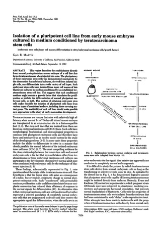
  • 1. Proc. Natl Acad. Sci. USA Vol. 78, No. 12, pp. 7634-7638, December 1981 Developmental Biology Isolation of a pluripotent cell line from early mouse embryos cultured in medium conditioned by teratocarcinoma stem cells (embryonic stem cells/inner cell masses/differentiation in vsitro/embryonal carcinoma cells/growth factors) GAIL R. MARTIN Department of Anatomy, University of California, San Francisco, California 94143 Communicated byJ. Michael Bishop, September 14, 1981 ABSTRACT This report describes the establishment directly NORMAL MOUSE TERATOMA from normal preimplantation mouse embryos of a cell line that forms teratocarcinomas when injected into mice. The pluripotency development differentiation of these embryonic stem cells was demonstrated conclusively by in utero in situ the observation that subclonal cultures, derived from isolated sin- gle cells, can differentiate into a wide variety of cell types. Such embryonic stem cells were isolated from inner cell masses of late blastocysts cultured in medium conditioned by an established ter- transfer to extra-uterine site atocarcinoma stem cell line. This suggests that such conditioned TERATOCARCINOMA medium might contain a growth factor that stimulates the prolif- BLASTOCYST injection into blastocyst eration or inhibits the differentiation of normal pluripotent em- l1 bryonic cells, or both. This method of obtaining embryonic stem cells makes feasible the isolation of pluripotent cells lines from various types of noninbred embryo, including those carrying mu- subcutaneous culture tant genes. The availability of such cell lines should make possible injection in vitro new approaches to the study of early mammalian development. Teratocarcinomas are tumors that arise with relatively high ef- ficiency when normal 1- to 7.5-day-old inbred mouse embryos are transplanted to an extra-uterine site in a histocompatible TERATOCARCINOMA host (1, 2). The stem cell lines that are isolated from them are STEM CELL LINE known as embryonal carcinoma cell (ECC) lines. Such cells have morphological, biochemical, and immunological properties in common with pluripotent embryonic cells and therefore have differentiation been used extensively as an in vitro model system for the study in vitro of the developing embryo (3-5). In some cases these properties include the ability to differentiate in vitro in a manner that closely parallels the normal behavior of the isolated embryonic DIFFERENTIATED CELL CULTURES inner cell mass (ICM) (6, 7). The most compelling evidence for the close relationship between the tumor stem cells and normal FIG. 1. Relationship between normal embryos and teratocarci- embryonic cells is the fact that stem cells taken either from ter- noma stem cells. Adapted from Martin (10). atocarcinomas or from embryonal carcinoma cell cultures can participate in the development of completely normal adult mice extra-embryonic site the signals they receive are apparently not when combined with embryonic cells by the technique of blas- conducive to completely normal embryogenesis. tocyst injection (8, 9). It is difficult to study the process by which teratocarcinoma These findings, schematically represented in Fig. 1, raise stem cells derive from normal embryonic cells as long as the questions about the origin of the teratocarcinoma stem cell. One transforming or selective events occur in vivo. As indicated by hypothesis is that the tumor stem cells arise as a consequence the dotted line in Fig. 1, it has long seemed logical to assume of a stable, but reversible, epigenetic change in normal pluri- that pluripotent stem cells capable of forming teratocarcinomas potent embryonic cells. Such "transformed" cells presumably might be isolated directly from embryos. Recently, Evans and continue proliferation in the undifferentiated state because neo- Kaufman (12) carried out experiments in which pregnant strain plastic conversion has reduced their efficiency of response to 129 female mice were subjected to a treatment, involving ova- the normal signals for differentiation (11). An alternative idea riectomy and appropriate hormonal stimulation, that prevents is that embryonal carcinoma cells are not transformed but rather normal implantation of blastocysts. Cells with the properties of represent a selected population of completely normal embry- teratocarcinoma stem cells were isolated from the embryos that onic cells that are programmed to divide until they receive the had been maintained in such "delay" in the reproductive tract. appropriate signals for differentiation; when the cells are in an Other attempts have been made to isolate cells with the prop- erties of teratocarcinoma stem cells directly from normal early The publication costs of this article were defrayed in part by page charge payment. This article must therefore be hereby marked "advertise- Abbreviations: ICM, inner cell mass; DME medium, Dulbecco's mod- ment" in accordance with 18 U. S. C. §1734 solely to indicate this fact. ified Eagle's medium; ESC, embryonic stem cell(s). 7634
  • 2. Developmental Biology: Martin Proc. Natl. Acad. Sci. USA 78 (1981) 7635 mouse embryos that have not been subjected to alteration in mycin C-treated STO fibroblasts (6). When colonies with a typ- vivo. The approaches employed included embryo culture in a ical embryonal carcinoma cell morphology were observed in the variety of media, and the use, as a starting material, of normal dissecting microscope, the culture was washed with phosphate- embryos at different stages of development and of giant em- buffered saline and treated with trypsin/EDTA/saline. By us- bryonic cell masses created by embryo aggregation. None of ing a micropipette, colonies were carefully pulled away from these approaches has led to the establishment of pluripotent cell the feeder cells and transferred, with some disaggregation, to cultures, although several differentiated cell lines have been fresh feeder layers. All colonies that proliferated were subse- isolated from early embryos (13). quently passaged in a similar manner at approximately weekly This report describes a method, involving culture in condi- intervals. As the cultures of embryo-derived cells became dens- tioned medium, for isolating and establishing pluripotent cell er, they were passaged to larger dishes of confluent feeder cells. lines with the properties of teratocarcinoma stem cells directly All subsequent handling of the cells, with respect to passage in from normal early mouse embryos in vitro. This method should the undifferentiated state, cloning, and studies of differentia- be useful not only for further elucidating the relationship be- tion in vitro and of tumor formation, was as previously described tween teratocarcinoma stem cells and their normal embryonic for PSA cells (6, 7, 15, 16). progenitors but also for generating new, genetically marked pluripotent cell lines that -can be used for studying various as- RESULTS pects of early mammalian development. Establishment of Embryonic Stem Cell Lines from Isolated Mouse ICMs. The work reported here began with the premise MATERIALS AND METHODS that teratocarcinoma stem -cells are derived from a small pop- Embryonal Carcinoma Cell Culture and Preparation of ulation of pluripotent stem cells in the peri-implantation em- Conditioned Medium. PSA-1 embryonal carcinoma cells were bryo that proliferate normally as a consequence of the produc- used to generate conditioned medium. These cells were main- tion of an endogenous factor that promotes growth, suppresses tained in the undifferentiated state by coculture with fibro- differentiation, or both. It was assumed that increasing the con- blastic STO feeder cells (6) in Dulbecco's modified Eagle's me- centration of such a factor in the growth medium by providing dium (DME medium) supplemented with 10% calf serum an exogenous supply might result in an expansion of this stem (GIBCO). To prepare conditioned medium the undifferentiated cell population in normal embryos in vitro. A corollary of this PSA-1-feeder cultures were disaggregated by treatment with idea is that once this population of stem cells is expanded it 0.05% trypsin and 0.02% EDTA in saline (trypsin/EDTA/sa- might produce a sufficiently high concentration of the factor to line). The resulting single cell suspension was first seeded at eliminate the need for an exogenous supply. Established tera- approximately 4 x 10⁷ cells per 10-cm tissue culture dish and tocarcinoma stem cell cultures seemed a logical source of such incubated for 30 min at 370C. The nonadherent PSA-1 cells an exogenous supply of growth factor in view of the hypothesis were then collected and seeded at approximately 107 cells per of Todaro and De Larco (17) that the autostimulatory growth 10-cm tissue culture dish. This "preplating" removes feeder factor(s) produced by certain tumor cells might be the product cells from the PSA-1 culture. After 2 days of growth at 370C the of a gene normally expressed by embryonic cells. serum-containing medium was removed from the PSA-1 cul- Medium conditioned by the PSA-1 embryonal carcinoma cell tures, and the cells were washed five times with phosphate- line was prepared and concentrated as described in Materials buffered saline and incubated in serum-free DME medium sup- and Methods. ICMs were isolated from normal late mouse blas- plemented with insulin at 10 pug/ml and transferrin at 5 4g/ tocysts, and approximately 30 of these were seeded on a con- ml. Forty-eight hours later this conditioned serum-free me- fluent fibroblastic feeder layer in this conditioned medium. dium was collected and the cell debris was removed by low- Within 1 week, four colonies of cells appeared. These showed speed centrifugation. Each 200 ml of conditioned medium was a remarkable resemblance to those formed when the pluripo- dialyzed against 10 liters of 20 mM NH4HCO3 for 2 days, during tent PSA-1 embryonal carcinoma cell line is cultured on a fi- which time the dialysis buffer was changed four to six times. The broblastic feeder layer (see Fig. 2). These embryo-derived col- dialyzed conditioned medium was then lyophilized, resus- onies were passaged to a fresh feeder layer in conditioned pended in 10 ml of serum-free DME medium, sterilized by medium, and within 1 week this secondary culture contained passage through a 0.2-,um-pore-diameter Millipore filter, and a very large number of embryonal carcinoma-like colonies. In supplemented with 10% calf serum and 0.1 mM 2-mercapto- contrast, control cultures of ICM-derived colonies grown and ethanol. This conditioned medium, thus concentrated approx- passaged in the absence ofconditioned medium did not contain imately 20-fold, can be stored at -700C without apparent loss continuously proliferating embryonal carcinoma-like cell colo- of activity. nies. The cells growing in conditioned medium were subse- Embryo and Embryonic Stem Cell Culture. Embryos were quently passaged at approximately weekly intervals until a mass obtained by mating superovulated random bred ICR female culture was obtained. As expected, conditioned medium was mice (Simenson Laboratories, Gilroy, CA) with SWR/J males not required for continued cell proliferation after five passages. (The Jackson Laboratory). Early blastocysts were flushed from These results are reproducible, because mass cultures of similar the uterus approximately 76 hr after detection of a copulation cells have also been obtained in two other experiments, both plug. Fully expanded late blastocysts were obtained by cultur- involving embryos of inbred genotypes [(C3H X C57BL/6)Fl ing the embryos overnight in DME medium supplemented hybrids]. In those experiments that have yielded positive re- with 10% Hyclone fetal calf serum (Sterile Systems, Logan, sults, individual ICMs apparently gave rise to embryonal car- UT). ICMs were isolated from these blastocysts by immuno- cinoma-like colonies with a frequency of approximately 1 in 8. surgery (14). As demonstrated below, the cells derived from ICMs' cul- All experiments involving ICM-derived cells were carried tured in conditioned medium have all the essential features of out in DME medium supplemented with 10% calf serum and teratocarcinoma stem cells. Such cells were termed embryonic 0.1 mM 2-mercaptoethanol. When conditioned medium was stem cells (ESC) to denote their origin directly from embryos used, the concentrated material described above was diluted and to distinguish them from embryonal carcinoma cells (ECC) 1:4 in this culture medium. Isolated ICMs were seeded in a 35- derived from teratocarcinomas. The specific cell line described mm tissue culture dish containing a confluent layer of mito- here was designated ESC-ICR.
  • 3. 7636 Developmental Biology: Martin Proc. Natl. Acad. Sci. USA 78 (1981) The mass cultures of ESC-ICR cells have a growth rate sim- SWR/J PSA-1 ESC-ICR ilar to that of PSA-1 cells and are morphologically identical to undifferentiated PSA-1 cells, both in the presence and absence of fibroblastic feeder cells (see Fig. 2). In addition, like many embryonal carcinoma cell lines, ESC-ICR cells are diploid. GPI-la Preliminary examination of G-banded chromosome spreads in- dicates that at least some ofthe cells in the culture have a normal G P1- lb female karyotype, with the exception of a possible deletion of the distal portion of one of the two X chromosomes. In view of the fact that ESC-ICR cells are morphologically indistinguishable from the PSA-1 teratocarcinoma stem cells FIG. 3. Separation of electrophoretic variants of glucosephosphate used to condition the medium in which they were grown, it was isomerase (GPI). Extracts of livers from SWR/J mice (homozygous for obviously necessary to demonstrate that the former did not arise Gpi-lb) and of cultures of PSA-1 cells (homozygous for Gpi-la) are by accidental contamination of the embryo cultures with PSA- compared with extracts of cultures of ESC-ICR cells. Expression of the 1 cells. The embryos from which the ESC-ICR cells were iso- faster electrophoretic form by ESC-ICR cells demonstrates that they lated were obtained by mating random-bred ICR female mice are not derived from PSA-1 cells. with males ofthe inbred SWR/J strain. Random-bred ICR mice 5 X 10⁶ ESC-ICR cells. In most cases a typical teratocarcinoma, segregate the glucosephosphate isomerase alleles Gpi-la and containing derivatives of all three primary germ layers, formed Gpi-lb, whereas SWR/J mice are homozygous for Gpi-lb. (ICR within 6 weeks. These results indicated that the ESC-ICR cells X SWR/J)Fl hybrids must therefore be either Gpi-lb homo- zygotes or Gpi-la/b heterozygotes. In contrast, the PSA-1 cell are similar to embryonal carcinoma cells in their ability to form line (and all other embryonal carcinoma cell lines currently tumors and suggested that the embryo-derived cells are plu- being cultured in this laboratory) are homozygous for Gpi-la. ripotent. To prove this, single ESC-ICR cells were isolated in Therefore, if the ESC-ICR cells produce the electrophoretically microdrops and subelonal cultures were established from them faster form ofthe enzyme glucosephosphate isomerase encoded (19). Although the cloning efficiency of the ESC-ICR cells was by the Gpi-lb gene, they could not have arisen by contami- rather low (approximately 1% as compared with at least 10% for nation of the embryo cultures with established embryonal car- PSA-1 cells), four single cell clones were obtained. Each clone cinoma cells. The results of electrophoresis and staining for glu- was tested for its ability to differentiate by injection into athymic cosephosphate isomerase activity (18), shown in Fig. 3, indicate mice, and each was found to form typical teratocarcinomas (Fig. that the ESC-ICR cells produce the faster form of the enzyme, 4) containing several differentiated cell types, including en- thus demonstrating their origin from (ICR X SWR/J) embryos. doderm and connective and epithelial tissues, as well as undif- Tumorigenicity and Pluripotency of the ESC. To determine ferentiated stem cells. These results conclusively demonstrate whether embryonic stem cells are capable of tumor formation, that individual cells in the ESC culture are pluripotent. athymic mice were injected subcutaneously with approximately Embryonic stem cells and their clonal derivatives were also found to be capable of differentiation in vitro when cultured under conditions appropriate for the differentiation of PSA em- PSA-1 ESC-ICR bryonal carcinoma cells (6, 7, 16). When the ESC-ICR cells, which are maintained in the undifferentiated state by coculture with fibroblastic feeder cells, are plated in the absence offeeder cells they form homogeneous rounded aggregates that, when placed in suspension, differentiate an outer layer of endoderm- like cells. These two-layered structures are known as embryoid bodies because of their similarity to the fetus-forming ICM of the normal mouse embryo (6). Further differentiation of the ESC-ICR cells could be obtained by allowing the embryoid FIG. 2. Morphological similarity of embryo-derived ESC-ICR cells to PSA-1 embryonal carcinoma cells. (Upper) Cells growing on a fi- FIG. 4. Section of a tumor formed by a clonal derivative of the broblastic feeder layer. (Lower) Mass cultures of the cells seeded in the ESC-ICR cell line. The tumor contains a variety of differentiated cell absence of feeder cells. (Phase-contrast microscopy; approximately types, including cartilage (Ca) and epithelial tissue (Ep). (Stained with x 250.) hematoxylin and eosin; approximately x 100.)
  • 4. Developmental Biology: Martin Proc. Natd Acad. Sci. USA 78 (1981) 7637 FIG. 5. Differentiation of ESC-ICR cells in vitro. A variety of cell types are apparent during the 6 weeks after the reattachment to tissue culture dishes of embryoid bodies formed by ESC-ICR cells. (Upper) Phase-contrast microscopy of live cells. (Approximately x 160). (A) Giant cells, (B) neuron-like cells, (C) endodermal cells. (Lower) (D) Section of plastic-embedded culture showing cartilage. (Approximately x 100.) (E) Live cells forming tubules. (Approximately x 35.) (F) Section of area shown in E after embedding in plastic. Tubules are filled with a granular, acellular deposit. (Approximately x 100.) bodies to reattach to a collagen-coated tissue culture substra- at present about this factor, it seems unlikely that it is commonly tum. Within a few days of replating, several morphologically produced by other cell types; fibroblastic feeder layers are gen- distinct cell types are observed migrating away from the at- erally considered potent conditioning agents, but in these ex- tached embryoid body cores, and within 6 weeks such cultures periments ICMs cultured on STO feeder layers did not give rise contain a wide variety of differentiated cell types. Fig. 5 illus- to ESC cells unless teratocarcinoma-conditioned medium was trates some of these, including giant cells, neuron-like cells, present in the early phases of the culture procedure. Obviously endodermal cells, cartilage, and a series of highly structured many questions remain to be answered about the teratocarci- tubules. noma-derived factor. Its purification will be difficult because the biological assay for its activity involves the growth of cells DISCUSSION from isolated mouse embryonic ICMs. It is nevertheless inter- The observations described above demonstrate that ICMs iso- esting to speculate on its possible relationship to other known lated from normal mouse blastocysts and cultured in medium tumor-derived growth factors (17, 21). Ultimately the infor- conditioned by an established embryonal carcinoma cell line can mation that is obtained about this factor should help to elucidate give rise to cultures of cells with the characteristics of mouse the mechanism by which growth and differentiation are regu- teratocarcinoma stem cells. These properties include cell mor- lated during embryonic development. phology, pluripotency, and the ability to form typical terato- The culture method described here, as well as the one re- carcinomas when injected into mice. In addition, it has been cently reported by Evans and Kaufman (12), also has immediate found that the embryonic stem cells described here express the practical value for the isolation of new pluripotent stem cell SSEA-1 cell surface antigen (20) common to teratocarcinoma lines. It provides a means of circumventing the need for "con- stem cells and early embryos but not expressed by -most dif- verting" an embryo to a tumor in vivo. This makes feasible the ferentiated cell types (data not shown). isolation of pluripotent cells from embryos that cannot directly By using this culture method, it should now be possible to form teratocarcinomas when they are transplanted to an extra- examine in vitro the way in which normal early embryonic cells uterine site. These include all noninbred embryos because, for give rise to teratocarcinoma stem cells. The results ofsuch stud- reasons that are not yet understood, stem-cell-containing ter- ies may ultimately serve to resolve the controversy surrounding atocarcinomas can be obtained only when an embryo is trans- the identity of teratocarcinoma stem cell progenitors in the early planted to a histocompatible host; the alternative approach of embryo and the question ofwhether the tumor stem cells have obtaining stem-cell-containing teratocarcinomas by transplant- undergone a process of neoplastic transformation. ing embryos to immunodeficient athymic mice has not been These experiments were undertaken on the premise that generally successful (2, 22). Many interesting mutations that medium conditioned by teratocarcinoma stem cells might con- affect early embryonic development are not maintained in tain a factor, perhaps identical to a normal endogenous embry- inbred stocks of mice, and it has therefore not been feasible to onic growth factor, capable of stimulating the proliferation of obtain teratocarcinomas from embryos carrying these mutant a small population of pluripotent cells in the normal embryo. genes. In this context it is noteworthy that the pluripotent ESC The success of the approach reported here suggests that this line described here was isolated from embryos of a noninbred working hypothesis has some validity. Although little is known genotype and thus is derived from an embryo that would not
  • 5. 7638 Developmental Biology: Martin Proc. Natl. Acad. Sci. USA 78 (1981) otherwise give rise to a teratocarcinoma. Given these results, 7. Martin, G. R., Wiley, L. M. & Damjanov, I. (1977) Dev. Biol 61, it seems likely that there will soon be available pluripotent, 230-244. embryo-derived cell lines with specific genetic alterations that 8. Mintz, B. & Ilimensee, K. (1975) Proc. Natl Acad. Sci. USA 72, 3585-3589. should make possible a variety of new approaches to the study 9. Papaioannou, V. E. (1979) in Cell Lineage, Stem Cells and Cell of early mammalian development. Determination, ed. Le Douarin, N. (Elsevier/North-Holland, New York), pp. 141-155. Note Added in Proof. The method of ESC isolation described here has 10. Martin, G. R. (1977) in Cell Interactions in Differentiation, eds. now been used to establish cell cultures from embryos homozygous for Karkinen-Jaaskelainen, M., Saxen, L. & Weiss, L. (Academic, the t"5 mutation of the T/t-complex. New York), pp. 59-75. 11. Mintz, B. (1978) Harvey Lect. 71, 193-245. 12. Evans, M. J. & Kaufman, M. H. (1981) Nature (London) 292, The author is grateful to Max Roth-and Marianne Gallup for technical 154-156. assistance, to Joanne Fujii and Leslie Lock for technical advice, to David 13. Sherman, M.I. (1975) Cell 5, 343-349. Akers for his help in the production ofthe figures, and to Dr. G. Steven 14. Solter, D. & Knowles, B. B. (1975) Proc. Nati Acad. Sci. USA 72, Martin for helpful suggestions during the preparation of this manu- 5099-5102. script. The work described here was supported by Grant 1 RO1 15. Martin, G. R. & Evans, M. J. (1975) in Teratomas and Differ- CA25966 from the National Institutes of Health. The author is a recip- entiation, eds. Sherman, M. I. & Solter, D. (Academic, New ient of a Faculty Research Award from. the American Cancer Society. York), pp. 169-187. 16. Martin, G. R. & Evans, M. J. (1975) Cell 6,467-474. 17. Tbdaro, G. J. & De Larco, J. E. (1980) in Control Mechanisms in 1. Stevens, L.C. (1970) Dev. Biol 21, 364-382. Animal Cells, eds. Jimenez de Asua, L., Levi-Montalcini, R., 2. Solter, D., Damjanov, I. & Koprowski, H. (1975) in The Early Shields, R.. & lacobelli, S. (Raven, New York), pp. 223-243. Development of Mammals, eds. Balls, M; & Wild, A. E. (Cam- 18. Fujii, J. T. & Martin, G. R. (1980) Dev. Biol. 74, 239-244. bridge Univ. Press, Cambridge, England), pp. 243-264. 19. Macpherson, I. (1973) in Tissue Culture Methods and Applica- 3. Hogan, B. L. M. (1977) in Biochemistry of Cell Differentiation tions, eds. Kruse, P. F. & Patterson, M. K. (Academic, New II, ed. Paul, J. (Univ. Park, Baltimore), Vol. 15, pp. 333-376. York), pp. 241-244. 4. Solter, D. & Damjanov, I. (1979) Methods Cancer Res. 28, 20. Solter, D. & Knowles, B. B. (1978) Proc. Nati Acad. Sci. USA 75, 277-332. 5565-5569. 5. Martin, G. R. (1980) Science 209, 768-776. 21. Todaro, C. J., Fryling, C. & De Larco, J. E. (1980) Proc. Nati 6. Martin, G. R. & Evans, M. J. (1975) Proc. Natl Acad. Sci. USA Acad. Sci. USA 77, 5258-5262. 72, 1441-1445. 22. Solter, D. & Damjanov, I. (1979) Nature (London) 278, 554-555.