SlideShare uma empresa Scribd logo
1 de 38
DORSAL HORN
IML
VENTRAL
HORN
The spinal ganglion -
location of neurons that
convey somatosensory
information to dorsal
horn. It’s peripheral
process associated
with a receptor and a
central process that
enters the CNS.
Ventral horn -
origin of
somatomotor fibers
innervating
skeletal muscles
Intermediate gray
(IML; T1-L2 and S2-4)
- location of
preganglionic
visceromotor neurons
innervating smooth and
cardiac muscles.
SPINAL
GANGLION
SPINAL
GANGLION Spinal cord - grey
matter deep to
white matter
Brainstem - grey
matter and white
matter intermingle
with one another
Brainstem - no
dorsal, intermediate
or ventral horns
Neuron groups are
organized into
nuclei
Brainstem:
1. Midbrain
2. Pons
3. Medulla
1
2
3
Most cranial nerves are associated with the brainstem (continuous with
spinal cord) except CN I and CN II.
Cranial nerve somatomotor nuclei are the location of neuronal cell bodies for
somatomotor fibers that innervate skeletal muscles of the face and neck.
The somatomotor cranial nerve nuclei are equivalent to the ventral horn of
the spinal cord.
Medulla
Cranial nerve sensory ganglia (equivalent to the spinal ganglia) are located
outside the brain (it may be a distinct ganglion or it may be many neurons
dispersed throughout a region). They have a peripheral process that is
associated with a receptor.
Its central process terminates in a cranial nerve sensory nucleus (equivalent
to the dorsal horn).
CN III, VII, IX and X have parasympathetic nucleus (equivalent to intermediate
gray matter of spinal cord levels S2-4). Preganglionic parasympathetic fibers
travel initially with the cranial nerve in which it is associated.
Preganglionic fibers synapse in parasympathetic ganglion located in head
(except vagus nerve’s parasympathetic component). Postganglionic
parasympathetic fibers from ganglion innervate smooth or cardiac muscles.
CN I, CN II and CN VIII are
purely sensory
CN IV, CN VI, CN XI, CN XII
are purely motor (not really
true)
CN III, CN V, CN VI, CN VII,
CN IX, CN X are mixed
(somatomotor,
somatosensory,
parasympathetic)
Sensory ganglion - dispersed
neurons located in olfactory
mucosa in roof of nasal cavity
Peripheral processes: located
at surface of olfactory mucosa
Central processes: pass
through cribriform plate to
terminate in olfactory bulb
(dorsal horn equivalent)
Olfactory bulb’s axons form
olfactory tract that terminates in
cortex.
Olfactory Nerve (Cranial Nerve I)
Light pass through pupil
anterior and posterior chambers
to retina. It passes through all
layers of retina until it strikes the
most posterior layer, which
contains the rods and cones.
They transduce light into a
neural signal.
bipolar cells - receive input
from rods and cones; allow for
convergence of information
from multiple receptor cells.
ganglion cells - receive input
from bipolar cells
-cell bodies of origin (sensory
ganglion) for optic nerve
-central processes of the sensory
ganglia.
-terminate in lateral geniculate
nucleus (dorsal horn equivalent)
of thalamus.
Optic Nerve (Cranial Nerve II)
Optic nerve - central processes of ganglion cell
that pass through the optic canal in posterior
wall of orbit to enter middle cranial fossa.
Optic chiasm - crossing of nasal (medial) half
of the optic nerve to the contralateral side;
-temporal (lateral) half remains uncrossed.
Optic tracts - caudal continuation of optic fibers
to the thalamus
Optic Nerve (Cranial Nerve II)
Oculomotor nucleus (ventral
horn equivalent) - cell
bodies of origin for nerves
innervating five extraocular
muscles.
Oculomotor nerve splits into:
- superior division innervates:
Superior Rectus
Levator Palpebra Superioris
-inferior division innervates:
Medial Rectus
Inferior Rectus
Inferior Oblique
Accessory oculomotor nucleus
(Edinger-Westphal nucleus)-
first cell body of two neuron
chain responsible for
parasympathetic innervation of
two smooth muscles
Oculomotor Nerve (Cranial Nerve III)
SUPERIOR ORBITAL FISSURE
Oculomotor fibers (both the somatomotor and preganglionic parasympathetic
fibers) leave the cranial cavity through the superior orbital fissure to enter the
orbit.
Trochlear Nerve
(Cranial Nerve IV)
Trochlear nucleus
(ventral horn
equivalent) is the
location of the cell
bodies of origin
- only cranial nerve
to leave from
brainstem’s dorsal
(posterior) side.
- innervates one
extraocular skeletal
muscle, superior
oblique
- Leaves cranial
cavity through
superior orbital
fissure
Trigeminal Nerve (Cranial
Nerve V): principal sensory
nerve of the head.
Trigeminal ganglion – cell
bodies of origin for sensory
fibers (spinal ganglion
equivalent located on petrous
ridge of temporal bone)
- Peripheral processes:
convey somatosensory
information from the face
- Central processes:
terminate in spinal
trigeminal nucleus of
brainstem.
Sensory component is
divided into:
Ophthalmic (V1)
Maxillary (V2)
Mandibular (V3)
Trigeminal motor nucleus-
(ventral horn equivalent)
neuronal cell bodies of origin
for fibers innervating eight
skeletal muscles.
Ophthalmic Division-area of forehead to the lateral corners of the eyes and onto the bridge of the nose
Maxillary Division-area between lateral corners of eyes to corners of the mouth
Mandibular Division-area of the mandible (only division to contain motor fibers)
SOMATOSENSORY INNERVATION PATTERN FOR THE FACE
Ophthalmic Division-area of
forehead to the lateral corners
of the eyes and onto the bridge
of the nose.
- Leaves cranial cavity through
superior orbital fissure to
enter orbit.
- peripheral processes divides
into:
frontal nerve
supraorbital nerve
supratrochlear nerve
lacrimal nerve
nasociliary nerve
Maxillary Division-area between lateral
corners of eyes to corners of mouth
- Leaves cranial cavity by passing
through foramen rotundum to enter
pterygopalatine fossa.
Its peripheral processes divides into:
1.infraorbital n.
2.greater and lesser palatine nn.
(descending palatine nerves)
3. zygomatic n. (cheek)
4.posterior superior alveolar n.
5.sphenopalatine n.(nasal cavity)
Mandibular Division-innervates the area of mandible; only division to contain motor fibers.
- Leaves cranial cavity by passing through foramen ovale to enter infratemporal fossa.
Its peripheral sensory
processes branch into :
1. inferior alveolar n.
2. lingual n.
3. buccal n.
4. auriculotemporal n.
5. motor nerves to
skeletal muscles.
Trigeminal motor nucleus: location of
somatomotor neuronal cell bodies of
origin for fibers innervating eight
muscles: muscles of mastication
(masseter, temporalis, medial pterygoid,
lateral pterygoid), mylohyoid, anterior
belly of digastric, tensor tympani and
tensor veli palatini.
Abducens Nerve
(Cranial Nerve VI)
Abducens nucleus
(ventral horn
equivalent)- cell bodies
of origin
- responsible for
innervating one
extraocular muscle,
lateral rectus.
- Exits cranial cavity by
passing through the
superior orbital fissure
to enter the orbit.
FACIAL NERVE (CN VII)
- contains somatomotor, somatosensory & preganglionic parasympathetic fibers.
- Location of cell bodies for components of facial nerve:
1.Facial nucleus-somatomotor nucleus (ventral horn equivalent) innervating: muscles of facial
expression & stylohyoid, posterior belly of the digastric and stapedius.
2. Superior salivary nucleus- visceral motor nucleus (intermediate gray equivalent) containing
first parasympathetic cell bodies of two neuron chain innervating lacrimal gland, submandibular
gland, sublingual gland, nasal mucous glands and oral mucous glands.
3. Geniculate ganglion,
somatosensory ganglion (spinal
ganglion equivalent) located in
bend of facial canal in temporal
bone posterior to middle ear.
Peripheral processes innervate
portions of the external ear and
taste receptors on the anterior 2/3
of the tongue; central processes
terminate in nucleus solitarius
(taste) and spinal trigeminal
nucleus (external ear).
FACIAL NERVE (CN VII)
Internal auditory meatus: exit/entrance of facial nerve from cranial cavity. Internal opening
of facial canal (posterior to the middle ear) in temporal bone.
Facial canal has an initial horizontal course and then a vertical course through the temporal
bone. In the horizontal part that the geniculate ganglion is located.
Stylomastoid foramen -
inferior opening of facial
canal; exit point of
somatomotor and
somatosensory fibers
leaving the skull.
★ Two additional
openings in the facial
canal before the
stylomastoid foramen
that serve as the exit
points for other parts of
the facial nerve.
INTERNAL AUDITORY MEATUS
The internal auditory meatus, which is on the posterior
edge of the petrous ridge of the temporal bone, is the
opening for the entrance/exit of the facial nerve.
Once it passes through the internal auditory meatus, the
facial nerve takes a complicated course in the facial canal,
which is inside the temporal bone.
Internal View
External View
In facial canal different components split off:
1)greater petrosal nerve (preganglionic parasympathetic fibers from the superior salivary
nucleus for lacrimal, nasal and oral mucosa glands) leaves the facial nerve at the geniculate
ganglion to move rostrally.
2)chorda tympani (preganglionic parasympathetic fibers for submandibular and sublingual
glands [superior salivary nucleus] and taste fibers [geniculate ganglion] for anterior 2/3
of tongue) leave the facial
nerve distal to geniculate
ganglion and enter the
middle ear cavity to move
rostrally.
3)somatomotor
fibers (facial nucleus)
to skeletal muscles &
somatosensory fibers
to external ear exit
the temporal bone at
the stylomastoid
foramen.
The greater petrosal nerve leaves the facial nerve at the geniculate ganglion to move
rostrally.
The chorda tympani leaves the facial nerve at the midpoint of the facial canal and takes a
rostral course through the middle ear cavity.
The remainder of the facial nerve continues through the facial canal until it exits on the
anterior side of the skull at the stylomastoid foramen.
Somatic motor component of facial nerve branches in parotid gland into:
1. temporal
2. zygomatic
3. buccal
4. mandibular
5. cervical
Vestibulocochlear Nerve (Cranial
Nerve VIII)
The two ganglia,
vestibular ganglion (associated
with the semicircular canals) &
dispersed neurons that comprise the
spiral ganglion (associated with the
cochlea) in the inner ear These are
the neuronal cell bodies of origin
for CN VIII.
Vestibular ganglion cells: convey
balance and orientation
Spiral ganglion cells: convey
auditory sense
Peripheral processes of the
vestibular ganglia are associated
with receptors in the semicircular
canals and the central processes end
in vestibular nuclei of the brainstem.
Peripheral processes of the spiral
ganglia are associated with
receptors in the cochlea and the
central processes end in cochlear
nuclei of the brainstem.
Central processes pass
through the internal
auditory meatus to
enter the cranial cavity.
GLOSSOPHARYNGEAL NERVE (CN IX)
- Primarily a sensory nerve, although it has a
small somatomotor & visceromotor
components.
-Sensory information from posterior third of
tongue, mucosa of pharynx, middle
ear, auditory tube, and blood pressure &
chemoreceptors associated with common
carotid artery.
- Somatomotor innervation: stylopharyngeus
of pharynx
-Visceromotor (parasympathetic) innervation:
parotid gland.
-Leaves cranial cavity thru jugular foramen
Location of cell bodies:
1) Nucleus Ambiguus: cell bodies of origin for somatomotor fibers that innervate stylopharyngeus
2) Inferior Salivary Nucleus: cell bodies of origin for visceromotor fibers that are involved in
innervation of the parotid gland
3) Inferior Ganglion of the
Glossopharyngeal nerve
(at the jugular foramen).
Peripheral processes carry:
1) special sense (taste) & somatosensory
information from the posterior 1/3 of tongue
2) Somatosensory information from the
pharynx
3) Somatosensory information from the
middle ear, auditory tube & mastoid air cells
4) General viscerosensory information from
the carotid body (chemoreception) & carotid
sinus (baroreception).
Central processes from the inferior
ganglion of CN IX terminate in:
Nucleus solitarius – taste fibers from
posterior third of tongue, carotid body
and carotid sinus.
Spinal trigeminal nucleus – general
sensory from posterior third of tongue,
pharynx, middle ear, auditory tube &
mastoid air cells
Spina
l
VAGUS NERVE (CN X)
- contains somatomotor, visceromotor
and somatosensory components
- somatomotor innervates skeletal
muscles of larynx, pharynx and
accessory muscles of pharynx and soft
palate
- visceromotor component innervates
smooth and cardiac muscles of thoracic
cavity and smooth muscle of the
abdomen up to splenic flexure
- sensory component innervates laryngeal
mucosa, meninges of posterior cranial
fossa, external ear and comparable
regions innervated by visceromotor
innervation
- Leaves cranial cavity through jugular
foramen
Nucleus Ambiguus: neuronal cell bodies that innervate skeletal muscles including: pharyngeal constrictor
muscles, intrinsic muscles of larynx (external laryngeal & recurrent laryngeal nn.), levator veli palatini
(soft palate) and palatoglossus, palatopharyngeus, salpingopharyngeus
Dorsal Motor Nucleus of the Vagus: first neuronal cell bodies responsible for parasympathetic innervation
of smooth muscle of digestive system (to splenic flexure), respiratory system and heart.
Spinal trigeminal
nucleus
Inferior Ganglion of the vagus nerve (nodose ganglion): innervate mucosa of larynx, respiratory system,
heart and digestive system.
Its central processes (axons) terminate in the nucleus solitarius of brainstem.
Superior Ganglion: innervate external auditory meatus, meninges of the posterior cranial fossa and
external surface of tympanic membrane. Its central fibers terminate in spinal trigeminal nucleus of
brainstem.
Spinal trigeminal
nucleus
(Spinal) Accessory Nerve (CN XI)
Location: C1-C5 of spinal cord is
in the posterior portion of the
anterior (ventral) horn.
- fibers leave spinal cord between
the posterior (dorsal) and anterior
(ventral) roots to enter cranial cavity
through foramen magnum.
- fibers then exit cranial cavity
through the jugular foramen.
- innervates the
sternocleidomastoid and trapezius.
Hypoglossal nucleus:
neuronal cell bodies for the
hypoglossal nerve.
- Exits cranial cavity through
hypoglossal canal of
occipital bone
- In neck it loops around
occipital artery to enter lateral
side of the tongue
- innervates:
Intrinsic and extrinsic
muscles (hyoglossus,
styloglossus & genioglossus)
of tongue
HYPOGLOSSAL NERVE (CN XII)
LEARNING OBJECTIVES FOR CRANIAL NERVES
By the end of this unit you must have mastered for each cranial nerve (where appropriate):
1) all nuclei or ganglia of origin for its fibers
2) specific region of the brain or spinal cord its fibers are associated
3) complete pathway, any bony openings it passes through, and all its terminal targets
4) identify as motor only, sensory only, both motor and sensory and autonomic component if any
5) for sensory fibers: brain nuclei for termination of central fibers
6) for autonomic component, the nucleus of origin, bony openings, path of its preganglionic fibers, parasympathetic ganglion,
path of its postganglionic fibers, terminal targets.
7) for the sympathetic fibers, cell bodies of origin, path of its fibers, sympathetic ganglion, pathway of its postganglionic fibers to
the target structures.
8) Describe the functional deficits that will be manifested if any cranial nerve is lesioned at any point along its course.
Specific objective for the 12 Cranial nerves:
1) Olfactory nerves-cell bodies of origin, bony openings and its path to its termination in olfactory bulb
termination of olfactory tract axons
2) Optic nerve-cell bodies of origin, path of its fibers, bony opening and thalamic nucleus for termination of its central fibers
location and consequences of optic chiasm
3) Oculomotor nerve-cell bodies of origin, path of its fibers
cell bodies of origin and path to striated muscles for its superior and inferior divisions
autonomic cell bodies of origin and complete path of its autonomic fibers
location of ciliary ganglion; its contribution to short ciliary nerves and target structures for its postganglionic fibers
know the location that its parasympathetic fibers join and leave specific parts of the nerve
4) Trochlear nerve-cell bodies of origin, its path, bony opening, muscle it innervates
5) Trigeminal nerve-cell bodies of origin, paths and bony openings of its subdivisions
ophthalmic division area of innervation
major branches
branches of the frontal nerve
associated autonomic ganglia
components and cell bodies of origin for long ciliary nerves
components and cell bodies of origin for short ciliary nerves
maxillary division area of innervation
branches in the pterygopalatine fossa, oral cavity, maxillary sinus and nasal cavity
associated autonomic ganglion
mandibular division area of innervation
sensory branches and muscles innervated
associated autonomic ganglia
Know the location that autonomic fibers “hitch a ride” on specific parts of the nerve and when they leave the nerve to innervate
a structure
6) Abducens nerve-cell bodies of origin, its bony opening and path to the muscle it innervates
7) Facial nerve-cell bodies of origin, bony openings, path of its fibers to its terminal targets
sensory targets and somatic muscles innervated by the facial nerve
autonomic cell bodies of origin and complete path of fibers to target structures
components and cell bodies of origin of the chorda tympani
components and cell bodies of origin for the nerve of the pterygoid canal
8) Vestibulocochlear nerve-cell bodies of origin, its path, bony openings and structures innervated
9) Glossopharyngeal nerve-cell bodies of origin, its path, bony openings and structures innervated.
cell bodies of origin for innervation of striated muscles
autonomic cell bodies of origin, path of its fibers and target structures
cell bodies of origin and sensory regions innervated
components and cell bodies of origin for tympanic nerve
components and cell bodies of origin for lesser petrosal nerve
contribution to pharyngeal plexus
10) Vagus nerve-cell bodies of origin, bony opening, path of its fibers and structures innervated.
cell bodies of origin and striated muscles innervated
cell bodies of origin and smooth muscle structures innervated
cell bodies of origin and sensory regions innervated
contribution to pharyngeal plexus
11) Accessory nerve-cell bodies of origin, its path, bony openings and terminal structures innervated
12) Hypoglossal nerve-cell bodies of origin, its path, bony openings and terminal structures innervated
13) Sympathetic innervation-cell bodies of origin for preganglionic and postganglionic fibers, most common pathways to
terminal target sites.
location and types of fibers in carotid plexus
path and terminal targets of sympathetic components of long and short ciliary nerves
components, path and terminal targets of deep petrosal nerve
sympathetic path for innervation of submandibular, sublingual and parotid glands

Mais conteúdo relacionado

Mais procurados

Hypoglossal nerve
Hypoglossal nerveHypoglossal nerve
Hypoglossal nerveAmyEmtage
 
CEREBELLUM- with clinical and radiology
CEREBELLUM- with clinical and radiologyCEREBELLUM- with clinical and radiology
CEREBELLUM- with clinical and radiologylalit yadav
 
Surgical anatomy of cranial nerves pallav
Surgical anatomy of cranial nerves pallavSurgical anatomy of cranial nerves pallav
Surgical anatomy of cranial nerves pallavpallavkedia
 
Cranial nerves
Cranial nervesCranial nerves
Cranial nervesAhlamt
 
Cranial nerve copy .pptx seminar
Cranial nerve copy .pptx seminar Cranial nerve copy .pptx seminar
Cranial nerve copy .pptx seminar Dr. Mitali Thamke
 
Accessory nerve
Accessory nerveAccessory nerve
Accessory nerveAmyEmtage
 
facial nerve (7th cranial nerve)
facial nerve (7th cranial nerve)facial nerve (7th cranial nerve)
facial nerve (7th cranial nerve)D.A.B.M
 
150 cranial nerves Anatomy
150 cranial nerves  Anatomy150 cranial nerves  Anatomy
150 cranial nerves Anatomyddert
 
Midbrain-Neuroradiology
Midbrain-NeuroradiologyMidbrain-Neuroradiology
Midbrain-NeuroradiologyParvathy Nair
 
Cranial nerves xi and xii
Cranial nerves xi and xiiCranial nerves xi and xii
Cranial nerves xi and xiiNeurologyKota
 
Anatomy of brainstem and its clinical significance
Anatomy of brainstem and its clinical significanceAnatomy of brainstem and its clinical significance
Anatomy of brainstem and its clinical significanceSnehasis Ghosh
 
Thalamus-Anatomy,Physiology,Applied aspects
Thalamus-Anatomy,Physiology,Applied aspectsThalamus-Anatomy,Physiology,Applied aspects
Thalamus-Anatomy,Physiology,Applied aspectsRanadhi Das
 
Glossopharyngeal nerve & its pathology ppt
Glossopharyngeal nerve & its pathology pptGlossopharyngeal nerve & its pathology ppt
Glossopharyngeal nerve & its pathology pptD Venkatesh Kumar
 

Mais procurados (20)

Cranial nerves
Cranial nerves Cranial nerves
Cranial nerves
 
Hypoglossal nerve
Hypoglossal nerveHypoglossal nerve
Hypoglossal nerve
 
CEREBELLUM- with clinical and radiology
CEREBELLUM- with clinical and radiologyCEREBELLUM- with clinical and radiology
CEREBELLUM- with clinical and radiology
 
olfactory nerve
 olfactory nerve olfactory nerve
olfactory nerve
 
Surgical anatomy of cranial nerves pallav
Surgical anatomy of cranial nerves pallavSurgical anatomy of cranial nerves pallav
Surgical anatomy of cranial nerves pallav
 
Facial nerve
Facial nerveFacial nerve
Facial nerve
 
Cranial nerves
Cranial nervesCranial nerves
Cranial nerves
 
Cranial nerve copy .pptx seminar
Cranial nerve copy .pptx seminar Cranial nerve copy .pptx seminar
Cranial nerve copy .pptx seminar
 
Anatomy of Facial Nerve
Anatomy of Facial NerveAnatomy of Facial Nerve
Anatomy of Facial Nerve
 
the cranial nerves
the cranial nerves the cranial nerves
the cranial nerves
 
Accessory nerve
Accessory nerveAccessory nerve
Accessory nerve
 
facial nerve (7th cranial nerve)
facial nerve (7th cranial nerve)facial nerve (7th cranial nerve)
facial nerve (7th cranial nerve)
 
150 cranial nerves Anatomy
150 cranial nerves  Anatomy150 cranial nerves  Anatomy
150 cranial nerves Anatomy
 
Neuroanatomy...Cranial nerve anatomy
Neuroanatomy...Cranial nerve anatomyNeuroanatomy...Cranial nerve anatomy
Neuroanatomy...Cranial nerve anatomy
 
Midbrain-Neuroradiology
Midbrain-NeuroradiologyMidbrain-Neuroradiology
Midbrain-Neuroradiology
 
Vagus nerve
Vagus nerveVagus nerve
Vagus nerve
 
Cranial nerves xi and xii
Cranial nerves xi and xiiCranial nerves xi and xii
Cranial nerves xi and xii
 
Anatomy of brainstem and its clinical significance
Anatomy of brainstem and its clinical significanceAnatomy of brainstem and its clinical significance
Anatomy of brainstem and its clinical significance
 
Thalamus-Anatomy,Physiology,Applied aspects
Thalamus-Anatomy,Physiology,Applied aspectsThalamus-Anatomy,Physiology,Applied aspects
Thalamus-Anatomy,Physiology,Applied aspects
 
Glossopharyngeal nerve & its pathology ppt
Glossopharyngeal nerve & its pathology pptGlossopharyngeal nerve & its pathology ppt
Glossopharyngeal nerve & its pathology ppt
 

Destaque (20)

Cranial nerves
Cranial nervesCranial nerves
Cranial nerves
 
Lecture localanesthesia
Lecture localanesthesiaLecture localanesthesia
Lecture localanesthesia
 
Nervio Vago/Wilson Pawels/idiomainglés
Nervio Vago/Wilson Pawels/idiomainglésNervio Vago/Wilson Pawels/idiomainglés
Nervio Vago/Wilson Pawels/idiomainglés
 
Oxytocin and heart rate variability
Oxytocin and heart rate variabilityOxytocin and heart rate variability
Oxytocin and heart rate variability
 
The Cranial nerve
The Cranial nerve The Cranial nerve
The Cranial nerve
 
Cranial Nerves
Cranial NervesCranial Nerves
Cranial Nerves
 
Nervous system final
Nervous system finalNervous system final
Nervous system final
 
Cranial nerves
Cranial nerves Cranial nerves
Cranial nerves
 
Cranial Nerves
Cranial NervesCranial Nerves
Cranial Nerves
 
Oculomotor Nerve
Oculomotor NerveOculomotor Nerve
Oculomotor Nerve
 
Suboccipital triangle
Suboccipital triangleSuboccipital triangle
Suboccipital triangle
 
Flaps in otolaryngology
Flaps in otolaryngology Flaps in otolaryngology
Flaps in otolaryngology
 
Facial nerve
Facial nerveFacial nerve
Facial nerve
 
olfactory nerve
olfactory nerveolfactory nerve
olfactory nerve
 
Olfaction and pathway
Olfaction and  pathwayOlfaction and  pathway
Olfaction and pathway
 
Cranial nerves 3,4,6-Neuroradioology
Cranial nerves 3,4,6-NeuroradioologyCranial nerves 3,4,6-Neuroradioology
Cranial nerves 3,4,6-Neuroradioology
 
Embryology 18 ear
Embryology 18   earEmbryology 18   ear
Embryology 18 ear
 
Upper limb nerve injuries
Upper limb nerve injuriesUpper limb nerve injuries
Upper limb nerve injuries
 
Cranial nerve
Cranial nerveCranial nerve
Cranial nerve
 
Activity 7 - Brain & Cranial Nerves
Activity 7 - Brain & Cranial NervesActivity 7 - Brain & Cranial Nerves
Activity 7 - Brain & Cranial Nerves
 

Semelhante a Spinal Cord and Brainstem Anatomy

Neuroanatomy seminar with special reference to trigeminal,facial and glossoph...
Neuroanatomy seminar with special reference to trigeminal,facial and glossoph...Neuroanatomy seminar with special reference to trigeminal,facial and glossoph...
Neuroanatomy seminar with special reference to trigeminal,facial and glossoph...Karishma Sirimulla
 
Cranial Nerves 5,7,8.ppt
Cranial Nerves 5,7,8.pptCranial Nerves 5,7,8.ppt
Cranial Nerves 5,7,8.pptmunnam37
 
Cranial and spinal nerves
Cranial and spinal nervesCranial and spinal nerves
Cranial and spinal nervesDomina Petric
 
Anatomy nazeen batch cranial nerves
Anatomy nazeen batch cranial nervesAnatomy nazeen batch cranial nerves
Anatomy nazeen batch cranial nervesMBBS IMS MSU
 
Nerves and the Nervous System
Nerves and the Nervous SystemNerves and the Nervous System
Nerves and the Nervous Systemkebnance
 
cranialnerve gokul.........................ppt
cranialnerve gokul.........................pptcranialnerve gokul.........................ppt
cranialnerve gokul.........................pptsacootcbe
 
Cranial Nerve V Trigemial
Cranial Nerve V Trigemial Cranial Nerve V Trigemial
Cranial Nerve V Trigemial Anam_ Khan
 
Trigeminal nerve
Trigeminal nerve Trigeminal nerve
Trigeminal nerve abhik28
 
Neuroanatomy._2..Cranial.nerves.2017.pptx
Neuroanatomy._2..Cranial.nerves.2017.pptxNeuroanatomy._2..Cranial.nerves.2017.pptx
Neuroanatomy._2..Cranial.nerves.2017.pptxmoonasadi99
 
The Cranial nerves
The Cranial nerves The Cranial nerves
The Cranial nerves mac os
 
Glossopharyngeal nerve
Glossopharyngeal nerveGlossopharyngeal nerve
Glossopharyngeal nerveRAJAT SINGHAL
 
cranial nerve for BPT students
cranial nerve for BPT studentscranial nerve for BPT students
cranial nerve for BPT studentsAnwesh Pradhan
 

Semelhante a Spinal Cord and Brainstem Anatomy (20)

Neuroanatomy seminar with special reference to trigeminal,facial and glossoph...
Neuroanatomy seminar with special reference to trigeminal,facial and glossoph...Neuroanatomy seminar with special reference to trigeminal,facial and glossoph...
Neuroanatomy seminar with special reference to trigeminal,facial and glossoph...
 
Cranial Nerves 5,7,8.ppt
Cranial Nerves 5,7,8.pptCranial Nerves 5,7,8.ppt
Cranial Nerves 5,7,8.ppt
 
Cranial and spinal nerves
Cranial and spinal nervesCranial and spinal nerves
Cranial and spinal nerves
 
Anatomy nazeen batch cranial nerves
Anatomy nazeen batch cranial nervesAnatomy nazeen batch cranial nerves
Anatomy nazeen batch cranial nerves
 
Facial nerve
Facial nerve Facial nerve
Facial nerve
 
Nerves and the Nervous System
Nerves and the Nervous SystemNerves and the Nervous System
Nerves and the Nervous System
 
cranialnerve gokul.........................ppt
cranialnerve gokul.........................pptcranialnerve gokul.........................ppt
cranialnerve gokul.........................ppt
 
Oral biology
Oral biologyOral biology
Oral biology
 
Oral biology
Oral biologyOral biology
Oral biology
 
Cranial Nerve V Trigemial
Cranial Nerve V Trigemial Cranial Nerve V Trigemial
Cranial Nerve V Trigemial
 
Trigeminal nerve
Trigeminal nerve Trigeminal nerve
Trigeminal nerve
 
Neuroanatomy._2..Cranial.nerves.2017.pptx
Neuroanatomy._2..Cranial.nerves.2017.pptxNeuroanatomy._2..Cranial.nerves.2017.pptx
Neuroanatomy._2..Cranial.nerves.2017.pptx
 
Trigeminal nerve
Trigeminal nerveTrigeminal nerve
Trigeminal nerve
 
The Cranial nerves
The Cranial nerves The Cranial nerves
The Cranial nerves
 
Glossopharyngeal nerve
Glossopharyngeal nerveGlossopharyngeal nerve
Glossopharyngeal nerve
 
Nervous system1
Nervous system1Nervous system1
Nervous system1
 
Cranial Nerve V
Cranial Nerve VCranial Nerve V
Cranial Nerve V
 
CRANIAL NERVES-OVERVIEW.pptx
CRANIAL NERVES-OVERVIEW.pptxCRANIAL NERVES-OVERVIEW.pptx
CRANIAL NERVES-OVERVIEW.pptx
 
cranial nerve for BPT students
cranial nerve for BPT studentscranial nerve for BPT students
cranial nerve for BPT students
 
1.facial nerve
1.facial nerve1.facial nerve
1.facial nerve
 

Último

Culture and Health Disorders Social change.pptx
Culture and Health Disorders Social change.pptxCulture and Health Disorders Social change.pptx
Culture and Health Disorders Social change.pptxDr. Dheeraj Kumar
 
epilepsy and status epilepticus for undergraduate.pptx
epilepsy and status epilepticus  for undergraduate.pptxepilepsy and status epilepticus  for undergraduate.pptx
epilepsy and status epilepticus for undergraduate.pptxMohamed Rizk Khodair
 
Basic principles involved in the traditional systems of medicine PDF.pdf
Basic principles involved in the traditional systems of medicine PDF.pdfBasic principles involved in the traditional systems of medicine PDF.pdf
Basic principles involved in the traditional systems of medicine PDF.pdfDivya Kanojiya
 
SYNDESMOTIC INJURY- ANATOMICAL REPAIR.pptx
SYNDESMOTIC INJURY- ANATOMICAL REPAIR.pptxSYNDESMOTIC INJURY- ANATOMICAL REPAIR.pptx
SYNDESMOTIC INJURY- ANATOMICAL REPAIR.pptxdrashraf369
 
Introduction to Sports Injuries by- Dr. Anjali Rai
Introduction to Sports Injuries by- Dr. Anjali RaiIntroduction to Sports Injuries by- Dr. Anjali Rai
Introduction to Sports Injuries by- Dr. Anjali RaiGoogle
 
Wessex Health Partners Wessex Integrated Care, Population Health, Research & ...
Wessex Health Partners Wessex Integrated Care, Population Health, Research & ...Wessex Health Partners Wessex Integrated Care, Population Health, Research & ...
Wessex Health Partners Wessex Integrated Care, Population Health, Research & ...Wessex Health Partners
 
Informed Consent Empowering Healthcare Decision-Making.pptx
Informed Consent Empowering Healthcare Decision-Making.pptxInformed Consent Empowering Healthcare Decision-Making.pptx
Informed Consent Empowering Healthcare Decision-Making.pptxSasikiranMarri
 
Presentation on General Anesthetics pdf.
Presentation on General Anesthetics pdf.Presentation on General Anesthetics pdf.
Presentation on General Anesthetics pdf.Prerana Jadhav
 
COVID-19 (NOVEL CORONA VIRUS DISEASE PANDEMIC ).pptx
COVID-19  (NOVEL CORONA  VIRUS DISEASE PANDEMIC ).pptxCOVID-19  (NOVEL CORONA  VIRUS DISEASE PANDEMIC ).pptx
COVID-19 (NOVEL CORONA VIRUS DISEASE PANDEMIC ).pptxBibekananda shah
 
Biomechanics- Shoulder Joint!!!!!!!!!!!!
Biomechanics- Shoulder Joint!!!!!!!!!!!!Biomechanics- Shoulder Joint!!!!!!!!!!!!
Biomechanics- Shoulder Joint!!!!!!!!!!!!ibtesaam huma
 
call girls in munirka DELHI 🔝 >༒9540349809 🔝 genuine Escort Service 🔝✔️✔️
call girls in munirka  DELHI 🔝 >༒9540349809 🔝 genuine Escort Service 🔝✔️✔️call girls in munirka  DELHI 🔝 >༒9540349809 🔝 genuine Escort Service 🔝✔️✔️
call girls in munirka DELHI 🔝 >༒9540349809 🔝 genuine Escort Service 🔝✔️✔️saminamagar
 
Presentation on Parasympathetic Nervous System
Presentation on Parasympathetic Nervous SystemPresentation on Parasympathetic Nervous System
Presentation on Parasympathetic Nervous SystemPrerana Jadhav
 
call girls in Dwarka Sector 21 Metro DELHI 🔝 >༒9540349809 🔝 genuine Escort Se...
call girls in Dwarka Sector 21 Metro DELHI 🔝 >༒9540349809 🔝 genuine Escort Se...call girls in Dwarka Sector 21 Metro DELHI 🔝 >༒9540349809 🔝 genuine Escort Se...
call girls in Dwarka Sector 21 Metro DELHI 🔝 >༒9540349809 🔝 genuine Escort Se...saminamagar
 
Case Report Peripartum Cardiomyopathy.pptx
Case Report Peripartum Cardiomyopathy.pptxCase Report Peripartum Cardiomyopathy.pptx
Case Report Peripartum Cardiomyopathy.pptxNiranjan Chavan
 
April 2024 ONCOLOGY CARTOON by DR KANHU CHARAN PATRO
April 2024 ONCOLOGY CARTOON by  DR KANHU CHARAN PATROApril 2024 ONCOLOGY CARTOON by  DR KANHU CHARAN PATRO
April 2024 ONCOLOGY CARTOON by DR KANHU CHARAN PATROKanhu Charan
 
Glomerular Filtration rate and its determinants.pptx
Glomerular Filtration rate and its determinants.pptxGlomerular Filtration rate and its determinants.pptx
Glomerular Filtration rate and its determinants.pptxDr.Nusrat Tariq
 
Glomerular Filtration and determinants of glomerular filtration .pptx
Glomerular Filtration and  determinants of glomerular filtration .pptxGlomerular Filtration and  determinants of glomerular filtration .pptx
Glomerular Filtration and determinants of glomerular filtration .pptxDr.Nusrat Tariq
 
call girls in Connaught Place DELHI 🔝 >༒9540349809 🔝 genuine Escort Service ...
call girls in Connaught Place  DELHI 🔝 >༒9540349809 🔝 genuine Escort Service ...call girls in Connaught Place  DELHI 🔝 >༒9540349809 🔝 genuine Escort Service ...
call girls in Connaught Place DELHI 🔝 >༒9540349809 🔝 genuine Escort Service ...saminamagar
 
Statistical modeling in pharmaceutical research and development.
Statistical modeling in pharmaceutical research and development.Statistical modeling in pharmaceutical research and development.
Statistical modeling in pharmaceutical research and development.ANJALI
 

Último (20)

Culture and Health Disorders Social change.pptx
Culture and Health Disorders Social change.pptxCulture and Health Disorders Social change.pptx
Culture and Health Disorders Social change.pptx
 
epilepsy and status epilepticus for undergraduate.pptx
epilepsy and status epilepticus  for undergraduate.pptxepilepsy and status epilepticus  for undergraduate.pptx
epilepsy and status epilepticus for undergraduate.pptx
 
Basic principles involved in the traditional systems of medicine PDF.pdf
Basic principles involved in the traditional systems of medicine PDF.pdfBasic principles involved in the traditional systems of medicine PDF.pdf
Basic principles involved in the traditional systems of medicine PDF.pdf
 
SYNDESMOTIC INJURY- ANATOMICAL REPAIR.pptx
SYNDESMOTIC INJURY- ANATOMICAL REPAIR.pptxSYNDESMOTIC INJURY- ANATOMICAL REPAIR.pptx
SYNDESMOTIC INJURY- ANATOMICAL REPAIR.pptx
 
Introduction to Sports Injuries by- Dr. Anjali Rai
Introduction to Sports Injuries by- Dr. Anjali RaiIntroduction to Sports Injuries by- Dr. Anjali Rai
Introduction to Sports Injuries by- Dr. Anjali Rai
 
Wessex Health Partners Wessex Integrated Care, Population Health, Research & ...
Wessex Health Partners Wessex Integrated Care, Population Health, Research & ...Wessex Health Partners Wessex Integrated Care, Population Health, Research & ...
Wessex Health Partners Wessex Integrated Care, Population Health, Research & ...
 
Informed Consent Empowering Healthcare Decision-Making.pptx
Informed Consent Empowering Healthcare Decision-Making.pptxInformed Consent Empowering Healthcare Decision-Making.pptx
Informed Consent Empowering Healthcare Decision-Making.pptx
 
Presentation on General Anesthetics pdf.
Presentation on General Anesthetics pdf.Presentation on General Anesthetics pdf.
Presentation on General Anesthetics pdf.
 
COVID-19 (NOVEL CORONA VIRUS DISEASE PANDEMIC ).pptx
COVID-19  (NOVEL CORONA  VIRUS DISEASE PANDEMIC ).pptxCOVID-19  (NOVEL CORONA  VIRUS DISEASE PANDEMIC ).pptx
COVID-19 (NOVEL CORONA VIRUS DISEASE PANDEMIC ).pptx
 
Biomechanics- Shoulder Joint!!!!!!!!!!!!
Biomechanics- Shoulder Joint!!!!!!!!!!!!Biomechanics- Shoulder Joint!!!!!!!!!!!!
Biomechanics- Shoulder Joint!!!!!!!!!!!!
 
call girls in munirka DELHI 🔝 >༒9540349809 🔝 genuine Escort Service 🔝✔️✔️
call girls in munirka  DELHI 🔝 >༒9540349809 🔝 genuine Escort Service 🔝✔️✔️call girls in munirka  DELHI 🔝 >༒9540349809 🔝 genuine Escort Service 🔝✔️✔️
call girls in munirka DELHI 🔝 >༒9540349809 🔝 genuine Escort Service 🔝✔️✔️
 
Presentation on Parasympathetic Nervous System
Presentation on Parasympathetic Nervous SystemPresentation on Parasympathetic Nervous System
Presentation on Parasympathetic Nervous System
 
call girls in Dwarka Sector 21 Metro DELHI 🔝 >༒9540349809 🔝 genuine Escort Se...
call girls in Dwarka Sector 21 Metro DELHI 🔝 >༒9540349809 🔝 genuine Escort Se...call girls in Dwarka Sector 21 Metro DELHI 🔝 >༒9540349809 🔝 genuine Escort Se...
call girls in Dwarka Sector 21 Metro DELHI 🔝 >༒9540349809 🔝 genuine Escort Se...
 
Epilepsy
EpilepsyEpilepsy
Epilepsy
 
Case Report Peripartum Cardiomyopathy.pptx
Case Report Peripartum Cardiomyopathy.pptxCase Report Peripartum Cardiomyopathy.pptx
Case Report Peripartum Cardiomyopathy.pptx
 
April 2024 ONCOLOGY CARTOON by DR KANHU CHARAN PATRO
April 2024 ONCOLOGY CARTOON by  DR KANHU CHARAN PATROApril 2024 ONCOLOGY CARTOON by  DR KANHU CHARAN PATRO
April 2024 ONCOLOGY CARTOON by DR KANHU CHARAN PATRO
 
Glomerular Filtration rate and its determinants.pptx
Glomerular Filtration rate and its determinants.pptxGlomerular Filtration rate and its determinants.pptx
Glomerular Filtration rate and its determinants.pptx
 
Glomerular Filtration and determinants of glomerular filtration .pptx
Glomerular Filtration and  determinants of glomerular filtration .pptxGlomerular Filtration and  determinants of glomerular filtration .pptx
Glomerular Filtration and determinants of glomerular filtration .pptx
 
call girls in Connaught Place DELHI 🔝 >༒9540349809 🔝 genuine Escort Service ...
call girls in Connaught Place  DELHI 🔝 >༒9540349809 🔝 genuine Escort Service ...call girls in Connaught Place  DELHI 🔝 >༒9540349809 🔝 genuine Escort Service ...
call girls in Connaught Place DELHI 🔝 >༒9540349809 🔝 genuine Escort Service ...
 
Statistical modeling in pharmaceutical research and development.
Statistical modeling in pharmaceutical research and development.Statistical modeling in pharmaceutical research and development.
Statistical modeling in pharmaceutical research and development.
 

Spinal Cord and Brainstem Anatomy

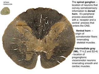
  • 1. DORSAL HORN IML VENTRAL HORN The spinal ganglion - location of neurons that convey somatosensory information to dorsal horn. It’s peripheral process associated with a receptor and a central process that enters the CNS. Ventral horn - origin of somatomotor fibers innervating skeletal muscles Intermediate gray (IML; T1-L2 and S2-4) - location of preganglionic visceromotor neurons innervating smooth and cardiac muscles. SPINAL GANGLION
  • 2. SPINAL GANGLION Spinal cord - grey matter deep to white matter Brainstem - grey matter and white matter intermingle with one another Brainstem - no dorsal, intermediate or ventral horns Neuron groups are organized into nuclei
  • 3. Brainstem: 1. Midbrain 2. Pons 3. Medulla 1 2 3 Most cranial nerves are associated with the brainstem (continuous with spinal cord) except CN I and CN II.
  • 4. Cranial nerve somatomotor nuclei are the location of neuronal cell bodies for somatomotor fibers that innervate skeletal muscles of the face and neck. The somatomotor cranial nerve nuclei are equivalent to the ventral horn of the spinal cord. Medulla
  • 5. Cranial nerve sensory ganglia (equivalent to the spinal ganglia) are located outside the brain (it may be a distinct ganglion or it may be many neurons dispersed throughout a region). They have a peripheral process that is associated with a receptor. Its central process terminates in a cranial nerve sensory nucleus (equivalent to the dorsal horn).
  • 6. CN III, VII, IX and X have parasympathetic nucleus (equivalent to intermediate gray matter of spinal cord levels S2-4). Preganglionic parasympathetic fibers travel initially with the cranial nerve in which it is associated. Preganglionic fibers synapse in parasympathetic ganglion located in head (except vagus nerve’s parasympathetic component). Postganglionic parasympathetic fibers from ganglion innervate smooth or cardiac muscles.
  • 7. CN I, CN II and CN VIII are purely sensory CN IV, CN VI, CN XI, CN XII are purely motor (not really true) CN III, CN V, CN VI, CN VII, CN IX, CN X are mixed (somatomotor, somatosensory, parasympathetic)
  • 8. Sensory ganglion - dispersed neurons located in olfactory mucosa in roof of nasal cavity Peripheral processes: located at surface of olfactory mucosa Central processes: pass through cribriform plate to terminate in olfactory bulb (dorsal horn equivalent) Olfactory bulb’s axons form olfactory tract that terminates in cortex. Olfactory Nerve (Cranial Nerve I)
  • 9. Light pass through pupil anterior and posterior chambers to retina. It passes through all layers of retina until it strikes the most posterior layer, which contains the rods and cones. They transduce light into a neural signal. bipolar cells - receive input from rods and cones; allow for convergence of information from multiple receptor cells. ganglion cells - receive input from bipolar cells -cell bodies of origin (sensory ganglion) for optic nerve -central processes of the sensory ganglia. -terminate in lateral geniculate nucleus (dorsal horn equivalent) of thalamus. Optic Nerve (Cranial Nerve II)
  • 10. Optic nerve - central processes of ganglion cell that pass through the optic canal in posterior wall of orbit to enter middle cranial fossa. Optic chiasm - crossing of nasal (medial) half of the optic nerve to the contralateral side; -temporal (lateral) half remains uncrossed. Optic tracts - caudal continuation of optic fibers to the thalamus Optic Nerve (Cranial Nerve II)
  • 11. Oculomotor nucleus (ventral horn equivalent) - cell bodies of origin for nerves innervating five extraocular muscles. Oculomotor nerve splits into: - superior division innervates: Superior Rectus Levator Palpebra Superioris -inferior division innervates: Medial Rectus Inferior Rectus Inferior Oblique Accessory oculomotor nucleus (Edinger-Westphal nucleus)- first cell body of two neuron chain responsible for parasympathetic innervation of two smooth muscles Oculomotor Nerve (Cranial Nerve III)
  • 12. SUPERIOR ORBITAL FISSURE Oculomotor fibers (both the somatomotor and preganglionic parasympathetic fibers) leave the cranial cavity through the superior orbital fissure to enter the orbit.
  • 13. Trochlear Nerve (Cranial Nerve IV) Trochlear nucleus (ventral horn equivalent) is the location of the cell bodies of origin - only cranial nerve to leave from brainstem’s dorsal (posterior) side. - innervates one extraocular skeletal muscle, superior oblique - Leaves cranial cavity through superior orbital fissure
  • 14. Trigeminal Nerve (Cranial Nerve V): principal sensory nerve of the head. Trigeminal ganglion – cell bodies of origin for sensory fibers (spinal ganglion equivalent located on petrous ridge of temporal bone) - Peripheral processes: convey somatosensory information from the face - Central processes: terminate in spinal trigeminal nucleus of brainstem. Sensory component is divided into: Ophthalmic (V1) Maxillary (V2) Mandibular (V3) Trigeminal motor nucleus- (ventral horn equivalent) neuronal cell bodies of origin for fibers innervating eight skeletal muscles.
  • 15. Ophthalmic Division-area of forehead to the lateral corners of the eyes and onto the bridge of the nose Maxillary Division-area between lateral corners of eyes to corners of the mouth Mandibular Division-area of the mandible (only division to contain motor fibers) SOMATOSENSORY INNERVATION PATTERN FOR THE FACE
  • 16. Ophthalmic Division-area of forehead to the lateral corners of the eyes and onto the bridge of the nose. - Leaves cranial cavity through superior orbital fissure to enter orbit. - peripheral processes divides into: frontal nerve supraorbital nerve supratrochlear nerve lacrimal nerve nasociliary nerve
  • 17. Maxillary Division-area between lateral corners of eyes to corners of mouth - Leaves cranial cavity by passing through foramen rotundum to enter pterygopalatine fossa. Its peripheral processes divides into: 1.infraorbital n. 2.greater and lesser palatine nn. (descending palatine nerves) 3. zygomatic n. (cheek) 4.posterior superior alveolar n. 5.sphenopalatine n.(nasal cavity)
  • 18. Mandibular Division-innervates the area of mandible; only division to contain motor fibers. - Leaves cranial cavity by passing through foramen ovale to enter infratemporal fossa. Its peripheral sensory processes branch into : 1. inferior alveolar n. 2. lingual n. 3. buccal n. 4. auriculotemporal n. 5. motor nerves to skeletal muscles.
  • 19. Trigeminal motor nucleus: location of somatomotor neuronal cell bodies of origin for fibers innervating eight muscles: muscles of mastication (masseter, temporalis, medial pterygoid, lateral pterygoid), mylohyoid, anterior belly of digastric, tensor tympani and tensor veli palatini.
  • 20. Abducens Nerve (Cranial Nerve VI) Abducens nucleus (ventral horn equivalent)- cell bodies of origin - responsible for innervating one extraocular muscle, lateral rectus. - Exits cranial cavity by passing through the superior orbital fissure to enter the orbit.
  • 21. FACIAL NERVE (CN VII) - contains somatomotor, somatosensory & preganglionic parasympathetic fibers. - Location of cell bodies for components of facial nerve: 1.Facial nucleus-somatomotor nucleus (ventral horn equivalent) innervating: muscles of facial expression & stylohyoid, posterior belly of the digastric and stapedius. 2. Superior salivary nucleus- visceral motor nucleus (intermediate gray equivalent) containing first parasympathetic cell bodies of two neuron chain innervating lacrimal gland, submandibular gland, sublingual gland, nasal mucous glands and oral mucous glands. 3. Geniculate ganglion, somatosensory ganglion (spinal ganglion equivalent) located in bend of facial canal in temporal bone posterior to middle ear. Peripheral processes innervate portions of the external ear and taste receptors on the anterior 2/3 of the tongue; central processes terminate in nucleus solitarius (taste) and spinal trigeminal nucleus (external ear).
  • 22. FACIAL NERVE (CN VII) Internal auditory meatus: exit/entrance of facial nerve from cranial cavity. Internal opening of facial canal (posterior to the middle ear) in temporal bone. Facial canal has an initial horizontal course and then a vertical course through the temporal bone. In the horizontal part that the geniculate ganglion is located. Stylomastoid foramen - inferior opening of facial canal; exit point of somatomotor and somatosensory fibers leaving the skull. ★ Two additional openings in the facial canal before the stylomastoid foramen that serve as the exit points for other parts of the facial nerve.
  • 23. INTERNAL AUDITORY MEATUS The internal auditory meatus, which is on the posterior edge of the petrous ridge of the temporal bone, is the opening for the entrance/exit of the facial nerve. Once it passes through the internal auditory meatus, the facial nerve takes a complicated course in the facial canal, which is inside the temporal bone. Internal View External View
  • 24. In facial canal different components split off: 1)greater petrosal nerve (preganglionic parasympathetic fibers from the superior salivary nucleus for lacrimal, nasal and oral mucosa glands) leaves the facial nerve at the geniculate ganglion to move rostrally. 2)chorda tympani (preganglionic parasympathetic fibers for submandibular and sublingual glands [superior salivary nucleus] and taste fibers [geniculate ganglion] for anterior 2/3 of tongue) leave the facial nerve distal to geniculate ganglion and enter the middle ear cavity to move rostrally. 3)somatomotor fibers (facial nucleus) to skeletal muscles & somatosensory fibers to external ear exit the temporal bone at the stylomastoid foramen.
  • 25. The greater petrosal nerve leaves the facial nerve at the geniculate ganglion to move rostrally. The chorda tympani leaves the facial nerve at the midpoint of the facial canal and takes a rostral course through the middle ear cavity. The remainder of the facial nerve continues through the facial canal until it exits on the anterior side of the skull at the stylomastoid foramen.
  • 26. Somatic motor component of facial nerve branches in parotid gland into: 1. temporal 2. zygomatic 3. buccal 4. mandibular 5. cervical
  • 27. Vestibulocochlear Nerve (Cranial Nerve VIII) The two ganglia, vestibular ganglion (associated with the semicircular canals) & dispersed neurons that comprise the spiral ganglion (associated with the cochlea) in the inner ear These are the neuronal cell bodies of origin for CN VIII. Vestibular ganglion cells: convey balance and orientation Spiral ganglion cells: convey auditory sense Peripheral processes of the vestibular ganglia are associated with receptors in the semicircular canals and the central processes end in vestibular nuclei of the brainstem. Peripheral processes of the spiral ganglia are associated with receptors in the cochlea and the central processes end in cochlear nuclei of the brainstem. Central processes pass through the internal auditory meatus to enter the cranial cavity.
  • 28. GLOSSOPHARYNGEAL NERVE (CN IX) - Primarily a sensory nerve, although it has a small somatomotor & visceromotor components. -Sensory information from posterior third of tongue, mucosa of pharynx, middle ear, auditory tube, and blood pressure & chemoreceptors associated with common carotid artery. - Somatomotor innervation: stylopharyngeus of pharynx -Visceromotor (parasympathetic) innervation: parotid gland. -Leaves cranial cavity thru jugular foramen
  • 29. Location of cell bodies: 1) Nucleus Ambiguus: cell bodies of origin for somatomotor fibers that innervate stylopharyngeus 2) Inferior Salivary Nucleus: cell bodies of origin for visceromotor fibers that are involved in innervation of the parotid gland
  • 30. 3) Inferior Ganglion of the Glossopharyngeal nerve (at the jugular foramen). Peripheral processes carry: 1) special sense (taste) & somatosensory information from the posterior 1/3 of tongue 2) Somatosensory information from the pharynx 3) Somatosensory information from the middle ear, auditory tube & mastoid air cells 4) General viscerosensory information from the carotid body (chemoreception) & carotid sinus (baroreception). Central processes from the inferior ganglion of CN IX terminate in: Nucleus solitarius – taste fibers from posterior third of tongue, carotid body and carotid sinus. Spinal trigeminal nucleus – general sensory from posterior third of tongue, pharynx, middle ear, auditory tube & mastoid air cells Spina l
  • 31. VAGUS NERVE (CN X) - contains somatomotor, visceromotor and somatosensory components - somatomotor innervates skeletal muscles of larynx, pharynx and accessory muscles of pharynx and soft palate - visceromotor component innervates smooth and cardiac muscles of thoracic cavity and smooth muscle of the abdomen up to splenic flexure - sensory component innervates laryngeal mucosa, meninges of posterior cranial fossa, external ear and comparable regions innervated by visceromotor innervation - Leaves cranial cavity through jugular foramen
  • 32. Nucleus Ambiguus: neuronal cell bodies that innervate skeletal muscles including: pharyngeal constrictor muscles, intrinsic muscles of larynx (external laryngeal & recurrent laryngeal nn.), levator veli palatini (soft palate) and palatoglossus, palatopharyngeus, salpingopharyngeus Dorsal Motor Nucleus of the Vagus: first neuronal cell bodies responsible for parasympathetic innervation of smooth muscle of digestive system (to splenic flexure), respiratory system and heart. Spinal trigeminal nucleus
  • 33. Inferior Ganglion of the vagus nerve (nodose ganglion): innervate mucosa of larynx, respiratory system, heart and digestive system. Its central processes (axons) terminate in the nucleus solitarius of brainstem. Superior Ganglion: innervate external auditory meatus, meninges of the posterior cranial fossa and external surface of tympanic membrane. Its central fibers terminate in spinal trigeminal nucleus of brainstem. Spinal trigeminal nucleus
  • 34. (Spinal) Accessory Nerve (CN XI) Location: C1-C5 of spinal cord is in the posterior portion of the anterior (ventral) horn. - fibers leave spinal cord between the posterior (dorsal) and anterior (ventral) roots to enter cranial cavity through foramen magnum. - fibers then exit cranial cavity through the jugular foramen. - innervates the sternocleidomastoid and trapezius.
  • 35. Hypoglossal nucleus: neuronal cell bodies for the hypoglossal nerve. - Exits cranial cavity through hypoglossal canal of occipital bone - In neck it loops around occipital artery to enter lateral side of the tongue - innervates: Intrinsic and extrinsic muscles (hyoglossus, styloglossus & genioglossus) of tongue HYPOGLOSSAL NERVE (CN XII)
  • 36. LEARNING OBJECTIVES FOR CRANIAL NERVES By the end of this unit you must have mastered for each cranial nerve (where appropriate): 1) all nuclei or ganglia of origin for its fibers 2) specific region of the brain or spinal cord its fibers are associated 3) complete pathway, any bony openings it passes through, and all its terminal targets 4) identify as motor only, sensory only, both motor and sensory and autonomic component if any 5) for sensory fibers: brain nuclei for termination of central fibers 6) for autonomic component, the nucleus of origin, bony openings, path of its preganglionic fibers, parasympathetic ganglion, path of its postganglionic fibers, terminal targets. 7) for the sympathetic fibers, cell bodies of origin, path of its fibers, sympathetic ganglion, pathway of its postganglionic fibers to the target structures. 8) Describe the functional deficits that will be manifested if any cranial nerve is lesioned at any point along its course. Specific objective for the 12 Cranial nerves: 1) Olfactory nerves-cell bodies of origin, bony openings and its path to its termination in olfactory bulb termination of olfactory tract axons 2) Optic nerve-cell bodies of origin, path of its fibers, bony opening and thalamic nucleus for termination of its central fibers location and consequences of optic chiasm 3) Oculomotor nerve-cell bodies of origin, path of its fibers cell bodies of origin and path to striated muscles for its superior and inferior divisions autonomic cell bodies of origin and complete path of its autonomic fibers location of ciliary ganglion; its contribution to short ciliary nerves and target structures for its postganglionic fibers know the location that its parasympathetic fibers join and leave specific parts of the nerve 4) Trochlear nerve-cell bodies of origin, its path, bony opening, muscle it innervates
  • 37. 5) Trigeminal nerve-cell bodies of origin, paths and bony openings of its subdivisions ophthalmic division area of innervation major branches branches of the frontal nerve associated autonomic ganglia components and cell bodies of origin for long ciliary nerves components and cell bodies of origin for short ciliary nerves maxillary division area of innervation branches in the pterygopalatine fossa, oral cavity, maxillary sinus and nasal cavity associated autonomic ganglion mandibular division area of innervation sensory branches and muscles innervated associated autonomic ganglia Know the location that autonomic fibers “hitch a ride” on specific parts of the nerve and when they leave the nerve to innervate a structure 6) Abducens nerve-cell bodies of origin, its bony opening and path to the muscle it innervates 7) Facial nerve-cell bodies of origin, bony openings, path of its fibers to its terminal targets sensory targets and somatic muscles innervated by the facial nerve autonomic cell bodies of origin and complete path of fibers to target structures components and cell bodies of origin of the chorda tympani components and cell bodies of origin for the nerve of the pterygoid canal 8) Vestibulocochlear nerve-cell bodies of origin, its path, bony openings and structures innervated
  • 38. 9) Glossopharyngeal nerve-cell bodies of origin, its path, bony openings and structures innervated. cell bodies of origin for innervation of striated muscles autonomic cell bodies of origin, path of its fibers and target structures cell bodies of origin and sensory regions innervated components and cell bodies of origin for tympanic nerve components and cell bodies of origin for lesser petrosal nerve contribution to pharyngeal plexus 10) Vagus nerve-cell bodies of origin, bony opening, path of its fibers and structures innervated. cell bodies of origin and striated muscles innervated cell bodies of origin and smooth muscle structures innervated cell bodies of origin and sensory regions innervated contribution to pharyngeal plexus 11) Accessory nerve-cell bodies of origin, its path, bony openings and terminal structures innervated 12) Hypoglossal nerve-cell bodies of origin, its path, bony openings and terminal structures innervated 13) Sympathetic innervation-cell bodies of origin for preganglionic and postganglionic fibers, most common pathways to terminal target sites. location and types of fibers in carotid plexus path and terminal targets of sympathetic components of long and short ciliary nerves components, path and terminal targets of deep petrosal nerve sympathetic path for innervation of submandibular, sublingual and parotid glands