
Recomendados
Mais conteúdo relacionado
Semelhante a Asms2004 Alternate Scanning Lcms
Semelhante a Asms2004 Alternate Scanning Lcms (20)
Último
Último (20)
Asms2004 Alternate Scanning Lcms
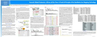
- 1. ASMS Towards Global Proteomics, All Ions all the Time: A Proof-of-Principle of the Qualitative Ion Mapping Technology 2004 Jeffrey C. Silva1, Keith Richardson2, Phillip Young2, Richard Denny2, Kieran Neeson2, Therese McKenna2, Craig Dorschel1, Guo-Zhong Li1, Marc Gorenstein1, Timothy Riley1, and Scott Geromanos1 1 Waters Corporation, Milford, MA USA, 2Waters Corporation, Manchester, UK Figure 5. As noted in the data processing section, Protein Expression System Informatics Table 1. Introduction Data Processing Figure 3. EE 080603_497HL_QuanExpSerial_03 2: TOF MS ES+ uses this principle of chromatographic apex alignment as a first filter to remove Delta b-ions y"-ions MSE Protein 100 73.42 BPI 3.53e3 Description Sequence Actual MH+ (ppm) (b1……..bn-1) (y”1…….y”n-1) Traditional tandem mass spectrometry (MS/MS) is a method of mass spectrometry A basic premise of the Protein Expression System Informatics software is that the PLG 2.0 dB Sequence 7.13 21.89 22.11 28.11 38.52 42.88 60.94 ions from the elevated energy spectra that may have a retention time similar to, Q96KJ4 Similar to pre-pro-megakarycyte potentiating factor MEVSGDPGIPALHR 1478.7420 2.6 0000000000000 1000000000001 (peptide fragments) 20.82 whereby intact peptides (parent ions) generated from an enzymatic digest of a exact mass MS data acquired at low energy is primarily composed of peptide 46.73 but not coincident with a parent ion of interest (see Poster WPJ 161, Goren- Q9H8J4 CDNA FLJ13564 fis clone PLACE1008201 AGFNSYAELLTHR 1478.7387 0.3 000000000000 100000000000 24.54 33.60 protein sample are mass filtered and collisionally dissociated to yield product or precursor ion information and the exact mass MS data acquired at elevated en- % 6.20 51.80 60.29 stein et al. “Statistical Study of Ions and Peptides Found In LC/MS/MS Analysis of Q9NVE3 CDNA FLJ10787 fis clone NT2RP4000481 ESDDGVVVYVAPTK 1478.7373 -0.6 0000000000000 0000000000000 78.49 fragment ions which are subsequently mass analyzed. The resulting product ion ergy is composed primarily of corresponding peptide product or fragment ion in- Candidate Candidate Peptide Sequence Peptide Protein 7.77 78.99 106.10 Human Serum Digest” and Poster TPR 354, Li et al.). Figure 8 illustrates this Q9BXU7 Ubiquitin carboxyl-terminal hydrolase 26 MTSGNISVSWPATK 1478.7308 -5.0 0000000000000 0000000000000 spectra can be used to determine the sequence of the parent peptide and, Peptide b1 bn Score Assignment 5.13 9.91 67.86 81.71 89.55 95.18 103.24 108.09 formation. It is also understood that fragment ions associated with a particular Mass y”n y”1 19.04 58.51 112.30 117.66 119.79 “cleaning” principle more graphically. The top panel shows the combined ele- P02787 Serotransferrin precursor (Siderophilin) MYLGYEYVTAIR 1478.7348 -2.3 01110000000 10111110111 through a database search, the identity of the originating protein. This method of parent ion are produced simultaneously with the parent ions as they elute from the + Accession #1 0 10.00 20.00 30.00 40.00 50.00 60.00 70.00 80.00 90.00 100.00 110.00 120.00 Time 130.00 vated energy spectra of all ions that elute at 72.425 minutes. The middle panel O60610 Diaphanous protein homolog 1 NSETFPTILEEAK 1478.7373 -0.6 000000000000 000100000000 O60522 Colon cancer antigen NY-CO-45 (Fragment) SALPYENIDSEIK 1478.7373 -0.6 000000000000 000000000000 analysis can be automated and it is generally performed in conjunction with a reverse phase column. 080603_497HL_QuanExpSerial_03 812 (72.425) Cm (812) 267.1570 2: TOF MS ES+ 235 shows the elevated energy spectra after being cleaned of ions that do not co- ++++++ Accession #2 100 P50440 Glycine amidinotransferase, mitochondrial precursor WLSMNVLMLDEK 1478.7382 0.0 00000000000 00000000000 real time liquid chromatography separation system. Although this methodology is apex with the low energy doubly charged peptide, m/z 872.54, at 71.96 min- Q9HB07 MYG1 protein AMDLVQEEFLQR 1478.7308 -5.0 00000000000 10000000000 quite powerful, it has been shown to have limitations when characterizing com- Protein Expression System Informatics filters the data acquired in the alternating ++ Accession #3 utes. Similarly, the bottom panel shows the elevated energy spectra after removal Q9H099 Hypothetical 78 5 kDa protein DLWHAAFFLSGSK 1478.7427 3.0 000000000000 000000000000 plex protein mixtures. Many of these limitations are associated with the serial na- scan experiment in a three-step process. The first step carefully aligns chroma- % 136.0944 295.1492 1071.7020 of ions that do not co-apex with the low energy doubly charged peptide, m/z ++ Accession #4 Q9UNI1 Elastase 1 precursor NSWPSQISLQYR 1478.7387 0.3 00010000000 10000000000 ture of the MS/MS data acquisition process which limits both the qualitative and tographic retention time to associate fragment ions to each appropriate precursor 687.4291 809.5603 1184.7798 1185.7744 739.96, at 72.60 minutes. P21266 Glutathione S transferase Mu 3 VDIIENQVMDFR 1478.7308 -5.0 00000000000 10000000000 670.4338 988.6307 1478.9341 the quantitative reproducibility of the information generated. ion (Figure 1). The second step compares the exact mass of each measured ++ Accession #5 366.1824 1186.7974 1386.7921 1613.9867 1745.0836 In Table 1, the qualitative identification data filtering steps illustrated in Figures P55884 Eukaryotic translation initiation factor 3 subunit 9 (eIF-3 eta) YVVTSVSWWSHK 1478.7427 3.0 01100000000 00000000000 0 m/z peptide precursor ion to a database containing the accurate mass of every pep- ++++++ Accession #6 100 200 300 400 500 600 700 800 900 1000 1100 1200 1300 1400 1500 1600 1700 1800 1900 2 and 3 have been applied to the 739.96 doubly charged peptide ion eluting A new method of protein characterization of proteolytically-generated peptides tide (including one missed cleavage) associated with every known protein in the at 72.60 minutes along with its chromatographically associated fragment ions. Table 2. + Accession #7 Delta b-ions y"-ions has been developed which is based on coupling the alternate scanning of pep- proteome under study. Typically, peptides which are within an acceptable mass Figure 6. Each of the proteins listed in Table 1 contains a tryptic peptide sequence that Description Sequence Actual MH+ (ppm) (b1……….…..bn-1) (y”1……….y”n-1) tides at low and elevated energy1,2 with a robust accurate mass LC/MS data ac- measurement tolerance (within +/- 5 ppm) are identified in a subset peptide data 080603_497HL_QuanExpSerial_03 1: TOF MS ES+ has an accurate mass within plus or minus 5ppm of the 739.96 measured mass. O14775 Guanine nucleotide-binding protein beta subunit 5 MATEGLHENETLASLK 1743.8582 -2.0 011000000000000 100000000000100 Cs 1, 1744.12 71.96 1744.115 0.05Da quisition mode3. This mode of data acquisition is very high in duty cycle and pro- 100 base (Figure 2). The accurate mass of each y” and b fragment ion is calculated 157 In Tables 1 and 2, columns 5 and 6 indicate those corresponding b- and y”- Q9P2G7 KIAA1380 protein DSSEIPGALWHIYAGK 1743.8701 4.8 010000000000000 100000000000000 vides exact mass MS analysis of both the precursor and product ions for every de- for each of the peptides in this subset database and the measured exact mass Results and Discussion % 0 ions for each peptide, that are found in the elevated energy data to within plus or P13796 L-plastin (Lymphocyte cytosolic protein 1) GDEEGVPAVVIDMSGLR 1743.8582 -2.0 0010000000000000 0000000010000000 080603_497HL_QuanExpSerial_03 1: TOF MS ES+ tectable peptide. Chromatographic retention time is used to associate fragments fragment ion information obtained in the first processing step is compared to this Cs 2, 1744.12 71.96 872.541 0.05Da Q96GG7 Interferon regulatory factor 1 LLEQSEWQPTNVDGK 1743.8548 -4.0 00000000000000 10100100000000 Figures 4 and 5 illustrate typical results from an alternating low and elevated 100 6.87e3 minus 10 ppm mass accuracy and plus or minus 0.05 minutes retention time. A to their corresponding precursors as a unique attribute of the Waters® Protein Ex- theoretical accurate mass information (Figure 3). A probability-based search % “0” at a given amino acid position indicates the corresponding fragment ion was Q9H473 Golgi-spec. brefeldin A-resistance guanine nef-1 NEEIVMPEEQTGLVR 1743.8582 -2.0 00000000000000 00000000000000 collision energy LC/MS analysis of non-depleted human serum tryptic digest. The 0 73.32 pression System technology. Proprietary Protein Expression System Informatics model that takes into account the number, type, and mass accuracy of the meas- 080603_497HL_QuanExpSerial_03 Cs 1, 1630.01 1: TOF MS ES+ not found within the specified search tolerances, and a “1” at a given amino acid Q9H0K9 Hypothetical 78 6 kDa protein SHPLQLTDDGGFSEIK 1743.8548 -4.0 001000000000000 100000000000000 upper panel of Figure 4 illustrates the low energy base peak intensity chroma- 100 72.68 1630.014 0.05Da 94.4 software has been developed which allows this information to be processed in a ured fragment ions is used to determine the best peptide sequence match. % position indicates the corresponding fragment ion was found within the specified O75018 Leucocyte Immunoglobulin-like Receptor 6A/B TASHPQDYTVENLIR 1743.8660 2.5 00000001000000 00000000000000 togram and the lower panel shows a low energy exact mass spectrum extracted P01842 Ig lambda chain C regions YAASSYLSLTPEQWK 1743.8588 -1.6 01111111110000 11111111111010 quantitative and qualitative manner. This poster will focus on the qualitative identi- 0 search tolerances. The measured exact mass fragment ion data associated with at the highlighted retention time of 72.390 minutes. Figure 5 illustrates the same 080603_497HL_QuanExpSerial_03 Cs 2, 1630.01 72.68 1: TOF MS ES+ 815.51 0.05Da Q9GZU1 CDNA FLJ22449 fis clone HRC09609 GFLLQNEFVGFMWR 1743.8676 3.4 0000000000000 0000000000000 fication capabilities of this software. Poster TPR 357 (Geromanos et al., Figure 1. 100 3.75e3 the 739.96 peptide parent ion has been compared to the theoretical y” and b information for the elevated energy function with the example spectrum extracted % O43736 Integral membrane protein 2A (E25 protein) GGEPNFLPVTEEADIR 1743.8548 -4.0 000000010000000 000000000000000 “Towards Global Proteomics by Analysis of Exact Mass Retention Time Pairs: A fragment ions associated with each of the candidate parent ions in Table 1. The one scan later at 72.425 minutes. While many peptide ions co-elute during this 0 080603_497HL_QuanExpSerial_03 1: TOF MS ES+ Q9Y4J7 Fibroblast growth factor 2-interacting factor NYGILADATEQVGQHK 1743.8660 2.5 000000000000000 110000000000000 proof-of-principle of the Quantitative Ion Mapping Technology”) addresses the 100 Cs 1, 1478.95 72.60 1478.95 0.05Da measured fragment ions associated with the MYLGYEYVTAIR clearly provide con- moment in time, only a subset of the peptide ions apex at the same moment in 144 O94935 KIAA0851 protein THLGLIMDGWNSMIR 1743.8669 3.0 00000000100000 00100010000000 quantitative capabilities of the software and Poster TPY 458 (C. Dorschel et % vincing evidence, with twelve matching fragment ions, to indicate that the 739.96 time. Figure 6 shows the low energy exact mass chromatograms for singly and 0 O94808 Glucosamine-fructose-6P. aminotransferase 2 WATHGVPSAVNSHPQR 1743.8674 3.3 000001000000000 010001100000000 al., “Protocols to Assure Reproducible Quantitative and Qualitative Analysis of 080603_497HL_QuanExpSerial_03 Cs 2, 1478.95 1: TOF MS ES+ doubly charged peptide originates from the Serotransferrin precursor pro- Collect all precursor ions doubly charge state ions for 4 example peptides that co-elute in the chroma- 100 72.60 739.957 0.05Da Tryptic Digests of Complex Protein Mixtures for Global Proteomic Experiments”) 4.57e3 tein. Table 2, illustrates a similar analysis of the 872.54 doubly charged parent Table 3. tographic region around 72 minutes. The singly and doubly charged ions associ- % % Coverage Description Median Mass Error % Coverage Description Median Mass Error highlights the reproducibility of the Protein Expression System platform. 0 ion and its chromatographically associated fragment ions. In this example, there 61 Zinc alpha 2 glycoprotein precursor 1.1 58 Hemoglobin alpha chain 0.2 ated with the same peptide clearly apex at the same chromatographic retention 080603_497HL_QuanExpSerial_03 100 70.82 Cs 1, 1311.92 1: TOF MS ES+ 1311.915 0.05Da 12 26 Vitamin D-binding protein precursor Very low density lipoprotein receptor 3.4 3.7 34 52 Heat shock 70 kDa protein 1 Haptoglobulin-2 6.3