SlideShare uma empresa Scribd logo
1 de 3
Baixar para ler offline
LIFE CYCLE OF FUNARIA
    (Division: Bryophyta, Class: Bryopsida, Order: Funariales, Family: Funariaceae, Genus: Funaria)

Occurrence: Funaria is a terrestrial moss which grows as dense green patches, in moist shady conditions
such as damp soil, shady banks, damp tree trunks and walls.

Structure of Gametophyte: It shows two parts – a prostrate underground protonema and an erect leafy
gametophores.
• The gametophores is differentiated into rhizoids, axis or stem and leaves.
• Rhizoids are multicellular, branched with oblique walls.
• The stem is erect, 1-3 cm tall, branched. Branches are extra-axillary.
• Stems and branches covered with spirally arranged leaves. Leaves are simple, sessile with a midrib.
   They are crowded at the tip of the stem to form a bud-like head.

Anatomy of stem: T.S. of stem shows 3 distinct parts: An outer single layered epidermis with green cells.
The middle region is cortex which is multilayered. Outer cells of cortex are thick walled.
The central region is called central cylinder. It consists of vertically elongated thin walled narrow, dead
cells without protoplasm. They are the conducting cells and also called hydroids.

Anatomy of Leaf: The leaf consists of a single layer of chlorophyllous cells. The mid rib part is many cells
in thickness. Within the midrib narrow thick-walled conducting cells are present.


           Gametophore                                       T. S. of stem and leaf
Vegetative Reproduction:
Multiplication by protonema: Fragments of leaf and stem can give rise to protonema which produces
new plant under suitable conditions.
Bulbils or tubers: bulbils develop in the rhizoids.
Gemmae: develop in the terminal cells of the protonemal branches.

 Sexual Reproduction: It is advanced oogamous type. Antheridia and archegonia are produced in
clusters at the tips of special branches called antheridial or male branches and archegonial or female
branches in same (monoecious) plant.
Structure of antheridial branch and antheridia: Antheridial branch or male flower is produced at the tip
of the main stem of the gametophyte. It is surrounded by cluster of leaves called perigonial leaves.
 • Antheridia are arranged in clusters, intermixed with sterile multicellular hairs called paraphyses.
      Paraphyses are protective in function.
 • Mature antheridia are club shaped with a long, multicellular stalk and a globular body.
 • The body is covered with a single layered jacket which surrounds numerous androgonial cells.
 • The androgonial cells develop into antherozoids or sperms.
 • Each sperm is spirally coiled, elongated structure with a pair of flagella. Mature sperms are released
      from the antheridium by the separation of the jacket cells.


 Structure of archegonial branch and archegonia: The archegonial branches develop from the base of
 the male branches. They are covered with large leaves called perichaetial leaves. Cluster of archegonia
 are found at the tip of the each
 branch.
 • The mature archegonium is flask
     shaped with a basal multicellular
     stalk, long twisted neck and a
     swollen venter.
 • The neck is covered with 6
     vertical rows of neck cells and
     cover cells. It encloses more than
     10 neck canal cells.
 • The venter is covered by 2-3
     layers of cells. It encloses a
     venter canal cell and a basal egg
     cell.Intermixed with the
     archegonia, long hair-like,
     multicellular paraphyses are
     present.

 Structure of Sporophyte: The sporophyte is seen at the apex of the archegonial or female branch. A
 mature sporophyte shows three distinct parts – the foot, seta and capsule.
 • The foot is a small conical structure embedded in the female tissue. It helps in attaching the
    sporophyte to the gametophyte and absorption of nutrients from it.
•   Seta is a long, slender and twisted structure. It carries the capsule at its tip.
•   The capsule is the fertile structure of the sporophyte. In an L.S., it shows three distinct regions – the
    apophysis, theca and operculum or lid.
•   Apophysis is the basal green, photosynthetic part of capsule. It has a central strand of conducting
    tissue.
•   The theca is the urn shaped fertile, middle part of the capsule. It has a central strand of tissue called
    columella which is cone shaped.
•   Surrounding the columella is the spore sac. It is covered by outer spore sac wall which is 3-4 layers
    in thickness. The inner wall is just one cell thick. Spore sac consists of diploid archesporial cells
    which after meiosis develops into haploid spores.
•   Outer to the spore sac is an air space consisting of many air cavities. These cavities are traversed by
    cells called trabeculae.
•   Outside the air space is the capsule wall which is many layers thick. The outer layer is the epidermis.
    Inner to the epidermis is the two layered hypodermis of colourless cells. Within the hypodermis is the
    spongy layers of green cells.
•   Operculum is the terminal lid-like portion of the capsule. It is 4-5 layers thick. Inner to the
    operculum is a structure called peristome. It consists of two sets of curved teeth like structures, lying
    one above the other. Each set has 16 teeth. The outer peristomal teeth are hygroscopic and help in
    the dehiscence of the capsule and dispersal of spores.
•   When capsule is mature, it dries up, the operculum is blown up. Spores are discharged by the
    hygroscopic movement of peristome teeth.


Germination of the spores: During favorable conditions, spores
germinate to form a multicellular, filamentous, branched structure called
protonema. It consists of green prostrate branches called chloronemal
branches and colourless branches called rhizoidal branches. Green Bud
like structures develop on the chloronemal branches which then grow and
develop into the leafy gametophores.


            ©Dr. Jayakara Bhandary M. SGL in Botany, GAS College, Karwar-581301

Mais conteúdo relacionado

Mais procurados (20)

Structure, reproduction, life history and systematic position of Lycopodium
Structure, reproduction, life history and systematic position of LycopodiumStructure, reproduction, life history and systematic position of Lycopodium
Structure, reproduction, life history and systematic position of Lycopodium
 
Vaucheria
VaucheriaVaucheria
Vaucheria
 
Gnetum
 Gnetum Gnetum
Gnetum
 
Oedogonium
OedogoniumOedogonium
Oedogonium
 
General characters of pteridophytes
General characters of pteridophytesGeneral characters of pteridophytes
General characters of pteridophytes
 
Anthoceros
AnthocerosAnthoceros
Anthoceros
 
Marsilea.pptx
Marsilea.pptxMarsilea.pptx
Marsilea.pptx
 
Marchantia
MarchantiaMarchantia
Marchantia
 
General Characters of Rhodophyceae & Life Cycle of Polysiphonia SMG
 General Characters of Rhodophyceae &  Life Cycle of Polysiphonia   SMG General Characters of Rhodophyceae &  Life Cycle of Polysiphonia   SMG
General Characters of Rhodophyceae & Life Cycle of Polysiphonia SMG
 
Rhynia
RhyniaRhynia
Rhynia
 
Notothylas by p.n
Notothylas by p.nNotothylas by p.n
Notothylas by p.n
 
cycas.pptx
cycas.pptxcycas.pptx
cycas.pptx
 
Selaginella
SelaginellaSelaginella
Selaginella
 
Funaria ( bryophytes)
Funaria ( bryophytes)Funaria ( bryophytes)
Funaria ( bryophytes)
 
Classification of pteridophytes
Classification of pteridophytesClassification of pteridophytes
Classification of pteridophytes
 
Morphological, anatomical and reproductive characters of Marchantia
Morphological, anatomical and reproductive characters of MarchantiaMorphological, anatomical and reproductive characters of Marchantia
Morphological, anatomical and reproductive characters of Marchantia
 
Lycopodium
LycopodiumLycopodium
Lycopodium
 
Riccia
RicciaRiccia
Riccia
 
Anthoceros
AnthocerosAnthoceros
Anthoceros
 
Life cycle of gnetum
Life cycle of gnetumLife cycle of gnetum
Life cycle of gnetum
 

Destaque (20)

Bio 211 report
Bio 211 reportBio 211 report
Bio 211 report
 
Bryophytes
BryophytesBryophytes
Bryophytes
 
Powders
PowdersPowders
Powders
 
Classification of pteridophyta
Classification of pteridophytaClassification of pteridophyta
Classification of pteridophyta
 
MIB1 - Pteridophytes
MIB1 - PteridophytesMIB1 - Pteridophytes
MIB1 - Pteridophytes
 
Ppt funaria
Ppt funariaPpt funaria
Ppt funaria
 
Marchantia
MarchantiaMarchantia
Marchantia
 
computer ppt by Shivanshi Dwivedi
 computer ppt by Shivanshi Dwivedi  computer ppt by Shivanshi Dwivedi
computer ppt by Shivanshi Dwivedi
 
Bryophyte
BryophyteBryophyte
Bryophyte
 
Pteridophytes
PteridophytesPteridophytes
Pteridophytes
 
Lycopodium clavatum
Lycopodium clavatumLycopodium clavatum
Lycopodium clavatum
 
Pteridophytes
PteridophytesPteridophytes
Pteridophytes
 
Stelarevolution 120401081016-phpapp02
Stelarevolution 120401081016-phpapp02Stelarevolution 120401081016-phpapp02
Stelarevolution 120401081016-phpapp02
 
Plant tissues, 9 cbse
Plant tissues, 9 cbsePlant tissues, 9 cbse
Plant tissues, 9 cbse
 
Bryophytes
BryophytesBryophytes
Bryophytes
 
The Gymnosperms
The GymnospermsThe Gymnosperms
The Gymnosperms
 
Vascular Plants: Pteridophyta (part 1)
Vascular Plants: Pteridophyta (part 1)Vascular Plants: Pteridophyta (part 1)
Vascular Plants: Pteridophyta (part 1)
 
Stelar evolution in Pteridophytes
Stelar evolution in PteridophytesStelar evolution in Pteridophytes
Stelar evolution in Pteridophytes
 
Pteridophyte ppt
Pteridophyte pptPteridophyte ppt
Pteridophyte ppt
 
Plant Kingdom
Plant KingdomPlant Kingdom
Plant Kingdom
 

Semelhante a Life cycle of_funaria

Life-cycle-of-Sphagnum.pdf
Life-cycle-of-Sphagnum.pdfLife-cycle-of-Sphagnum.pdf
Life-cycle-of-Sphagnum.pdfVainyaVerma
 
Funaria botany
Funaria botanyFunaria botany
Funaria botanyMANSI
 
PSILOTUM : structure, morphology, anatomy, reproduction , life cycle etc.
PSILOTUM : structure, morphology, anatomy,  reproduction , life cycle etc.PSILOTUM : structure, morphology, anatomy,  reproduction , life cycle etc.
PSILOTUM : structure, morphology, anatomy, reproduction , life cycle etc.Silpa Selvaraj
 
MARSILEA notes in detail for II year Botany.ppt
MARSILEA  notes in detail for II year Botany.pptMARSILEA  notes in detail for II year Botany.ppt
MARSILEA notes in detail for II year Botany.pptaigil2
 
selaginella species characters and distribution .pptx
selaginella species characters and distribution .pptxselaginella species characters and distribution .pptx
selaginella species characters and distribution .pptxSHUBHAMDOUND1
 
Kr bryophyta classification
Kr bryophyta classificationKr bryophyta classification
Kr bryophyta classificationDrKavitaRambal
 

Semelhante a Life cycle of_funaria (20)

Bryopsida – funaria
Bryopsida – funariaBryopsida – funaria
Bryopsida – funaria
 
Life-cycle-of-Sphagnum.pdf
Life-cycle-of-Sphagnum.pdfLife-cycle-of-Sphagnum.pdf
Life-cycle-of-Sphagnum.pdf
 
Type polytrichum
Type polytrichumType polytrichum
Type polytrichum
 
Marchantia jb
Marchantia jbMarchantia jb
Marchantia jb
 
Funaria botany
Funaria botanyFunaria botany
Funaria botany
 
Life cycle of_sphagnum
Life cycle of_sphagnumLife cycle of_sphagnum
Life cycle of_sphagnum
 
Psilotum
PsilotumPsilotum
Psilotum
 
Marchantia
MarchantiaMarchantia
Marchantia
 
Pteridophytes
PteridophytesPteridophytes
Pteridophytes
 
PSILOTUM : structure, morphology, anatomy, reproduction , life cycle etc.
PSILOTUM : structure, morphology, anatomy,  reproduction , life cycle etc.PSILOTUM : structure, morphology, anatomy,  reproduction , life cycle etc.
PSILOTUM : structure, morphology, anatomy, reproduction , life cycle etc.
 
MARSILEA notes in detail for II year Botany.ppt
MARSILEA  notes in detail for II year Botany.pptMARSILEA  notes in detail for II year Botany.ppt
MARSILEA notes in detail for II year Botany.ppt
 
Plant Kingdom III
Plant Kingdom IIIPlant Kingdom III
Plant Kingdom III
 
selaginella species characters and distribution .pptx
selaginella species characters and distribution .pptxselaginella species characters and distribution .pptx
selaginella species characters and distribution .pptx
 
CYCAS 1.pdf
CYCAS 1.pdfCYCAS 1.pdf
CYCAS 1.pdf
 
THUJA.pptx
THUJA.pptxTHUJA.pptx
THUJA.pptx
 
Funaria
FunariaFunaria
Funaria
 
Furneria
FurneriaFurneria
Furneria
 
Kr bryophyta classification
Kr bryophyta classificationKr bryophyta classification
Kr bryophyta classification
 
Adiantum .pptx
Adiantum .pptxAdiantum .pptx
Adiantum .pptx
 
99997024.pdf
99997024.pdf99997024.pdf
99997024.pdf
 

Mais de Jayakara Bhandary (20)

Science writing
Science writingScience writing
Science writing
 
Jeevavaividhya1
Jeevavaividhya1Jeevavaividhya1
Jeevavaividhya1
 
Mg parichaya
Mg parichayaMg parichaya
Mg parichaya
 
Mangrove flora of karnataka
Mangrove flora of karnatakaMangrove flora of karnataka
Mangrove flora of karnataka
 
Engler & prantl system of classification
Engler & prantl system of classificationEngler & prantl system of classification
Engler & prantl system of classification
 
Mangroves of Honnavar
Mangroves of Honnavar Mangroves of Honnavar
Mangroves of Honnavar
 
Ecotourismin in honnavar
Ecotourismin in honnavarEcotourismin in honnavar
Ecotourismin in honnavar
 
Viroids, Prions and Mycoplasma
Viroids, Prions and Mycoplasma Viroids, Prions and Mycoplasma
Viroids, Prions and Mycoplasma
 
Morphology of fruits
Morphology of fruitsMorphology of fruits
Morphology of fruits
 
Fertilisation in _angiosperms
Fertilisation in _angiospermsFertilisation in _angiosperms
Fertilisation in _angiosperms
 
Genetic recombination in_bacteria-jb
Genetic recombination in_bacteria-jbGenetic recombination in_bacteria-jb
Genetic recombination in_bacteria-jb
 
Microsporogenesis
MicrosporogenesisMicrosporogenesis
Microsporogenesis
 
Question _bank-sem_1
Question  _bank-sem_1Question  _bank-sem_1
Question _bank-sem_1
 
Kud bsc botany-syllabus_sem5&6
Kud bsc botany-syllabus_sem5&6Kud bsc botany-syllabus_sem5&6
Kud bsc botany-syllabus_sem5&6
 
Embryogenesis
EmbryogenesisEmbryogenesis
Embryogenesis
 
Biotechnology karnataka puc
Biotechnology karnataka pucBiotechnology karnataka puc
Biotechnology karnataka puc
 
Biotechnology karnataka puc
Biotechnology karnataka pucBiotechnology karnataka puc
Biotechnology karnataka puc
 
Life cycle of_funaria-jb
Life cycle of_funaria-jbLife cycle of_funaria-jb
Life cycle of_funaria-jb
 
Life cycle of_riccia-jb
Life cycle of_riccia-jbLife cycle of_riccia-jb
Life cycle of_riccia-jb
 
Gene ineractions jb
Gene ineractions jbGene ineractions jb
Gene ineractions jb
 

Life cycle of_funaria

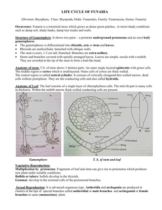
  • 1. LIFE CYCLE OF FUNARIA (Division: Bryophyta, Class: Bryopsida, Order: Funariales, Family: Funariaceae, Genus: Funaria) Occurrence: Funaria is a terrestrial moss which grows as dense green patches, in moist shady conditions such as damp soil, shady banks, damp tree trunks and walls. Structure of Gametophyte: It shows two parts – a prostrate underground protonema and an erect leafy gametophores. • The gametophores is differentiated into rhizoids, axis or stem and leaves. • Rhizoids are multicellular, branched with oblique walls. • The stem is erect, 1-3 cm tall, branched. Branches are extra-axillary. • Stems and branches covered with spirally arranged leaves. Leaves are simple, sessile with a midrib. They are crowded at the tip of the stem to form a bud-like head. Anatomy of stem: T.S. of stem shows 3 distinct parts: An outer single layered epidermis with green cells. The middle region is cortex which is multilayered. Outer cells of cortex are thick walled. The central region is called central cylinder. It consists of vertically elongated thin walled narrow, dead cells without protoplasm. They are the conducting cells and also called hydroids. Anatomy of Leaf: The leaf consists of a single layer of chlorophyllous cells. The mid rib part is many cells in thickness. Within the midrib narrow thick-walled conducting cells are present. Gametophore T. S. of stem and leaf Vegetative Reproduction: Multiplication by protonema: Fragments of leaf and stem can give rise to protonema which produces new plant under suitable conditions. Bulbils or tubers: bulbils develop in the rhizoids. Gemmae: develop in the terminal cells of the protonemal branches. Sexual Reproduction: It is advanced oogamous type. Antheridia and archegonia are produced in clusters at the tips of special branches called antheridial or male branches and archegonial or female branches in same (monoecious) plant.
  • 2. Structure of antheridial branch and antheridia: Antheridial branch or male flower is produced at the tip of the main stem of the gametophyte. It is surrounded by cluster of leaves called perigonial leaves. • Antheridia are arranged in clusters, intermixed with sterile multicellular hairs called paraphyses. Paraphyses are protective in function. • Mature antheridia are club shaped with a long, multicellular stalk and a globular body. • The body is covered with a single layered jacket which surrounds numerous androgonial cells. • The androgonial cells develop into antherozoids or sperms. • Each sperm is spirally coiled, elongated structure with a pair of flagella. Mature sperms are released from the antheridium by the separation of the jacket cells. Structure of archegonial branch and archegonia: The archegonial branches develop from the base of the male branches. They are covered with large leaves called perichaetial leaves. Cluster of archegonia are found at the tip of the each branch. • The mature archegonium is flask shaped with a basal multicellular stalk, long twisted neck and a swollen venter. • The neck is covered with 6 vertical rows of neck cells and cover cells. It encloses more than 10 neck canal cells. • The venter is covered by 2-3 layers of cells. It encloses a venter canal cell and a basal egg cell.Intermixed with the archegonia, long hair-like, multicellular paraphyses are present. Structure of Sporophyte: The sporophyte is seen at the apex of the archegonial or female branch. A mature sporophyte shows three distinct parts – the foot, seta and capsule. • The foot is a small conical structure embedded in the female tissue. It helps in attaching the sporophyte to the gametophyte and absorption of nutrients from it.
  • 3. Seta is a long, slender and twisted structure. It carries the capsule at its tip. • The capsule is the fertile structure of the sporophyte. In an L.S., it shows three distinct regions – the apophysis, theca and operculum or lid. • Apophysis is the basal green, photosynthetic part of capsule. It has a central strand of conducting tissue. • The theca is the urn shaped fertile, middle part of the capsule. It has a central strand of tissue called columella which is cone shaped. • Surrounding the columella is the spore sac. It is covered by outer spore sac wall which is 3-4 layers in thickness. The inner wall is just one cell thick. Spore sac consists of diploid archesporial cells which after meiosis develops into haploid spores. • Outer to the spore sac is an air space consisting of many air cavities. These cavities are traversed by cells called trabeculae. • Outside the air space is the capsule wall which is many layers thick. The outer layer is the epidermis. Inner to the epidermis is the two layered hypodermis of colourless cells. Within the hypodermis is the spongy layers of green cells. • Operculum is the terminal lid-like portion of the capsule. It is 4-5 layers thick. Inner to the operculum is a structure called peristome. It consists of two sets of curved teeth like structures, lying one above the other. Each set has 16 teeth. The outer peristomal teeth are hygroscopic and help in the dehiscence of the capsule and dispersal of spores. • When capsule is mature, it dries up, the operculum is blown up. Spores are discharged by the hygroscopic movement of peristome teeth. Germination of the spores: During favorable conditions, spores germinate to form a multicellular, filamentous, branched structure called protonema. It consists of green prostrate branches called chloronemal branches and colourless branches called rhizoidal branches. Green Bud like structures develop on the chloronemal branches which then grow and develop into the leafy gametophores. ©Dr. Jayakara Bhandary M. SGL in Botany, GAS College, Karwar-581301