
Recomendados
Mais conteúdo relacionado
Mais procurados
Mais procurados (20)
Semelhante a Genetic disorders Marfan's, albinism, hairy ears and hemophilia explained
Semelhante a Genetic disorders Marfan's, albinism, hairy ears and hemophilia explained (20)
Último
Último (20)
Genetic disorders Marfan's, albinism, hairy ears and hemophilia explained
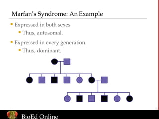
- 1. www.BioEdOnline.org Marfan’s Syndrome: An Example Expressed in both sexes. Thus, autosomal. Expressed in every generation. Thus, dominant. BioEd Online
- 2. www.BioEdOnline.org Marfan’s: Genotype the Normal Individuals Assign codes for the alleles. Code “m” for the recessive normal allele. Code “M” for the dominant allele for Marfan’s syndrome. Normal individuals must be “mm.” BioEd Online
- 3. www.BioEdOnline.org Marfan’s: Genotype the Affected Individuals Affected individuals must have at least one “M.” BioEd Online
- 4. www.BioEdOnline.org Marfan’s: Parent-Offspring Relationships Possibilities for #1 and #2: Heterozygote (Mm) or homozygous for “M?” If “MM,” all offspring from a normal mate should be affected. Therefore, both must be heterozygotes. BioEd Online
- 5. www.BioEdOnline.org Marfan’s: Parental Genotypes Known “M” must have come from the mother. The father can contribute only “m.” Thus, the remaining genotypes are “Mm.” BioEd Online
- 6. www.BioEdOnline.org Albinism: An Example Expressed in both sexes at approximately equal frequency. Thus, autosomal. Not expressed in every generation. Thus, recessive. BioEd Online
- 7. www.BioEdOnline.org Albinism: Genotype the Affected Individuals Assign codes for the alleles. Code “A” for the dominant normal allele. Code “a” for the recessive allele for albinism. Affected individuals must be homozygous for “a.” First generation parents must be “Aa” because they have normal phenotypes, but affected offspring. BioEd Online
- 8. www.BioEdOnline.org Albinism: Genotype the Normal Individuals Normal individuals must have at least one “A.” BioEd Online
- 9. www.BioEdOnline.org Albinism: Parent-Offspring Relationships #1 must transmit “a” to each offspring. The “A” in the offspring must come from the father. Normal father could be either heterozygous or homozygous for an “A.” ** BioEd Online
- 10. www.BioEdOnline.org Albinism: Parental Genotypes are Known Both parents are heterozygous. Normal offspring could have received an “A” from either parent, or from both. BioEd Online
- 11. www.BioEdOnline.org Albinism: One Parental Genotype is Known Only the genotype of the offspring expressing albinism are known. Normal offspring must have received an “a” from their affected father. BioEd Online
- 12. www.BioEdOnline.org Hairy Ears: An Example Only males are affected. All sons of an affected father have hairy ears. Thus, hairy ears is Y-linked. BioEd Online
- 13. www.BioEdOnline.org Hairy Ears: Female Sex Determination All females are XX. BioEd Online
- 14. www.BioEdOnline.org Hairy Ears: Male Sex Determination All males are XY. BioEd Online
- 15. www.BioEdOnline.org Hairy Ears: Gene on the Y Chromosome Code “H” indicates the allele on the Y chromosome for hairy ears. BioEd Online
- 16. www.BioEdOnline.org Hairy Ears: Wild-Type Allele for Normal Ears Code “+” indicates the allele on the Y chromosome for normal ears. BioEd Online
- 17. www.BioEdOnline.org Hemophilia: An Example In this pedigree, only males are affected, and sons do not share the phenotypes of their fathers. Thus, hemophilia is linked to a sex chromosome–the X. Expression of hemophilia skips generations. Thus, it is recessive. Extensive bruising of the left forearm and hand in a patient with hemophilia. BioEd Online
- 18. www.BioEdOnline.org Hemophilia: Expression of the Female Sex Chromosomes All females are XX. BioEd Online
- 19. www.BioEdOnline.org Hemophilia: Expression of Male Sex Chromosomes All males are XY. BioEd Online
- 20. www.BioEdOnline.org Hemophilia: Genotype the Affected Individuals Assign codes for the alleles. Code “H” for the recessive hemophilia allele. Code “+” for the wild-type normal allele. Affected individuals must have an “H” on an X chromosome. BioEd Online
- 21. www.BioEdOnline.org Hemophilia: Father-Daughter Relationship All daughters of an affected father receive an X chromosome with the “H” allele. BioEd Online
- 22. www.BioEdOnline.org Hemophilia: Genotyping the Normal Individuals Normal individuals must have at least one X chromosome with the wild-type allele, “+.” BioEd Online
- 23. www.BioEdOnline.org Hemophilia: Homozygous or Heterozygous? Only males affected Not Y-linked Skips a generation: recessive X-linked BioEd Online
Notas do Editor
- Marfan’s Syndrome: An Example Marfan’s Syndrome is an example of an inherited disorder that can be followed in a pedigree. People expressing Marfan’s Syndrome have hyper-elastic joints and elongated bones (among other features), as depicted in the photo. Note that in this pedigree, the expression of the trait has no sex-specific pattern. Both males and females express Marfan’s syndrome (depicted by black circles or squares) at approximately equal frequency. Thus, we can assume the gene that causes Marfan’s syndrome is autosomal. Next, note that the trait is expressed in every generation. Therefore, we assume that an allele for Marfan’s syndrome is dominant to a normal allele. An allele is an alternate form of a gene. In the this case, for example, one form (or allele) of the gene is expressed as Marfan’s Syndrome and the other allele produces normal joints and bone length. Reference Campbell, N. E. & Reece, J. B. (2002). Biology (6th ed.). San Francisco: Benjamin Cummings. Image Reference Young, M. (2005). Pedigree chart. Houston, TX: Baylor College of Medicine, Center For Educational Outreach.
- Marfan’s: Genotype the Normal Individuals The first step for working out the pedigree for an autosomal dominant trait is to genotype the normal individuals. Here, we are using an arbitrary coding system of “m” for the recessive normal allele and “M” for the dominant allele that confers Marfan’s syndrome. As noted in the previous slide, an allele is an alternate form of a gene. All normal individuals (purple circles and squares) must be homozygous for the “m” allele. Thus, we write the appropriate genotype below each normal individual in the pedigree. In general, genotype refers to the genetic makeup of an individual. Geneticists also use the term to refer to the process of identifying the alleles present in an individual. Phenotype refers to the observable characteristics of an organism. Reference Campbell, N. E. & Reece, J. B. (2002). Biology (6th ed.). San Francisco: Benjamin Cummings. Image Reference Young, M. (2005). Pedigree chart. Houston, TX: Baylor College of Medicine, Center For Educational Outreach.
- Genotype the Affected Individuals The next step is to genotype the affected individuals. Since only one “M” allele is sufficient to cause Marfan’s syndrome, the genotype of any individual expressing the trait must have at least one “M” (see the notations in black). Reference Campbell, N. E. & Reece, J. B. (2002). Biology (6th ed.). San Francisco: Benjamin Cummings. Image Reference Young, M. (2005). Pedigree chart. Houston, TX: Baylor College of Medicine, Center For Educational Outreach.
- Parent – Offspring Relationships Now we will begin to consider parent – offspring relationships. At first glance, we do not know if individual #1 is heterozygous or homozygous for the “M” allele. However, note that when she mates with a normal male, she produces some normal offspring. Thus, we can conclude that she is a heterozygote. If she were homozygous for “M,” then every one of her offspring would receive the allele for Marfan’s syndrome. The same logic applies to assigning the genotype to individual #2. Reference Campbell, N. E. & Reece, J. B. (2002). Biology (6th ed.). San Francisco: Benjamin Cummings. Image Reference Young, M. (2005). Pedigree chart. Houston, TX: Baylor College of Medicine, Center For Educational Outreach.
- Parental Genotypes Known We now can focus on families where the parental genotypes are known. We know the remaining affected individuals must have received their “M” from their mothers, since their fathers were normal (“mm”” genotypes). Thus, their other allele must have come from their fathers. Since the fathers are homozygous for “m,” each one of their offspring must have received an “m” allele. Reference Campbell, N. E. & Reece, J. B. (2002). Biology (6th ed.). San Francisco: Benjamin Cummings. Image Reference Young, M. (2005). Pedigree chart. Houston, TX: Baylor College of Medicine, Center For Educational Outreach.
- Albinism: An Example People expressing albinism lack pigmentation in their eyes and skin (among other features). As with the pedigree for Marfan’s syndrome, expression of the albino trait has no sex-specific pattern. Both males and females express albinism at approximately equal frequency (the black circles and squares). Thus, we can assume the gene that causes albinism is autosomal. Note that the trait is not expressed in every generation. Neither parent in the first generation was albino, but the offspring were. Also, individual #1 expressed albinism, but this trait was not found in any of her offspring. Therefore, we can assume that an allele for albinism is recessive to a normal allele. Reference Campbell, N. E. & Reece, J. B. (2002). Biology (6th ed.). San Francisco: Benjamin Cummings. Image Reference: Young, M. (2005). Pedigree chart. Houston, TX: Baylor College of Medicine, Center For Educational Outreach.
- Genotype the Affected Individuals The first step in working out the pedigree of an autosomal recessive trait is to genotype the affected individuals. Here, we are using an arbitrary coding system of “A” for the dominant normal allele and “a” for the recessive allele that confers albinism. Since both “a” alleles are required to cause albinism, the genotype of any individual expressing the trait must be homozygous for “a.” Because the parents of the first generation did not express albinism, we know they must have at least one “A” each. And since we know those same parents also produced affected offspring, we know they also must have at least one “a” each. Thus, the parents in the first generation must be heterozygotes. Reference Campbell, N. E. & Reece, J. B. (2002). Biology (6th ed.). San Francisco: Benjamin Cummings. Image Reference Young, M. (2005). Pedigree chart. Houston, TX: Baylor College of Medicine, Center For Educational Outreach.
- Genotype the Normal Individuals The next step is to genotype the normal individuals. Since only one “A” allele is sufficient for normal pigmentation, the genotype of any normal individual must have at least one “A” (see notations in black). Reference Campbell, N. E. & Reece, J. B. (2002). Biology (6th ed.). San Francisco: Benjamin Cummings. Image Reference Young, M. (2005). Pedigree chart. Houston, TX: Baylor College of Medicine, Center For Educational Outreach.
- Parent – Offspring Relationships Now, we will consider more of the parent – offspring relationships, starting with individual #1’s family. We already know that individual #1 is a homozygote for “a” because she expresses albinism. We also already have determined that her offspring must have at least one “A” because they all are normal. Thus, we can conclude that the offspring must have received their normal allele from their father, because the mother can contribute only an “a.” All offspring in this family are heterozygotes. We do not have enough information to determine, for sure, whether the father (individual #2) is a heterozygote or is homozygous for the normal allele. If we consider that albinism is a rare genetic trait, however, we could fairly safely guess that the father is homozygous for the normal allele because we expect heterozygotes to be rare in the population. Still, we cannot rule out that the father is carrying the “a” allele. Reference Campbell, N. E. & Reece, J. B. (2002). Biology (6th ed.). San Francisco: Benjamin Cummings. Image Reference Young, M. (2005). Pedigree chart. Houston, TX: Baylor College of Medicine, Center For Educational Outreach.
- Parental Genotypes are Known The genotype of parents in the first generation is known. In this case, we cannot determine whether the normal offspring are heterozygotes or are homozygous for the “A” allele. We know they must have at least one “A,” but we cannot tell which parent gave them the “A” or if they received an “A” allele from both parents. Reference Campbell, N. E. & Reece, J. B. (2002). Biology (6th ed.). San Francisco: Benjamin Cummings. Image Reference Young, M. (2005). Pedigree chart. Houston, TX: Baylor College of Medicine, Center For Educational Outreach.
- One Parental Genotype Known For the offspring genotypes from a mating between #1 and #2, we know at least one parental genotype. Even though we are unsure of the mother’s genotype, we know that the affected father must have passed on an “a” allele. Thus, the normal offspring must be heterozygotes. Reference Campbell, N. E. & Reece, J. B. (2002). Biology (6th ed.). San Francisco: Benjamin Cummings. Image Reference Young, M. (2005). Pedigree chart. Houston, TX: Baylor College of Medicine, Center For Educational Outreach.
- Hairy Ears Hairy ears is a rare condition with a simple genetic basis. Only males express the trait. Moreover, all sons have the same phenotype as their fathers, whether expressing hairy ears or not. Thus, we can conclude that the gene for hairy ears is on the Y-chromosome. Reference Campbell, N. E. & Reece, J. B. (2002). Biology (6th ed.). San Francisco: Benjamin Cummings. Image Reference Young, M. (2005). Pedigree chart. Houston, TX: Baylor College of Medicine, Center For Educational Outreach.
- Hairy Ears: Female Sex Determination In considering the expression of the sex chromosomes, all females must be XX. Reference Campbell, N. E. & Reece, J. B. (2002). Biology (6th ed.). San Francisco: Benjamin Cummings. Image Reference Young, M. (2005). Pedigree chart. Houston, TX: Baylor College of Medicine, Center For Educational Outreach.
- Hairy Ears: Male Sex Determination Under the human sex determination system, all males are XY. Reference Campbell, N. E. & Reece, J. B. (2002). Biology (6th ed.). San Francisco: Benjamin Cummings. Image Reference Young, M. (2005). Pedigree chart. Houston, TX: Baylor College of Medicine, Center For Educational Outreach.
- Hairy Ears: Gene on the Y Chromosome Here, we denote “H” for the hairy ear gene on the Y chromosome. Reference Campbell, N. E. & Reece, J. B. (2002). Biology (6th ed.). San Francisco: Benjamin Cummings. Image Reference Young, M. (2005). Pedigree chart. Houston, TX: Baylor College of Medicine, Center For Educational Outreach.
- Wild-Type Allele for Normal Ears It is customary to use a “+” to denote wild-type, or normal, alleles. Thus, “+” will indicate the wild-type allele on the Y chromosome for normal ears. The daughters do not need to be genotyped because they do not have Y chromosomes. Reference Campbell, N. E. & Reece, J. B. (2002). Biology (6th ed.). San Francisco: Benjamin Cummings. Image Reference Young, M. (2005). Pedigree chart. Houston, TX: Baylor College of Medicine, Center For Educational Outreach.
- Hemophilia Hemophilia is a condition of excessive bleeding caused by missing clotting factors in the blood. Hemophiliacs are prone to bruising, as illustrated in the photo here, and to other, potentially fatal, risk factors. In this pedigree, there is a trend for only males to express the trait strongly suggesting the role of sex chromosomes. However, the sons do not share the phenotypes of their fathers, so the Y chromosome is not a likely candidate. Thus, we can conclude that the gene for hemophilia is on the X chromosome. Since the trait skips generations, we can assume that an allele for hemophilia is recessive to an allele for normal blood clotting factors. Although rare, a female can be afflicted if she inherits an allele for hemophilia on both X chromosomes. Reference Campbell, N. E. & Reece, J. B. (2002). Biology (6th ed.). San Francisco: Benjamin Cummings. Image Reference Young, M. (2005). Pedigree chart. Houston, TX: Baylor College of Medicine, Center For Educational Outreach. Hemophilia A. Retrieved 08-10-2006 from http://medgen.genetics.utah.edu/index.html
- Expression of Sex Chromosomes In human expression of the sex chromosomes, all females must be XX. Reference Campbell, N. E. & Reece, J. B. (2002). Biology (6th ed.). San Francisco: Benjamin Cummings. Image Reference Young, M. (2005). Pedigree chart. Houston, TX: Baylor College of Medicine, Center For Educational Outreach.
- Hemophilia: Male Sex Chromosomes Under the human sex determination system, all males are XY. Reference Campbell, N. E. & Reece, J. B. (2002). Biology (6th ed.). San Francisco: Benjamin Cummings. Image Reference Young, M. (2005). Pedigree chart. Houston, TX: Baylor College of Medicine, Center For Educational Outreach.
- Hemophilia: Genotype the Affected Individuals The first step in working out the pedigree of a recessive trait is to genotype the affected individuals. Here, we are using a coding system and adding a “tag” of “H” for the recessive hemophilia allele and “+” for the wild-type allele for normal blood clotting factors. We know that affected males must have the “H” allele on the X chromosome they inherited from their mothers because they had to receive a Y chromosome from their fathers. Thus, we also know that the mother (see individual #1) of an affected son must have the “H” allele on one of her X chromosomes. Reference Campbell, N. E. & Reece, J. B. (2002). Biology (6th ed.). San Francisco: Benjamin Cummings. Image Reference Young, M. (2005). Pedigree chart. Houston, TX: Baylor College of Medicine, Center For Educational Outreach.
- Hemophilia: Father – Daughter Relationship Because of the father-daughter relationship, all daughters of a hemophiliac father must receive an X chromosome that carries the disease. When the father passes on his Y sex chromosome, he produces a son. Reference Campbell, N. E. & Reece, J. B. (2002). Biology (6th ed.). San Francisco: Benjamin Cummings. Image Reference Young, M. (2005). Pedigree chart. Houston, TX: Baylor College of Medicine, Center For Educational Outreach.
- Hemophilia: Genotyping the Normal Individuals Genotyping normal individuals is straightforward, since every normal individual must have at least one X chromosome with the wild-type normal allele tagged, “+.” Reference Campbell, N. E. & Reece, J. B. (2002). Biology (6th ed.). San Francisco: Benjamin Cummings. Image Reference Young, M. (2005). Pedigree chart. Houston, TX: Baylor College of Medicine, Center For Educational Outreach.
- Homozygous or Heterozygous? We do not have enough information to tell if the remaining normal females are homozygous or heterozygous. The mother in the first generation may carry the hemophilia allele on one of her X chromosomes, and may have “gotten lucky” by transmitting only wild-type alleles to her daughters. If she were a heterozygote, you would expect half of her daughters to have hemophilia, since they all receive a hemophilia allele from their father. Since hemophilia is a rare disease, heterozygotes carrying the disease should be rare in the population. Therefore, it is likely that the mother is homozygous for the wild-type allele, but we cannot know for sure. We know the daughters in the last generation must have at least one wild-type allele, which they could have gotten from either parent. Thus, we do not know if daughters received the father’s normal allele and the mother’s disease allele, or if they are homozygous for the wild-type allele. Reference Campbell, N. E. & Reece, J. B. (2002). Biology (6th ed.). San Francisco: Benjamin Cummings. Image Reference Young, M. (2005). Pedigree chart. Houston, TX: Baylor College of Medicine, Center For Educational Outreach.